Here, we describe the large-scale production of adipose-derived stromal/stem cell (ASC) spheroids using an automated pipetting system to seed the cell suspension, thus ensuring homogeneity of spheroid size and shape. These ASC spheroids can be used as building blocks for 3D bioprinting approaches.
Adipose-derived stromal/stem cells (ASCs) are a subpopulation of cells found in the stromal vascular fraction of human subcutaneous adipose tissue recognized as a classical source of mesenchymal stromal/stem cells. Many studies have been published with ASCs for scaffold-based tissue engineering approaches, which mainly explored the behavior of these cells after their seeding on bioactive scaffolds. However, scaffold-free approaches are emerging to engineer tissues in vitro and in vivo, mainly by using spheroids, to overcome the limitations of scaffold-based approaches.
Spheroids are 3D microtissues formed by the self-assembly process. They can better mimic the architecture and microenvironment of native tissues, mainly due to the magnification of cell-to-cell and cell-to-extracellular matrix interactions. Recently, spheroids are mainly being explored as disease models, drug screening studies, and building blocks for 3D bioprinting. However, for 3D bioprinting approaches, numerous spheroids, homogeneous in size and shape, are necessary to biofabricate complex tissue and organ models. In addition, when spheroids are produced automatically, there is little chance for microbiological contamination, increasing the reproducibility of the method.
The large-scale production of spheroids is considered the first mandatory step for developing a biofabrication line, which continues in the 3D bioprinting process and finishes in the full maturation of the tissue construct in bioreactors. However, the number of studies that explored the large-scale ASC spheroid production are still scarce, together with the number of studies that used ASC spheroids as building blocks for 3D bioprinting. Therefore, this article aims to show the large-scale production of ASC spheroids using a non-adhesive micromolded hydrogel technique spreading ASC spheroids as building blocks for 3D bioprinting approaches.
Spheroids are considered a scaffold-free approach in tissue engineering. ASCs are capable of forming spheroids by the self-assembly process. The spheroid's 3D microarchitecture increases the regenerative potential of ASCs, including the differentiation capacity into multiple lineages1,2,3. This research group has been working with ASC spheroids for cartilage and bone tissue engineering4,5,6. More importantly, spheroids are considered building-blocks in the biofabrication of tissues and organs, mainly due to their fusion capacity.
The use of spheroids for tissue formation depends on three main points: (1) the development of standardized and scalable robotic methods for their biofabrication7, (2) the systematic phenotyping of tissue spheroids8, (3) the development of methods for the assembly of 3D tissues9. These spheroids can be formed with different cell types and obtained through various methods, including hanging drop, reaggregation, microfluidics, and micromolds8,9,10. Each of these methods has advantages and disadvantages related to the homogeneity of size and shape of the spheroids, recovery of the spheroids after formation, the number of spheroids produced, process automation, labor intensity, and costs11.
In the micromold method, the cells are dispensed and deposited at the bottom of the micromold because of gravity. The non-adhesive hydrogel does not allow the cells to adhere to the bottom, and cell-to-cell interactions lead to the formation of a single spheroid per recession8,12. This biofabrication method generates spheroids of homogeneous and controlled size, can be robotized for large-scale production in a time-efficient manner with minimal effort, and has good cost-effectiveness-critical factors in the design of a biofabrication of tissue spheroid7,8. This method can be applied to form spheroids of any cell lineage to prepare a new tissue type with predictable, optimal, and controllable characteristics8.
Biofabrication is defined as "the automated generation of biologically functional products with structural organization …"13. Therefore, the automated production of spheroids is considered the first mandatory step for developing a biofabrication line, which continues in the 3D bioprinting process and finishes in the full maturation of the bioprinted tissue by spheroid fusion. In this study, to improve the scalability of ASC spheroid biofabrication, we use an automated pipetting system to seed the cell suspension, thus ensuring the homogeneity of spheroid size and shape. This paper shows that it was possible to produce a large number (thousands) of spheroids needed for 3D bioprinting approaches to biofabricate more complex tissue models.
The ASCs used in this study were previously isolated from healthy human donors and cryopreserved as described14 according to the Research Ethics Committee of Clementino Fraga Filho University Hospital, Federal University of Rio de Janeiro, Brazil (25818719.4.0000.5257). See Table of Materials for details regarding all the materials and equipment used in this study.
1. Trypsinization of ASC monolayer at passage three
2. Micromolded non-adherent agarose hydrogel fabrication
3. Biofabrication of ASC spheroids using the automated pipetting system
The automatic pipette system can seed the ASC cell suspension into 12 wells of one 12-well plate in 15 min. Using the 81 micromolded non-adherent hydrogel will produce 972 spheroids at the end of the protocol. Using the 256 micromolded non-adherent hydrogel will produce 3,072 spheroids at the end of the protocol. ASC spheroids were analyzed for the homogeneity of their size and shape. ASC spheroids from micromolds with 81 recessions showed homogeneous diameter during the culture period in contrast to ASC spheroids from micromolds with 256 recessions (Figure 1C,D). The ratio of the smallest and largest diameters (sphericity) was close to 1 in ASC spheroids from micromolds with 81 and 256 recessions (Figure 1E,F). The results from viability, morphology, and force (Figure 2) analyses provide evidence for the successful, large-scale production of ASC spheroids.
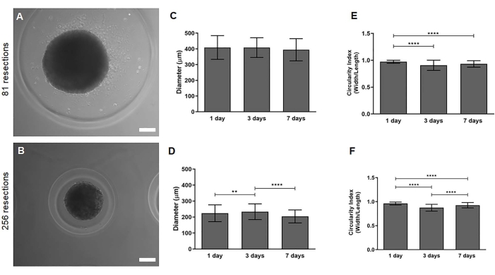
Figure 1: ASC spheroids showed high diameter reproducibility. Representative optical micrographs of ASC spheroids from micromolded non-adhesive hydrogels with (A) 81 circular recessions and (B) 256 circular recessions. (C,D) Spheroid diameter at 1, 3, and 7 days from five micromolded non-adhesive hydrogels with 81 and 256 circular recessions, respectively. (E,F) Ratio of the smallest and largest diameters from micromolded non-adhesive hydrogels with 81 and 256 circular recessions, respectively. To measure the smallest and largest diameters of the spheroids, images were acquired weekly using an optical microscope equipped with a digital camera. Width and length were measured using ImageJ software. A diameter ratio of each spheroid was obtained by dividing width by length (spheroid sphericity). A total of 85 spheroids were measured from micromolded, non-adhesive hydrogels with 81 circular recessions, and a total of 160 spheroids were measured from micromolded, non-adhesive hydrogels with 256 circular recessions. The data are expressed as mean ± standard deviation. The asterisks indicate values of P obtained by ANOVA nonparametric and unpaired followed by multiple comparisons (**P < 0.001; ****P < 0.0001). Scale bars = 100 µm. Abbreviation: ASC = adipose tissue stem/stromal cells. Please click here to view a larger version of this figure.
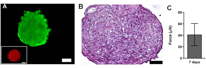
Figure 2: ASC spheroids showed high cell viability. (A) Fluorescence micrographs of viability assay of ASC spheroids at day 7 from micromolded, non-adhesive hydrogels with 256 circular recessions. Live and dead cells are observed in green and red, respectively. Death control is shown in the inset. (B) Section of an ASC spheroid stained by hematoxylin and eosin from micromolded, non-adhesive hydrogels with 81 circular recessions. The fixed spheroids samples were embedded in paraffin, and samples were cut into 5 µm thickness sections using a microtome. (C) Compressive resistance force measured in µN of ASC spheroids at day 7. The data were collected from five spheroids and expressed as mean ± standard deviation, according to previously described methodology5. The asterisks indicate the value of P obtained by two-way ANOVA, nonparametric and unpaired, followed by multiple comparisons. Scale bars = 50 µm. Abbreviation: ASC = adipose tissue stem/stromal cells. Please click here to view a larger version of this figure.
This paper presents the large-scale generation of ASC spheroids using an automated pipette system. The critical step of the protocol is to precisely set up the software to ensure the correct volume of cell suspension, speed, and distance for pipetting. The parameters described in the protocol were determined after a number of trials to optimize the dispensing of the ASC cell suspension into the wells of 12-well plates containing the micromolded, non-adherent hydrogels. The optimization was evaluated by measuring the diameter and sphericity of the spheroids. The area and height of the micromolded non-adherent hydrogels were used to calculate the ideal speed to dispense the cell suspension. If the cell suspension is seeded by the equipment at a different dispensing speed, this will directly affect the formation of the spheroids. Hence, these are crucial parameters to be optimized after trials with the equipment. This protocol has some limitations. Only seeding of the ASCs in the non-adherent hydrogel in the micromold can be automated, followed by the addition of cell culture medium. The features of the system are limited by the version of the equipment and the software. However, the main advantage of this reproducible approach is to produce up to 3,072 ASC spheroids-homogeneous in size and shape and viable-with minor risks of human errors or contamination. The large number of biofabricated ASC spheroids can be directly loaded into a 3D bioprinter to produce more complex tissue models.
In general, it is widely known that accurate and automated pipetting plays a central role in the automation of cell culture. Automated cell culture systems enable large-scale production and increase technical accuracy, reproducibility, and process efficiency15. Therefore, automated systems can facilitate large-scale production, which is common and beneficial for industrial environments16,17. However, there are few studies describing the development of protocols to automate 3D cell culture systems7,18,19,20,21.
Mehesz and collaborators7 and Meseguer-Ripolles and colleagues21 used a similar protocol to biofabricate ASC spheroids. In the study by Mehesz et al.7, the authors used the same automated pipetting system. However, their protocol produced only 96 spheroids, whereas the protocol developed in the present work was able to produce up to 3,072 ASC spheroids at once, homogeneous in size and shape. Meseguer-Ripolles et al.21 also used an automated pipetting system to produce up to 73 liver spheroids. Another approach being discussed to fabricate spheroids on a large scale nowadays is via microfluidics devices. However, the articles published so far have explored the technology to develop better devices that can produce the spheroids rather than increasing the scale for spheroid fabrication7,20.
Another method that can be used to fabricate a high number of spheroids is by using spinner flasks. However, one main issue regarding this technique is the difficulty in controlling the size and shape of the spheroids22,23. In addition, 3D bioprinting was also already applied to biofabricate spheroids on a large scale. In the study by Daly and Kelly19, the authors used an inkjet technique to produce a large number of spheroids in polycaprolactone microchambers. The spheroids were viable and homogeneous in size and shape.
The main application of the protocol described in this work is to biofabricate ASC spheroids on a large scale needed for 3D bioprinting. As discussed by Mironov and collaborators24, thousands of spheroids will be necessary to biofabricate complex tissues and organ models. The automated method described in this protocol will allow for the fabrication of a large number of ASC spheroids that can be loaded directly in the 3D bioprinter. It is also possible to differentiate these spheroids to a desired mesodermal pathway to engineer musculoskeletal tissues. De Moor and collaborators25 developed a high-throughput method to fabricate a large number of vascularized spheroids for future 3D bioprinting applications.
The equipment can be modified to improve the protocol. More recent versions of the equipment allow the use of a larger number of plates, which could enhance the number of ASC spheroids produced. This same recent version allows the accurate stacking of the plates, and this also would provide a better efficiency of the protocol. In conclusion, we successfully showed a protocol allowing the biofabrication up to 3,072 ASC spheroids homogeneous in size and shape in 15 min, and that this is considered the first step to build a biofabrication line for Tissue Engineering applications.
The authors have nothing to disclose.
We thank the National Institute of Metrology, Quality and Technology (INMETRO, RJ, Brazil) for the use of their facilities. This study was partially supported by the Carlos Chagas Filho Foundation for Research Support of the State of Rio de Janeiro (Faperj) (finance Code: E26/202.682/2018 and E-26/010.001771/2019, the National Council for Scientific and Technological Development (CNPq) (finance code: 307460/2019-3), and the Office of Naval Research (ONR) (finance code: N62909-21-1-2091). This work was partially supported by the National Center of Science and Technology on Regenerative Medicine-INCT Regenera (http://www.inctregenera.org.br/).
12-well plastic plate | Corning | 3512 | |
50 mL centrifuge tube | Corning | CLS430828 | |
EpMotion 5070 | Eppendorf | 5070000282 | |
epT.I.P.S. Motion | Eppendorf | 30015231 | |
ethylenediaminetetraacetic acid (EDTA) | Invitrogen | 15576028 | |
fetal bovine serum (FBS) | Gibco | 10082147 | |
Low Glucose Dulbecco's Modified Eagle Medium (DMEM LOW) | Gibco | 31600034 | |
MicroTissues 3D Petri Dish micro-mold spheroids – 16 x 16 array | Sigma | Z764000 | |
MicroTissues 3D Petri Dish micro-mold spheroids – 9 x 9 array | Sigma | Z764019 | |
phosphate saline buffer (PBS) | Sigma | 806552 | |
sodium chloride (NaCl) | Sigma | S8776 | |
tissue culture flask | Corning | 430720U | |
trypan | Lonza | 17-942E | |
trypsin | Gibco | 27250018 | |
ultrapure agarose | Invitrogen | 16500100 |