The present protocol describes the echocardiographic assessment of left ventricular morphology, function, and coronary blood flow in 7-day old neonate mice.
Echocardiography is a non-invasive procedure that enables the evaluation of structural and functional parameters in animal models of cardiovascular disease and is used to assess the impact of potential treatments in preclinical studies. Echocardiographic studies are usually conducted in young adult mice (i.e., 4-6 weeks of age). The evaluation of early neonatal cardiovascular function is not usually performed because of the small size of the mouse pups and the associated technical difficulties. One of the most important challenges is that the short length of the pups' limbs prevents them from reaching the electrodes in the echocardiography platform. Body temperature is the other challenge, as pups are very susceptible to changes in temperature. Therefore, it is important to establish a practical guide for performing echocardiographic studies in small mouse pups to help researchers detect early pathological changes and study the progression of cardiovascular disease over time. The current work describes a protocol for performing echocardiography in mouse pups at the early age of 7 days old. The echocardiographic characterization of cardiac morphology, function, and coronary flow in neonatal mice is also described.
The overall goal of this protocol is to examine cardiac morphology, function, and coronary artery flow in 7-day-old neonatal mouse pups using echocardiography. The rationale behind the development of this technique is to determine early changes in coronary flow and cardiac function in mouse models of cardiac disease1. The non-invasive nature of echocardiography is advantageous because it allows researchers to assess cardiovascular function under physiological conditions and provides researchers with a screening tool for the study of targeted therapies to treat cardiovascular diseases2,3. Traditionally, echocardiographic studies are conducted with young adult mice (4-6 weeks); however, some mice models (i.e., genetically modified models) already exhibit pathological changes and cardiac dysfunction at this age. Therefore, cardiac research using animal models has focused primarily on therapeutic agents that ameliorate or treat cardiac dysfunction. In contrast, more recently, research efforts have been redirected to focus on preventive measures and early interventions in cardiac diseases4.
Previous studies have described the use of echocardiography to measure cardiac function in models of myocardial infarction in neonatal mice5,6; however, these studies failed to measure coronary flow and, most importantly, failed to record an electrocardiogram (ECG) and heart rate (HR) data during the procedure, most likely due to the small size of the pups' limbs, which could not reach the electrode pads. We overcome this problem in this protocol by attaching aluminum foil to the limbs to enable them to reach the electrode pads and create an ECG circuit. Furthermore, this protocol describes and characterizes coronary artery flow in neonatal mice.
This study obtained B-mode and M-mode images in parasternal long and short axis views to measure structural and functional parameters2,3. The morphological parameters included left atrial dimensions, left ventricular (LV) dimensions, LV wall thickness, LV mass, and relative wall thickness (RWT). The functional parameters included ejection fraction (EF), fractional shortening (FS), cardiac output (CO), and velocity of circumferential fiber shortening (Vcf). Pulse wave (PW) Doppler was used to measure aortic flow in the parasternal short-axis (PSAX) view and to measure mitral blood flow in the apical four-chamber view. The apical four-chamber view was also used to perform Tissue Doppler at the septal part of the mitral valve annulus. Coronary flow at the left anterior descending (LAD) coronary artery was also examined using a modified parasternal long-axis (PLAX) view. Coronary flow reserve (CFR) was calculated after a stress challenge induced by increased isoflurane concentration.
The present protocol demonstrates that echocardiographic studies can be performed at a very early age in neonatal mice, thus allowing early recognition of cardiac pathologies and longitudinal follow-up studies of LV hemodynamics and coronary flow parameters in different mice models. This technique can be used to study the role of genetic alterations or pharmacological interventions in cardiac function at early postnatal ages. Moreover, the protocol provides a valuable tool for determining the onset of cardiac diseases early in life, thus enabling researchers to unlock the molecular mechanisms underlying the initial stages of cardiac diseases in different mouse models.
All experiments were approved by the Animal Care and Use Committee of the University of Illinois at Chicago. For the experiments, 7-day-old FVB/N mice were used. The protocol is divided into mouse preparation, echocardiography image acquisition, and post-imaging animal care.
1. Mouse preparation
2. Echocardiographic image acquisition and analyses
3. Post-imaging animal monitoring and care
This study used 7-day-old mouse pups to characterize cardiac morphology, function, and coronary artery flow. Mouse handling needs to be done with care, and the mouse platform must be adapted for the small size of the pups, as described in Figure 1. A representative image of the PLAX view is shown in Figure 2A and Supplementary Video 1. In this view, M-mode was used to measure the left atrium (LA) diameter (Figure 2B). The PSAX view (Supplementary Video 2) was used to measure the left ventricular chamber dimensions (Figure 3A), pulmonary flow (Figure 3B), and aortic flow (Figure 3C). The apical four-chamber view (Supplementary Video 3 and Figure 4A) was used to examine the blood flow velocities across the mitral valve (Figure 4B), as well as the myocardial relaxation and contraction velocities at the mitral valve annulus (Figure 4C).
The modified PLAX view was used to examine the LAD coronary artery flow parameters (Figure 5A,B and Supplementary Video 4), as previously described15,16,21. In Figure 5C, representative results of the diastolic peak CFV, mean CFV, and VTI are shown at a resting flow state (1.5% isoflurane) and 5 min after increasing isoflurane to 2.5% to induce maximal vasodilation. The increased values of these parameters (i.e., peak CFV, mean CFV, and VTI) 5 min after isoflurane increment confirm the expected response to hyperemia in the neonatal mice18. CFR was calculated as the ratio of diastolic peak CFV during maximal vasodilation induced by 2.5% isoflurane to diastolic peak CFV at a baseline of 1.5% isoflurane concentration18. All measurements and calculations were averaged across 3 consecutive cycles, and the representative results are shown in Table 1 and Table 2.
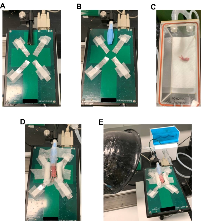
Figure 1: Echocardiographic platform setup and 7-day-old mouse pup preparation. (A) Aluminum foil strips are placed on the platform electrode pads and secured with tape. (B) The glove finger is cut and adapted to fit the isoflurane/oxygen nose cone. (C) The pup is placed in the isoflurane induction chamber, and the isoflurane delivery starts at 2.5% concentration. (D) The pup is placed in a supine position with paws touching the aluminum foil strips and secured with tape. Two rolls of gauze are used to keep the acoustic gel in place. (E) A heating lamp is placed close to the pup to maintain its body temperature. Please click here to view a larger version of this figure.
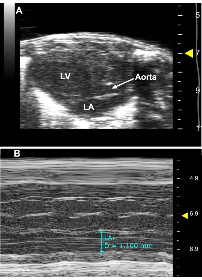
Figure 2: Parasternal long-axis (PLAX) view of the left ventricle. (A) B-mode images of the left ventricular chamber (LV), left atrium (LA), and the aorta. (B) M-mode is used to measure the LA diameter. Please click here to view a larger version of this figure.
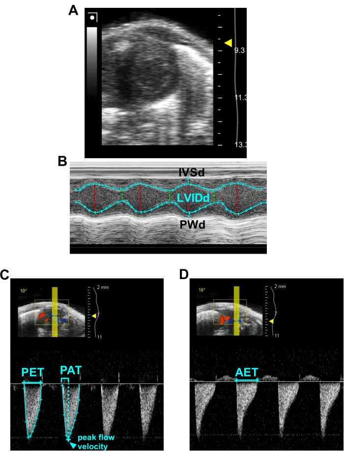
Figure 3: Parasternal short-axis (PSAX) view of the left ventricle. (A) B-mode images of the left ventricular chamber. (B) M-mode sample of the interventricular septum at diastole (IVSd), left ventricular internal diameter at diastole (LVIDd), and posterior wall thickness at diastole (PWd). (C) Representative images of the pulmonary peak flow velocity, pulmonary ejection time (PET), and pulmonary acceleration time (PAT). (D) Representative images of the aortic ejection time (AET). Please click here to view a larger version of this figure.
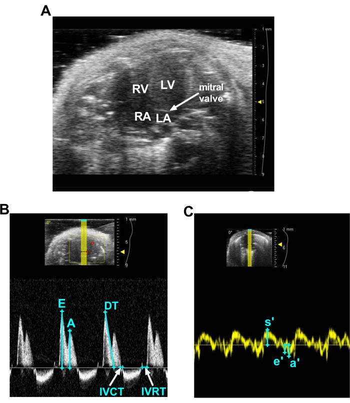
Figure 4: Apical four-chamber view. (A) B-mode image of the left ventricle (LV), right ventricle (RV), left atrium (LA), and right atrium (RA). (B) Representative images of the maximal blood inflow velocity in the early phase of diastole (E), maximal blood inflow velocity in the late phase of diastole (A), deceleration time (DT), isovolumetric contraction time (IVCT), and isovolumetric relaxation time (IVRT). (C) Tissue Doppler sample images of the peak myocardial relaxation velocity in the early diastolic filling (e'), late diastolic filling (a'), and peak systolic myocardial velocity (s'). Please click here to view a larger version of this figure.
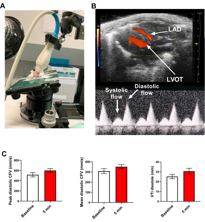
Figure 5: Modified parasternal long-axis view. (A) Platform and transducer position in modified parasternal long-axis view. (B) Visualization and recording of left anterior descending (LAD) coronary artery flow. LVOT = left ventricular outflow tract. (C) Peak coronary flow velocity (CFV), mean CFV, and velocity-time integral (VTI) in diastole are measured at 1.5% isoflurane (baseline) and 5 min after increasing isoflurane concentration to 2.5%; 7-day old mice, N = 7; data presented as mean ± SD. Please click here to view a larger version of this figure.
Echocardiographic parameters | WT (n = 7) | |
Mean ± SD | ||
Morphology | LA (mm) | 1.25 ± 0.11 |
PWd (mm) | 0.40 ± 0.06 | |
LVIDd (mm) | 1.98 ± 0.34 | |
LV Mass (g) | 10.92 ± 3.53 | |
RWT | 0.39 ± 0.09 | |
Systolic Function | HR (bpm) | 500.69 ± 40.04 |
EF(%) | 81.97 ± 10.76 | |
SV (ml) | 10.16 ± 3.44 | |
CO (ml/min) | 5.04 ± 1.53 | |
s’ (cm/s) | 16.16 ± 3.56 | |
Vcf (circ/s) | 10.50 ± 3.12 | |
Diastolic Function | E/A | 1.25 ± 0.11 |
E/e’ | 45.58 ± 11.44 | |
DT (s) | 23.97 ± 2.63 | |
IVRT (s) | 16.27 ± 2.11 |
Table 1: Echocardiographic assessment of left ventricular morphology and function in 7-day old mouse pups.
Coronary Flow Parameters | Baseline | 5 min | CFR | |
Isoflurane 1.5% | Isoflurane 2.5% | 5 min/baseline | ||
Diastole | Peak velocity (mm/s) | 516.58 ± 113.04 | 599.43 ± 101.34 | 1.18 ± 0.18 |
Mean velocity (mm/s) | 308.50 ± 63.44 | 351.50 ± 53.98 | ||
VTI (mm) | 25.23 ± 5.86 | 30.65 ± 7.75 | ||
Sytole | Peak velocity (mm/s) | 121.81 ± 40.52 | 163.13 ± 32.59* | |
Mean velocity (mm/s) | 84.82 ± 27.16 | 114.70 ± 21.84* | ||
VTI (mm) | 5.21 ± 1.84 | 7.76 ± 2.08* | ||
Heart rate (bpm) | 536.20 ± 128.90 | 540.80 ± 233.15 | ||
Respiratory rate (rpm) | 69.60 ± 15.89 | 38.80 ± 24.18 |
Table 2: Echocardiographic evaluation of coronary artery flow in 7-day old mouse pups. Seven-day old mice, N = 7; data presented as mean ± SD; the Student's t-test was used to analyze the data; *p < 0.05; CFR = coronary flow reserve; VTI = velocity time integral.
Supplementary Video 1: The parasternal long-axis view of the left ventricular outflow and left atrium. Please click here to download this Video.
Supplementary Video 2: The parasternal short-axis view of the left ventricular chamber. Please click here to download this Video.
Supplementary Video 3: The apical four-chamber view. Please click here to download this Video.
Supplementary Video 4: The modified parasternal long-axis view of left anterior descending coronary artery flow. Please click here to download this Video.
In the era of preventive medicine, early assessment of alterations in cardiovascular function is required to establish the onset of the disease and design appropriate interventional therapies. Mice are increasingly being used as preclinical models in cardiac research, and echocardiographic studies are typically conducted with young adult mice. However, to study the role of genetic alterations or pharmacological interventions in the early stages of cardiac diseases, echocardiographic imaging needs to be initiated earlier in life. Problematically, echocardiographic studies in neonate mice are technically challenging. In this study, we have established a protocol for performing echocardiographic measurements in mice as young as 7 days old. This is especially important for transgenic mouse models, in which the deletion or overexpression of a gene is believed to cause cardiovascular dysfunction. Early recognition of the cardiovascular abnormalities in these animal models allows researchers to design pharmacological treatments that prevent disease progression.
Due to the small size of the 7-day-old mice, some technical considerations in this protocol included maintaining their normal body temperatures and minimizing the length of the echocardiographic procedure. A heated platform, a heating lamp, and prewarmed acoustic gel were used to prevent hypothermia. Ideally, the animal's temperature should be monitored using a rectal probe; however, given the small size of the pups in this study, we were unable to use a rectal probe during the procedure. Moreover, hyperthermia is also a concern, and care needs to be taken to avoid the pups being in close proximity to the heating lamp. The duration of the echo procedure needs to be kept to less than 1 h to minimize major temperature variations and avoid the physiological effects of prolonged anesthesia22. Additionally, since the size of the echocardiographic probe is designed to image adult mice, using a thicker layer of acoustic gel is recommended to adjust the focal distance. It is also important to mention that the imaging system used in this study calculates respiration rate and heart rate from the ECG signal detected by the platform electrode pads (Table 2). As the ECG pads were extended to reach the pup's limbs using aluminum foil, the signal detected may have been distorted. Another problem encountered was that, by the end of the procedure, we noticed that the gel underneath the aluminum foil strips had dried out, which may have affected the conductivity and the ECG signal. Ideally, a platform with electrode pads that match the animal's size or needle electrodes that contact the pup's limbs should be used to obtain a more reliable ECG signal23,24.
The limitations of the current study include the higher isoflurane concentrations needed for the anesthesia of neonatal mice. This protocol used 1.5% isoflurane to perform echocardiographic analyses, including coronary flow dynamics. The isoflurane concentration was increased from 1.5% to 2.5% to induce hyperemia and evaluate CFR. In adult mice, resting coronary flow velocity assessment is performed at 1% isoflurane, and the hyperemic response is made at 2.5%18,25,26. However, in neonatal mice, 1% isoflurane is not sufficient to maintain an adequate level of anesthesia. Nevertheless, the shift from 1.5% to 2.5% isoflurane in neonatal mice increased peak CFV, mean CFV, and VTI (Figure 5C and Table 2), thus verifying isoflurane-induced coronary artery vasodilation. It is also important to mention that, in this protocol, a modified PLAX view was used to visualize and examine the LAD coronary flow parameters15,16,21; however, LAD can also be visualized using a modified PSAX16,19,21 or a modified apical four-chamber view16,21. In the present study, the modified PLAX gave us more consistent results in the correct visualization and assessment of LAD coronary flow and CFR in neonatal mice.
This article provides a practical guide for imaging and assessing cardiovascular function in neonatal mice. It must be considered that cardiac function parameters vary according to mice strain and age. In this study, we used FVB/N mice, and these results may be used as reference values for future studies with the same strain (Table 1 and Table 2).
The authors have nothing to disclose.
The authors thank Chad M. Warren, MS (University of Illinois at Chicago), for editing this manuscript. This work was supported by NIH/NHLBI K01HL155241 and AHA CDA849387 grants to PCR.
Depilating agent | Nair Hair Remover | ||
Electrode gel | Parker Laboratories | 15-60 | |
High Frequency Ultrasound | FUJIFILM VisualSonics, Inc. | Vevo 2100 | |
Isoflurane | MedVet | RXISO-250 | |
Linear array high frequency transducer | FUJIFILM VisualSonics, Inc. | MS550D | |
Mice breeding pair | Charles River Laboratories | FVB/N | Strain Code 207 |
Ultrasound Gel | Parker Laboratories | 11-08 | |
Vevo Lab Software | FUJIFILM VisualSonics, Inc. | Verison 5.5.1 |