This method exploits the contribution of the mitochondrial permeability transition pore to low-conductance proton leak to determine the voltage threshold for pore opening in neonatal fragile X syndrome mice with increased cardiomyocyte mitochondrial coenzyme Q content compared to wildtype control.
The mitochondrial permeability transition pore (mPTP) is a voltage-gated, nonselective, inner mitochondrial membrane (IMM) mega-channel important in health and disease. The mPTP mediates leakage of protons across the IMM during low-conductance opening and is specifically inhibited by cyclosporine A (CsA). Coenzyme Q (CoQ) is a regulator of the mPTP, and tissue-specific differences have been found in CoQ content and open probability of the mPTP in forebrain and heart mitochondria in a newborn mouse model of fragile X syndrome (FXS, Fmr1 knockout). We developed a technique to determine the voltage threshold for mPTP opening in this mutant strain, exploiting the role of the mPTP as a proton leak channel.
To do so, oxygen consumption and membrane potential (ΔΨ) were simultaneously measured in isolated mitochondria using polarography and a tetraphenylphosphonium (TPP+) ion-selective electrode during leak respiration. The threshold for mPTP opening was determined by the onset of CsA-mediated inhibition of proton leak at specific membrane potentials. Using this approach, differences in voltage gating of the mPTP were precisely defined in the context of CoQ excess. This novel technique will permit future investigation for enhancing the understanding of physiological and pathological regulation of low-conductance opening of the mPTP.
The mPTP mediates the permeability transition (PT), whereby the IMM becomes abruptly permeable to small molecules and solutes1,2. This striking phenomenon is a distinct departure from the characteristic impermeability of the IMM, which is fundamental for establishing the electrochemical gradient necessary for oxidative phosphorylation3. PT, unlike other mitochondrial transport mechanisms, is a high-conductance, nonspecific, and nonselective process, allowing the passage of a range of molecules up to 1.5 kDa4,5. The mPTP is a voltage-gated channel within the IMM whose opening alters ΔΨ, ATP production, calcium homeostasis, reactive oxygen species (ROS) production, and cell viability4.
At the pathologic extreme, uncontrolled and prolonged high-conductance opening of mPTP leads to the collapse of the electrochemical gradient, matrix swelling, depletion of matrix pyridine nucleotides, outer membrane rupture, release of intermembrane proteins (including cytochrome c), and ultimately, cell death4,6. Such pathological mPTP opening has been implicated in cardiac ischemia-reperfusion injury, heart failure, traumatic brain injury, various neurodegenerative diseases, and diabetes1,7. However, low-conductance mPTP opening is physiological in nature and, in contrast to high-conductance opening, does not lead to profound depolarization or mitochondrial swelling4.
Low-conductance opening of the pore restricts permeability to ~300 Da, allows the passage of protons independent of ATP synthesis, and is a potential source of physiological proton leak5. Physiologic mPTP opening causes a controlled decline in ΔΨ, increases electron flux through the respiratory transport chain, and results in a short burst or flash of superoxide, contributing to ROS signaling8. Regulation of such transient mPTP opening is important for calcium homeostasis and normal cellular development and maturation4,9,10,11. Transient pore opening in developing neurons, for example, triggers differentiation, while the closure of the mPTP induces maturation in immature cardiomyocytes4,5.
Although the functional significance of the mPTP in health and disease is well established, its precise molecular identity remains debated. Progress on the molecular structure and function of the mPTP has been comprehensively reviewed elsewhere12. Briefly, currently, high- and low- conductance states of the mPTP have been hypothesized to be mediated by distinct entities12. The leading candidates are the F1/F0 ATP synthase (ATP synthase) and adenine nucleotide transporter (ANT) for high- and low-conductance modes, respectively12.
Despite the lack of consensus regarding the exact identity of the pore-forming component of the mPTP, certain key characteristics have been detailed. A well-established feature of the mPTP is that it is regulated by the electrochemical gradient such that depolarization of the IMM leads to pore opening13. Prior work has shown that the redox state of vicinal thiol groups alters the voltage gating of the mPTP, such that oxidation opens the pore at relatively higher ΔΨs, and thiol group reduction results in closed mPTP probability14. However, the identity of the proteinaceous voltage sensor is unknown.
Various small molecules that modulate the open probability of the pore have been identified. For example, the mPTP can be stimulated to open with calcium, inorganic phosphate, fatty acids, and ROS and can be inhibited by adenine nucleotides (particularly ADP), magnesium, protons, and CsA5,12. The mechanisms of action of some of these regulators have been elucidated. Mitochondrial calcium triggers mPTP opening at least in part by binding to the β-subunit of the ATP synthase15. ROS can activate the mPTP by decreasing its affinity for ADP and enhancing its affinity for cyclophilin D (CypD), the best-studied proteinaceous mPTP activator16. The mechanism of activation of the mPTP by inorganic phosphate and fatty acids is less clear. As for endogenous inhibitors, ADP is thought to inhibit the mPTP by binding at the ANT or ATP synthase, while magnesium exerts its inhibitory effect by displacing calcium from its binding site15,17,18,19.
Low pH inhibits mPTP opening by protonating histidine 112 of the regulatory oligomycin sensitivity-conferring protein (OSCP) subunit of the ATP synthase12,20,21. The prototypical pharmacologic inhibitor of the mPTP, CsA, acts by binding CypD and preventing its association with OSCP22,23. Previous work has also shown that a variety of CoQ analogs interact with the mPTP, inhibiting it or activating it24. In recent work, we found evidence of a pathologically open mPTP, excessive proton leak, and inefficient oxidative phosphorylation due to a CoQ deficiency in forebrain mitochondria of newborn FXS mouse pups25.
Closure of the pore with exogenous CoQ blocked the pathologic proton leak and induced morphologic maturity of dendritic spines25. Interestingly, in the same animals, FXS cardiomyocytes had excessive CoQ levels and closed mPTP probability compared to wildtype controls26. Although the cause of these tissue-specific differences in CoQ levels is unknown, the findings underscore the concept that endogenous CoQ is likely a key regulator of the mPTP. However, there is a major gap in our knowledge because the mechanism of CoQ-mediated inhibition of the mPTP remains unknown.
Regulation of the mPTP is a critical determinant of cell signaling and survival4. Thus, detecting mPTP opening within mitochondria is key when considering specific pathophysiological mechanisms. Typically, the threshold for high-conductance pore opening is determined using calcium to trigger the permeability transition. Such calcium loading leads to the collapse of the membrane potential, rapid uncoupling of oxidative phosphorylation, and mitochondrial swelling27,28. We sought to develop a method to detect low-conductance mPTP opening in situ, without inducing it per se.
The approach exploits the role of the mPTP as a proton leak channel. To do so, Clark-Type and TPP+ ion-selective electrodes were employed to simultaneously measure oxygen consumption and membrane potential, respectively, in isolated mitochondria during leak respiration29. The threshold for mPTP opening was determined by the onset of CsA-mediated inhibition of proton leak at specific membrane potentials. Using this approach, differences in voltage gating of the mPTP in the context of CoQ excess were precisely defined.
Institutional Animal Care and Use Committee of Columbia University Medical Center approval was obtained for all methods described. FXS (Fmr1 KO) (FVB.129P2-Pde6b+ Tyrc-ch Fmr1tm1Cgr/J) and control (FVB) (FVB.129P2-Pde6b+ Tyrc-ch/AntJ) mice used as the model systems for this study were commercially acquired (see the Table of Materials). Five to eleven animals were used in each experimental group. Postnatal day 10 (P10) mice were used to model for a time point in human infancy.
1. Mitochondrial isolation from mouse heart
2. Mitochondrial oxygen (O2) consumption and ΔΨ
Typical O2 consumption and ΔΨ curves generated in these experiments are shown (Figure 1A,B). The logarithmic decline in the voltage signal with TPP+ calibration is shown at the start of each experiment. The absence of this logarithmic pattern may suggest a problem with the TPP+ selective electrode. Mitochondria typically generate ΔΨ immediately upon addition to respiratory buffer. ΔΨ can be interpreted from changes in TPP+ voltage based on the Nernst equation (logarithmic ratio of [TPP+ inside mitochondrial matrix] to [external TPP+])30. Note that physiological ΔΨ approximates the 2 μM TPP+ level of the standard curve. Voltage thresholds were thus defined relative to the 2 μM TPP+ standard (denoted as Δ mV TPP+ voltage signal).
For example, "high ΔΨ" was defined as ~Δ0 mV (at the 2 μM TPP+ level), an "intermediate ΔΨ" was set at ~Δ5 mV (below 2 μM TPP+), and "low ΔΨ" was set at ~Δ10 mV (below 2 μM TPP+) (Figure 1A,B). In pilot experiments, mitochondria at Δ0 mV exhibited 100% mPTP closed probability and those at Δ10 mV exhibited 100% open probability; representative graphs are shown in Figure 1A,B. The Δ5 mV threshold was thus arbitrarily chosen as an intermediate ΔΨ. Note that in these experiments, upon addition of mitochondria to the reaction chamber, mitochondria are in state 2 respiration where they are exposed to excess substrate. Mitochondria are not stimulated to enter state 3 with ADP; as a result, once oligomycin is added, they transition directly to state 4 oligomycin (leak respiration). As a result, an appreciable difference in oxygen consumption is typically not observed after oligomycin addition, suggesting that the ATP synthase contributes minimally to respiration during state 2 (Figure 1A,B). To evaluate the open or closed status of the mPTP, the O2 consumption rate and membrane potential just before and just after CsA addition are compared (Figure 1C). A decrease in the O2 consumption rate and an increase and stabilization of ΔΨ in response to CsA indicate closure of an open mPTP (blockade of pore-mediated proton leak) (Figure 1C, black curves).
When the mPTP is closed, there is no decrease in O2 consumption rate, and ΔΨ does not increase and stabilize but continues to fall (Figure 1C, red curves). The experimental results demonstrate similar closed and open mPTP probabilities at high and low ΔΨs, respectively, in FVB control and Fmr1 KO cardiac mitochondria (Figure 1D). These findings are consistent with known voltage-sensitive properties of the mPTP, where the pore tends to be closed at high ΔΨs and open at low ΔΨs13. Thus, using this method, we reliably demonstrated the usual voltage dependence of the mPTP in both strains. Interestingly, at the intermediate ΔΨ (Δ5 mV threshold), Fmr1 KO cardiac mitochondria demonstrated increased closed mPTP probability compared to FVB controls (Figure 1D). Thus, Fmr1 KO cardiac mitochondria demonstrated a shift in gating such that the pore required a lower ΔΨ for opening to be triggered. This is consistent with the previous finding that Fmr1 KOs have elevated levels of CoQ and a relatively closed mPTP26. Thus, the method identified an optimal ΔΨ threshold to resolve differences in mPTP opening. Importantly, defining this ΔΨ threshold will enable further investigation to better understand how CoQ regulates the mPTP and how CoQ deficiency leads to pathological pore opening in FXS.
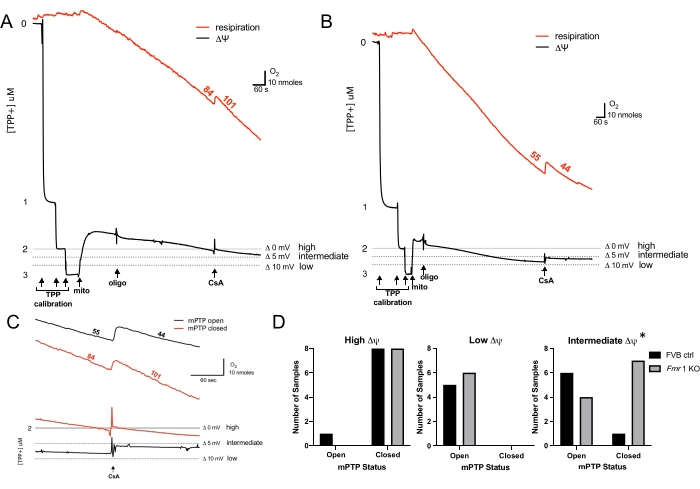
Figure 1: Detection of low-conductance mPTP open probability. (A, B) Representative curves of simultaneous O2 consumption (red, numbers are rates in nmol of O2 mL-1 min-1 mg protein-1) with ΔΨ (black, [TPP+]). Arrows indicate the addition of TPP+, mitochondria, oligomycin, and CsA. High, intermediate, and low voltage thresholds relative to the 2 μM TPP+ calibration level are shown. Addition of CsA at high (A) and low (B) voltage thresholds is shown. (C) Magnified section of O2 consumption (upper) and ΔΨ (lower) curves in (A) and (B) at the point of CsA addition, illustrating closed mPTP (red) at high ΔΨ (insensitive to CsA) and open mPTP (black) at low ΔΨ (sensitive to CsA). (D) Summary graphs of mPTP open and closed status at high, low, and intermediate ΔΨs are shown for FVB controls (black) and Fmr1 KOs (gray). Five to 11 animals were evaluated at each ΔΨ threshold; p values were calculated using a chi-squared test, *p < 0.05. Abbreviations: ΔΨ = mitochondrial membrane potential; mPTP = mitochondrial permeability transition pore; TPP+ = tetraphenylphosphonium; mito = mitochondria; oligo = oligomycin; CsA = cyclosporine A; ΔΨ = ; KO = knockout. Please click here to view a larger version of this figure.
Component | MI/BSA* | SB/BSA* | Density Gradient Diluent | RB/BSA* |
Mannitol | 225 mM | – | – | – |
Sucrose | 75 mM | 250 mM | 1 M | 200 mM |
HEPES | 5 mM | 5 mM | 50 mM | 5 mM |
EGTA | 1 mM | 0.1 mM | 10 mM | – |
KCl | – | – | – | 25 mM |
KH2PO4 | – | – | – | 2 mM |
MgCl2 | – | – | – | 5 mM |
pH** | 7.4 | 7.4 | – | 7.2 |
BSA*** | 0.10% | 0.10% | – | 0.02% |
AP5A*** | – | – | – | 30 mM |
Table 1: Buffers and solutions.
* Filter through 0.2 μm filters
**Adjust pH with 3 M potassium hydroxide as needed.
***Denotes buffer components that are added just prior to mitochondrial isolation.
Abbreviations: MI = mitochondrial isolation buffer; SB = sucrose buffer; RB = Respiration buffer; BSA = fatty acid-free bovine serum albumin; HEPES = 4-(2-hydroxyethyl)-1-piperazineethanesulfonic acid; EGTA = ethylene glycol-bis(β-aminoethyl ether)-N,N,N′,N′-tetraacetic acid; KCl = potassium chloride; KH2PO4 = potassium dihydrogen phosphate; MgCl2 = magnesium chloride; AP5A = P1,P5-diadenosine-5' pentaphosphate pentasodium.
This paper describes a method to assess the open probability of the mPTP. Specifically, the voltage threshold for low-conductance mPTP opening was determined by assessing the effect of CsA inhibition on proton leak over a range of ΔΨs. Using this technique, we could identify differences in voltage gating of the mPTP between FXS mice and FVB controls consistent with their differences in tissue-specific CoQ content. Critical to the success of this methodology is that mitochondria are freshly isolated prior to use and are of good quality. Mitochondria that are either damaged during isolation or too old will be unable to generate appropriate ΔΨ and will typically be uncoupled. Note that while matrix pH typically contributes to the proton motive force, nigericin is used to collapse ΔpH across the IMM in these experiments, as previously described in standard protocols assessing proton leak29. Therefore, in this case, the proton motive force is equivalent to ΔΨ, which simplifies the experimental approach.
Another important consideration is that some of the inhibitors used in the respiration mixture (such as rotenone, oligomycin, and CsA) are difficult to rinse away between experiments because they adhere to plastic surfaces of the reaction chamber, including the ion-selective electrodes. The chamber must be washed routinely with ethanol and a suspension of crudely prepared freeze-thawed mitochondria to "mop up" these inhibitors from the chamber and reduce the likelihood of inhibitor carry-over that can affect the reproducibility of experiments.
As mitochondria must be freshly prepared to maintain their integrity, this method is not amenable to high-throughput analyses. In addition, relatively large amounts of mitochondria are needed for each experiment. Typically, for P10 animals, enough mitochondria can be isolated from one organ to conduct a single experiment. Therefore, testing different conditions on the same batch of mitochondria is not usually possible. If experiments are performed with older animals, this limitation is less of an issue, given the relatively larger size of tissues and organs.
A number of well-described methods exist to study the open probability of the mPTP in isolated intact mitochondria. Techniques, such as mitochondrial matrix swelling and calcium loading or retention capacity assays, necessarily trigger high-conductance PT and thus do not permit the study of physiologic low-conductance opening that is of interest in these experiments28. The patch-clamp technique provides a powerful and direct method to study the voltage dependence of high- or low-conductance states of the mPTP31,32. However, it is a laborious technique, where only a single mitochondrion and a single experimental condition can be studied at a time, which makes comparative studies more difficult32,33.
In addition, the outer mitochondrial membrane is typically lysed, and the inner mitochondrial membrane is typically ruptured in the patch-clamp methods used for isolated mitochondria, which may result in the loss of mPTP regulatory factors that are not intimately associated with the IMM31. Low-conductance opening of the mPTP has also been studied using a variety of fluorescence techniques typically employing a tetramethylrhodamine ester (to assess changes in ΔΨ) either alone or in combination with calcein, whose intramitochondrial fluorescence is susceptible to cobalt quenching when the mPTP is open8,34. However, in fluorescence-based approaches, as changes in ΔΨ are expressed as relative changes in fluorescence, the approach typically does not allow for precise measurement of ΔΨ8,34.
This method provides a new alternative approach to assess the low-conductance opening of the mPTP with respect to ΔΨ. Using this approach, investigators can study the regulation of physiologic and pathologic mPTP voltage gating. This technique also holds promise to help understand the developmental role of the mPTP in organ maturation as it can be applied to various tissues and different stages of development. This approach can be readily applied to study voltage gating of the mPTP in FXS mouse forebrain tissue, which previously exhibited decreased CoQ levels25. Further, we propose that this technique can also be used to study how other agents, endogenous or exogenous, impact voltage gating of the mPTP. Thus, it will prove to be a useful tool to expand the understanding of the contribution of the mPTP to health and disease.
The authors have nothing to disclose.
This work is supported by the following grants: NIH/NIGMS T32GM008464 (K.K.G.), Columbia University Irving Medical Center Target of Opportunity Provost award to the Department of Anesthesiology (K.K.G.), Society of Pediatric Anesthesia Young Investigator Research Award (K.K.G.), and NIH/NINDS R01NS112706 (R.J.L.)
4-(2-hydroxyethyl)-1-piperazineethanesulfonic acid (HEPES) | Fisher Scientific | 15630080 | |
Adapted plunger assembly for pH or ion-selective electrodes for use with OXYT1 | PP systems | 941039 | |
BD Intramedic PE Tubing, PE 50, 0.023 in. 10 ft. | Fisher Scientific | 14-170-11B | to modify the length of the hamilton synringe as needed |
Bovine Serum Albumin (BSA). Fatty acid free | Sigma | A7030-10G | |
Dri-Ref Reference Electrode, 2 mm | World Precision Inst. LLC | DRIREF-2 | |
Electrode Holder for KWIK-Tips | World Precision Inst. LLC | KWIK-2 | ion selective electrode holder |
Ethylene glycol-bis(β-aminoethyl ether)-N,N,N′,N′-tetraacetic acid (EGTA) | Sigma | 324626 | |
FVB.129P2-Pde6b+ Tyrc-ch Fmr1tm1Cgr/J | Jackson Laboratory, Bar Harbor, ME | FXS mice, Fmr1 KO | |
FVB.129P2-Pde6b+ Tyrc-ch/AntJ | Jackson Laboratory, Bar Harbor, ME | FVB mice | |
Hamilton 80366 Standard Syringes, 10 uL, Cemented-Needle, 6/pk | Cole-Parmer | EW-07938-30 | microsyringe |
Hamilton 80500 Standard Microliter Syringes, 50 uL, Cemented-Needle | Cole-Parmer | EW-07938-02 | microsyringe |
Hansatech Instruments Oxytherm+ System (Respiration) Complete | PP systems | OXYTHERM+R | oxygen electrode and software |
Magnesium Chloride (MgCl2) | Sigma | 1374248 | |
Mannitol | Sigma | M9546-250G | |
P1,P5-diadenosine-5′ pentaphosphate pentasodium (AP5A) | Sigma | D4022-10MG | |
Percoll | Sigma | P1644 | medium for density gradient separation |
Potassium chloride (KCl) | Sigma | P3911 | |
Potassium dihydrogen phosphate (KH2PO4) | Sigma | 5.43841 | |
Sucrose | Sigma | S0389 | |
TPP+ Electrode Tips (3) | World Precision Inst. LLC | TIPTPP |