The present protocol describes the induction of experimental autoimmune encephalomyelitis in a mouse model using myelin oligodendrocyte glycoprotein and monitoring the disease process using a clinical scoring system. Experimental autoimmune encephalomyelitis-related symptoms are analyzed using mouse femur micro-computed tomography analysis and open field test to assess the disease process comprehensively.
Multiple sclerosis (MS) is a typical autoimmune disease of the central nervous system (CNS) characterized by inflammatory infiltration, demyelination, and axonal damage. Currently, there are no measures to cure MS completely, but multiple disease-modifying therapies (DMT) are available to control and mitigate disease progression. There are significant similarities between the CNS pathological features of experimental autoimmune encephalomyelitis (EAE) and MS patients. EAE has been widely used as a representative model to determine MS drugs’ efficacy and explore the development of new therapies for MS disease. Active induction of EAE in mice has a stable and reproducible effect and is particularly suitable for studying the effects of drugs or genes on autoimmune neuroinflammation. The method of immunizing C57BL/6J mice with myelin oligodendrocyte glycoprotein (MOG35-55) and the daily assessment of disease symptoms using a clinical scoring system is mainly shared. Given the complex etiology of MS with diverse clinical manifestations, the existing clinical scoring system can’t satisfy the assessment of disease treatment. To avoid the shortcomings of a single intervention, new indicators to assess EAE based on clinical manifestations of anxiety-like moods and osteoporosis in MS patients are created to provide a more comprehensive assessment of MS treatment.
Autoimmune diseases are a spectrum of disorders caused by the immune system's immune response to its own antigens resulting in tissue damage or dysfunction1. Multiple sclerosis (MS) is a chronic autoimmune disease of polyneuropathy in the central nervous system (CNS), characterized by inflammatory infiltration, demyelination, and neuronal axonal degeneration2,3. At present, MS has affected as many as 2.5 million people around the world, mostly young and middle-aged people aged 20-40, who are often the backbone of their families and society. This has caused considerable impact and harm to families and society2,4.
MS is a multifactorial disease with diverse and complex clinical manifestations. In addition to classic neurological disorders characterized by inflammatory infiltration and demyelination, MS often shows visual impairment, limb dyskinesia, and cognitive and emotional disorders5,6,7. If MS patients do not get the proper and correct treatment, half of them will live in wheelchairs after 20 years, and nearly half of them will experience depressive and anxiety symptoms, leading to much higher levels of suicidal ideation than the general population8,9.
Despite a long research period, the etiology of MS remains elusive, and the pathogenesis of MS has not yet been elucidated. Animal models of MS have allowed serving as testing tools to explore disease development and new therapeutic approaches, despite the significant differences between the rodent and human immune systems, while at the same time sharing some basic principles. Experimental autoimmune encephalomyelitis (EAE) is currently the ideal animal model for studying MS, which uses autoantigen immunity from myelin proteins to induce autoimmunity to CNS components in susceptible mice, with the addition of complete Freund's adjuvant (CFA) and pertussis toxin (PTX) to enhance the humoral immune response. Depending on the genetic background and immune antigens, different disease processes, including acute, relapsing-remitting, or chronic, are obtained to mimic various clinical forms of MS10,11,12. The relevant immunogens commonly used in the construction of EAE models come from self-CNS proteins, such as myelin basic protein (MBP), proteolipid protein (PLP), or myelin oligodendrocyte glycoprotein (MOG). MBP- or PLP-immunized SJL/L mice develop a relapsing-remitting course, and MOG triggers chronic progressive EAE in C57BL/6 mice11,12,13.
The main purpose of disease-modifying therapy (DMT) is to minimize disease symptoms and improve function6. Several drugs are used clinically to alleviate MS, but no drug has yet been used to completely cure it, revealing the necessity of synergistic treatment. C57BL/6 mice are currently the most commonly used to construct transgenic mice, and in this work, an EAE model induced by MOG35-55 in C57BL/6J mice with a 5-point scale was used to monitor the disease progression. EAE models also suffer from anxiety-like moods and bone loss, and the widely known demyelinating lesions. Here, the method to assess the symptoms of EAE from multiple perspectives using open-field test and micro-computed tomography (Micro-CT) analysis is also described.
The Animal Care Committee of Tongji University approved the present work, and all animal care guidelines were followed. Male or female C57BL/6J mice between 8-12 weeks of age were used for the experiments. It was ensured that the age and sex were the same in the experimental groups; otherwise, the susceptibility to the disease was affected. Mice were housed in a specific pathogen-free environment with alternating 12 h light and dark cycles under constant conditions (room temperature 23 ± 1 °C, humidity 50% ± 10%), with free access to mouse food and water.
1. Preparation of MOG35-55 emulsion
2. Preparation of pertussis toxin (PTX)
3. Establishment of EAE animal model
4. Clinical monitoring of mice
5. Open field test
NOTE: The experimental animals selected for this step are EAE mice in the early onset, peak, and remission periods. In addition, WT mice were used as control. It is to be noted that all mice were tested for anxiety-like behavior prior to modeling to exclude mice with anxiety disorders for EAE modeling. In addition, EAE mice in peak and remission periods with complete motor incapacity were excluded from the test.
6. Analysis of bone phenotype
After immunization of the mice, the bodyweight of the mice is recorded daily, and their clinical symptoms are evaluated according to the protocol described above (step 4). In C57BL/6J mice immunized with MOG peptide, because the location of the lesion is mainly confined to the spinal cord,the pathogenesis of EAE mice spreads from the tail end to the head. At the beginning of the disease, EAE mice exhibit weakness and drooping of the tail, followed by weakness of the hind limbs, uncoordinated movement, and paralysis. As the disease worsens, it gradually develops into the weakness of the forelimbs, paralysis, and in severe cases, causes difficulty in moving the mice and even near death. As shown in Figure 1A, the state diagram of the mice with different degrees of EAE pathology depicts an exemplary picture of a group of mice changing from asymptomatic to high-scoring EAE symptoms (score 4). It has also been mentioned earlier that the bodyweight of EAE mice is correlated with clinical symptoms. Compared with WT mice, weight loss in EAE mice may start to occur in the first few days after immunization, while the clinical symptoms of EAE mice usually begin on day 6-9 after immunization and will reach a peak on day 14-16. After this, the symptoms of EAE mice generally partially recover, and at the same time, the weight loss of the mice will be alleviated (Figure 1B,C). Thus, the course of EAE onset is usually divided into early-onset, peak, and remission periods, and the prediction of these time points is important in assessing outcome parameters. In general, to analyze the production of immune cells and cytokines at the site of EAE lesions, immune cells in the brain and spinal cord of EAE mice at the peak of the disease can be isolated and further processed, which can be analyzed by flow cytometry14,15. The spinal cord tissue at peak onset is also most suitable for preparing hematoxylin and eosin (H&E) staining and Luxol fast blue staining to further investigate the inflammatory cell infiltration and demyelination of the spinal cord14,16. For monitoring changes in the immune system at different onset times of EAE, spleen and lymph nodes at early onset are also essential options17,18. In addition, cells from the spleen or lymph nodes of MOG-immunized EAE mice are commonly used to construct transfer models, which are transferred to recipient mice after restimulation of MOG in vitro to induce passive immunization of EAE mice18.
MS is an autoimmune inflammatory induced demyelinating lesion of the central nervous system characterized by inflammatory demyelination and neuronal loss2,3. This disease is usually accompanied by psychiatric comorbidities, such as affective disorders, of which anxiety disorder is very common in MS patients, with up to 30% of MS patients suffering from anxiety9,19. Anxiety disorder is a psychiatric abnormality characterized by excessive emotional stress and worry. Open field test is often used to analyze behaviors for anxiety in rodents20,21. With the help of analyzing the exploration behaviors of EAE mice in open field tests during early onset, peak, and remission periods, it was found that EAE mice also had anxiety-like behaviors similar to those of MS patients (Figure 2A). In the open field test, anxious rodents tend to have reduced activity and increased stereotypic behavior, including a preference for being closer to corners, a bias toward the peripheral domain, and a lack of desire to explore the central area. Compared to WT mice, EAE mice had significantly lower walking distance and movement time in all three periods of the disease, even in the early onset of the disease when EAE mice did not yet have a motor impairment (Figure 2B,C). In addition, EAE mice pass significantly less distance and stay in the central area less than normal mice, and even move only in the peripheral area, showing obvious anxiety-like mood (Figure 2D,E).EAE mice still exhibit a strong anxiety-like mood when the onset is mild, that is, motor coordination. Some studies suggest that this may be attributed to mild neuroinflammation, which further affects neurotransmitter secretion22,23. Open field tests monitoring anxiety-like mood triggers in EAE mice could help researchers understand and treat MS psychiatric comorbidities.
With the progress of the disease, MS essentially eventually manifests as dyskinesia. Studies have found that MS patients have a higher susceptibility to osteoporosis and fracture, mainly due to bone mass loss, and that the severity of dyskinesia is strongly correlated with the patient's bone density24,25. A similar phenomenon can be observed by Micro-CT analysis with the help of the EAE animal model (Figure 3A,H). From the trabecular analysis data of femur in mice, EAE mice underwent a significant decrease in bone mineral density (BMD) compared to WT mice, which is an important indicator of the response to bone strength and an important basis for the diagnosis of osteoporosis (Figure 3B). Further analysis showed that trabecular bone loss occurred significantly in EAE mice compared with healthy WT mice and was accompanied by a reduction in trabecular connection density, trabecular numbers, and trabecular thickness. These are all characteristic of reduced bone mass, suggesting that EAE also causes trabecular bone loss in the femur of mice (Figure 3C–F).At the same time, the structural morphology of the bone trabeculae changed, and the spacing of the trabeculae increased significantly; the greater the spacing, the more osteoporotic the bone (Figure 3G). This is consistent with the notion that MS patients are prone to osteoporosis. In cortical bones of the femur diaphysis, the thickness of cortical bone in the EAE model was significantly less than that in normal mice (Figure 3I). In MS, decreased motility and increased muscular dystrophy are strongly associated with osteoporosis, fracture, and increased bone resorption due to reduced mechanical forces, gradually decreasing bone integrity, thereby increasing the risk of osteoporosis and fracture26. Micro-CT analysis of the femur in EAE mice can monitor bone health well, and the intervention is beneficial in controlling the condition of EAE.
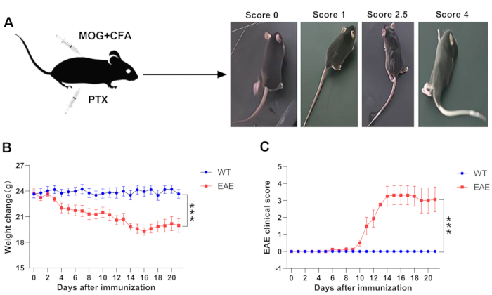
Figure 1: Monitoring of clinical symptoms of EAE. (A) The exemplary picture of the mice with different degrees of EAE pathology. (B) Weight change in WT and EAE mice. (C) Clinical score in WT and EAE mice. Data are given as mean ± SEM (n = 5), ***p < 0.001 versus WT mice,two-way ANOVA test. Please click here to view a larger version of this figure.
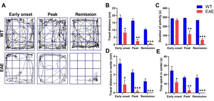
Figure 2: Anxiety-like behavior of EAE mice in the open field test. (A) Representative track plots of the open field test. Travel distance (B), duration of activity (C), travel distance in the center (D), and time spent in the center (E) of WT mice and EAE mice in early-onset, peak, and remission periods. Data are mean ± SEM (n = 3), *p < 0.05, **p < 0.01, ***p < 0.001 versus WT mice, Student's t-test. Please click here to view a larger version of this figure.
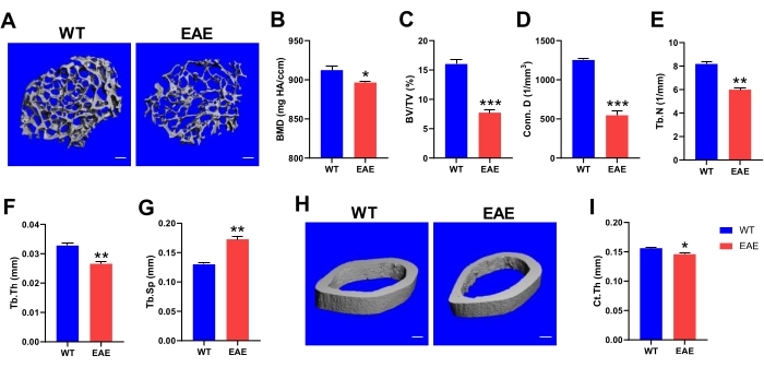
Figure 3: Micro-CT analysis of bone health in EAE mice. (A) Representative 3D images of femoral trabecular architecture (scale bars, 100 µm). Bone mineral density (B), bone volume/tissue volume ratio (C), connection density (D), numbers (E), thickness (F), and separation (G) of femoral trabecular were determined by Micro-CT analysis. (H) Representative 3D images of cortical bone (scale bars, 100 µm). (I) Cortical thickness obtained from microcomputed tomography data. Data are mean ± SEM (n = 3), *p < 0.05, **p < 0.01, ***p < 0.001 versus WT mice, Student's t-test. Please click here to view a larger version of this figure.
Score | Clinical sign | ||
0 | No clinical signs | ||
0.5 | Tail weakness, front of tail dropping | ||
1 | Tail completely paralyzed | ||
2 | Mild paralysis of hind limbs (weakness of both hind limbs or unilateral paralysis, uncoordinated walking, response to pinch) | ||
3 | Complete paralysis of hind limbs, hind limbs dragging and walking, hind limbs not responding to pinch | ||
4 | Paralysis of the hind limbs and weakness of the forelimbs | ||
5 | Near death or dying |
Table 1: Clinical scoring system.
Antigen | Strain | Characteristic | Application | Limitation | ||
PLP | SJL/J34 | Relapsing-remitting EAE | Study of the cellular and molecular events involved in clinical relapse | Limited to T-cell specific responses; Studying specific genes and pathways in the SJL/J strain is challenging | ||
MBP | SJL/J35 | Relapsing paralysis followed in partial or total recipients after recovery from acute paralysis | Study of neuroinflammation and immune system activation | Limited to T-cell specific responses; Studying specific genes and pathways in the SJL/J strain is challenging | ||
MOG35-55 | C57BL/631 | Primary progressive EAE; Chronic progressive EAE | Study of axonal damage mechanisms; Study of molecular disease mechanisms in transgenic and knockout models | Primary demyelination and myelin regeneration studies are of limited value | ||
Spinal cord homogenate | Biozzi ABH36 | Relapsing-remitting EAE | Study of myelin regeneration and neuroprotective therapy for MS | Not clinically progressive, with accumulation of neurological deficits over time; Limited to CD4 T-cell specific responses | ||
Theiler’s murine encephalomyelitis virus | Multiple strains of mice are highly susceptible, including SJL/J, SWR, PL/J12,31 | Viral models of inflammatory demyelination | Study of axonal injury and inflammation-induced demyelination | No MS-specific viral infections have been identified; Myelin regeneration is difficult to assess, demyelination and myelin regeneration occur simultaneously | ||
Cuprizone | Widely used in C57BL/6 mice, while other strains, such as the CD1 strain, are less susceptible to copper cuprizone-induced damage37,38 | Toxic model of demyelination and remyelination | Study of T cells-independent demyelination, especially myelin regeneration and basic myelin repair processes | Significant spontaneous myelin regeneration occurs after cessation of toxic injury; Demyelination is brain region-specific and not useful for studying spinal cord demyelination | ||
Lysolecithin | SJL/J12, C57BL/639, etc. | Toxic model of demyelination and remyelination | Study of T cells-independent demyelination, especially myelin regeneration and basic myelin repair processes | Lack of immune response observed during MS | ||
Ethidium bromide | C57BL/640, etc. | Toxic model of demyelination and remyelination | Study of focal demyelinating lesions and prediction of the kinetics of demyelination and myelin regeneration | Ethidium bromide damages all nucleolus containing cells; Limited correlation with MS |
Table 2: Different mouse models of MS.
MS is a demyelinating inflammatory disease of the CNS and is one of the most common neurological disorders causing chronic disability in young people, imposing a huge burden on families and society3,4. MS has always been classified as an organ-specific T cell-mediated autoimmune disease, inducing the autoimmune system to slowly erode CNS, which will involve multiple systems throughout the body27. Typical clinical symptoms include visual impairment, motor disorders, cognitive and emotional disturbances, etc6,7. MS is a devastating disease that will progressively worsen if not properly treated, leading to severe blindness and paralysis28. The pathogenesis of MS is not yet fully understood, as its clinical manifestations and pathological progression are heterogeneous, and therefore animal models are essential for the research and development of treatment of MS.
The most frequently studied animal model for MS is EAE, which mainly has two modeling methods: active immunization and passive immunization29. The former is widely adopted because of its simplicity, lack of mouse irradiation, and short cycle time. The EAE active immune model induces autoimmune responses in the CNS by immunizing rodents with self-myelin antigens or their subsequent peptides, usually manifesting as tail weakness and limb paralysis13. Specifically, the EAE model induced by MOG35-55 in C57BL/6J mice is characterized by alternating remission-relapse cycles, accompanied by CNS inflammation, demyelination, and axonal injury, which makes the MOG35-55-induced EAE the preferred model11,12. EAE can be well induced in mice sensitized by subcutaneous injection of MOG35-55 peptide, emulsified with immunopotentiator CFA enriched with MTB, and given an intraperitoneal injection of PTX with blood-brain barrier disrupting effect on days 0-230. The biggest challenge to the EAE model is the low incidence of weak symptoms, and several influencing factors are critical to the course of the experiment. (1) The age and environment of mice will affect the susceptibility to EAE. Therefore, mice of the same age need to be selected before the experiment. The mice should be weighted and randomly grouped before cage allocation, and the weight of each group needs to be kept close. Also, ensure that environmental conditions between independent experiments are comparable. (2) The concentration of PTX: The main role of PTX is to increase the permeability of the blood-brain barrier, which allows pathogenic T cells to enter the CNS to secrete relevant cytokines and promote the inflammatory response, ultimately leading to the hydrolysis of myelin proteins wrapped in the outer layer of nerve axons. The onset of disease in EAE mice was delayed with decreasing PTX administration concentration, and the severity of the disease would be weakened if PTX decreased by more than 60%. (3) The dissolution of MOG35-55 antigen: To avoid freezing and thawing, it is recommended to procure the lyophilized MOG35-55 antigen in separate packages, 4 mg/tube, to avoid freezing and thawing. Also, PTX is used for dissolution instead of sterile water because it is better to emulsify with CFA using PTX. (4) MOG35-55 antigen and CFA must be sufficiently emulsified by oscillating to form a water-in-oil structure, and the emulsion droplets do not disperse on the water surface for a long time, indicating good emulsification. (5) Two-point injection was used for mouse EAE modeling. The injection site is preferably located above the waist and below the neck, close to the spine, to avoid mice licking and biting the skin, resulting in the spillage of MOG35-55 emulsion.
However, the EAE model induced by the MOG35-55 antigen still has some limitations. As an inflammatory encephalopathy model of primary axonal injury mediated by CD4 T cells, the main lesion is massive axonal degeneration with secondary demyelination and less primary demyelination, which is different from the MS pathology31,32,33.The EAE model induced by MOG35-55 mainly reflects the immune response driven by CD4 T cells, while other immune cells such as CD8 T and B cells do not have a strong sense of participation in this model31. Another limiting factor is that EAE induced by MOG35-55 has a certain bias towards the immunological component of MS pathophysiology,and other animal models can be considered when considering studies focused on CNS pathologies, such as the study of demyelination and myelin regeneration processes34,35,36,37,38,39,40, as described in Table 2.
EAE is an effective predictor in imitating the autoimmune properties of MS, as well as its inflammation and demyelination mechanism12, which makes many scholars keen to study its immunomodulatory mechanisms, ignoring other clinical symptoms of MS. Studies have shown that MS-associated anxiety is attributed to pathophysiological processes such as immune dysregulation and brain inflammation22,23,41. Mental health issues, such as anxiety, are particularly common in people with MS, and anxiety disorder can impede the performance of basic daily tasks and significantly affect the quality of life42,43. Alterations in the locomotor ability of EAE mice may predict alterations in neural processes. In open-field experiments, anxiety-like behaviors occurred in EAE mice,including a decrease in exploratory behaviors that started early onset of the disease (essentially, test subjects with clinical scores <0.5 and locomotor ability almost as good as normal mice), suggesting that the onset of anxiety-like behaviors precedes motor impairment. In addition, EAE mice in the remission period did not improve anxiety-like behavior with increased motor ability. The emergence of the inflammatory response may be responsible for the mental impairment in EAE mice through neuronal activity, leading to changes in mood, and upregulation of inflammatory cytokine levels interferes with striatal synaptic function prior to the onset of motor impairment23,41. However, the open field test requires attention to the fact that the test room must be a quiet area, and the test mice need to acclimatize to the room for 1 h in advance to avoid excessive pressure caused by the new environment. Additional cleaning with ethanol is required between tests to remove odors and other substances left behind by the last test subject. The main limitation of the open field test is the interference caused by motor dysfunction in EAE mice, and the test for evaluating rodent anxiety-like behavior is based on the distance, time, and range of rodent activity in the reaction chamber, and cannot rule out the effect of motor dysfunction on anxiety-like behavior. Therefore, future studies need to take locomotor impairment into account.
Traditional methods to analyze bone tissue-related studies often use two-dimensional bone section microscopy, which can have greater difficulties in analyzing structural morphology, density distribution, orientation distribution, and other characteristics due to the influence of two-dimensional planes and inhomogeneity of biological specimens. Many studies have confirmed the accuracy and effectiveness of the Micro-CT system to describe bone morphology, and many studies44,45,46 have confirmed microstructure detection results. The three-dimensional imaging of bone tissue by Micro-CT allows for more intuitive and accurate bone histomorphometry results. It has been achieved to scan small animals and specimens without executing the animal or damaging the tissue specimen, allowing for detailed three-dimensional spatial structure information inside the tissue44. Studies have shown that MS patients are prone to bone mass loss and osteoporosis24,25; By scanning and imaging the femur of EAE mice with a Micro-CT system, an obvious deterioration of bone microarchitecture and decreased bone strength could be observed in the femur of EAE mice compared to WT mice that had developed osteoporosis. Micro-CT analysis is relatively simple to perform and relatively inexpensive, and Micro-CT live imaging can be used to monitor bone health in the future when administering treatment to EAE mice. However, the drawback is the absence of bone turnover markers assessed through blood samples or bone histomorphometry to observe fundamental changes in bone metabolic homeostasis in EAE mice47. Regulation of osteoblasts and osteoclasts could be considered within the study in the future.
MS is a multi-symptom disease, and the sole aim of disease management is to minimize disease symptoms. In the protocol mentioned above, the clinical symptoms of MS can be simulated by constructing the MOG35-55 induced EAE model. EAE mice showed clinical symptoms of motor dysfunction and anxiety-like behavior, and osteoporosis. The 5-point scoring system, open field test, and Micro-CT analysis described in this protocol can monitor the pathological symptoms of EAE mice from multiple perspectives and provide a reference scheme for the treatment of EAE. In the absence of effective disease-modifying drugs, a potential combination of synergistic therapy can provide the best opportunity to relieve symptoms and improve function.
The authors have nothing to disclose.
The authors acknowledge the support from the National Natural Science Foundation of China (32070768, 31871404, 31900658, 32270754) and the State Key Laboratory of Drug Research.
1 mL syringe(with 26 G needle) | Shanghai Kindly Medical Instruments Co., Ltd | 60017031 | |
2 mL microcentrifuge tube | HAIKELASI | KY-LXG2A | |
22 G needle | Shanghai Kindly Medical Instruments Co., Ltd | 60017208 | |
Complete Freund’s Adjuvant | Sigma | F5881 | Stored at 4 °C, 1 mg of heat-inactivated MTB (H37Ra) per mL |
Conditioned place preference system | Shanghai Jiliang Software Technology Co., Ltd | Animal behavior | |
Ethanol | Sinopharm Chemical Reagent Co., Ltd | 10009218 | Stored at RT |
Locomotion activity (open field) video analysis system | Shanghai Jiliang Software Technology Co., Ltd | DigBehv-002 | Animal behavior |
MOG35-55 peptide | Gill Biochemical Co., Ltd | GLS-Y-M-03590 | Stored at -20 °C |
Mycobacterium tuberculosis H37Ra | BD | 231141 | Stored at 4 °C |
Open field reaction chamber | Shanghai Jiliang Software Technology Co., Ltd | Animal behavior | |
Pertussis toxin | Calbiochem | 516560 | Stored at 4 °C |
Phosphate Buffered Saline | Made in our laboratory | ||
Scissor | Shanghai Medical Instrument (group) Co., Ltd | J21010 | |
Sealing film | Heathrow Scientific | HS 234526B | |
Sorvall Legend Micro 21R Microcentrifuge | Thermo Scientific | 75002447 | |
Steel ball | QIAGEN | 69975 | |
TissueLyser II | QIAGEN | 85300 | |
Tweezer | Shanghai Medical Instrument (group) Co., Ltd | JD1060 | |
μCT 35 desktop microCT scanner | Scanco Medical AG, Bassersdorf, Switzerland |