All human participants provided informed consent. The protocol was part of a study approved by the Institutional Review Board of the University of Central Florida. Inclusion criteria included ages 18 -45 and physically active according to ACSM guidelines (30 min of moderate to vigorous activity 5 days per week)19. Exclusion criteria included low back pain within the past year, current hip, upper, or lower extremity pain or injury, a year history of low back surgery or lower extremity surgery, self-reported balance disorder, muscular abnormalities, currently pregnant, or having an open wound in the abdominal area (Table 1).
1. Data collection instrument preparation
- Check the integrity of the ultrasound machine and sEMG machine (see Table of Materials).
- Turn on the ultrasound machine, press Patient, and add a new patient. Click New Patient and enter the patient identification number. Select MSK exam type > Abdominal preset and press Registrati. Press Exit.
- Turn on the tablet (see Table of Materials) and click the EMG recording application. Click the Menu button in the top left screen and scan for sensors. Take the sensor out of the base and hover over the activation button. Once the sensor is connected with the application, create a new folder with the participant's identification number.
NOTE: Ultrasound settings in Abdominal Preset, B-mode: B color = Tint Map D, Write Zoom Height = 4, Write Zoom Width = 4, Thermal Index = Tls, ATO Level = Low, Focus Number = 2, Focus Number CrossXBeam = 2, Focus Depth = 50, Depth (cm) = 3, Compression = 1, Focus Width = 1, Focus Width CrossXBeam = 1, Line Density = 3, Line Density CrossXBeam = 3, Suppression = 0, Frame Average = 4, From Average CrossXBeam = 2, CrossXBeam = 2, CrossXBeam # = Low, CrossXBeam Type = Mean, Edge Enhance = 3, B Steer = 0, Gray Scale Map = Gray Map C, Gain = 34, Dynamic Range = 69, Rejection = 0, Frequency (MHz) = 12. Ultrasound settings in Abdominal Preset, M-mode: sweep speed = 0, M color = Tint Map C, Dop Display Format = Vert ½ B, Rejection = 0, Compression = 1, Gray Scale Map = Gray Map D, M Gain (delta from B) = 0.
2. Surface electromyography preparation (see Table of Materials)
- Determine the external oblique sensor location by palpating the right iliac crest and right inferior rib while the participant is supine hook-lying (Figure 1). Place the sensor 3 cm anterior from the midway point between the lower rib and the iliac crest, parallel to the muscle fibers20.
- Shave, clean, and debride the area of the skin where the sensor (see Table of Materials) will be placed. Add adhesive (see Table of Materials) to the sensor and secure onto skin.
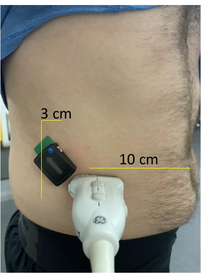
Figure 1: Examination location of the lateral abdominal wall. The sEMG sensor is placed 3 cm anterior from the midway point between the lower rib and the iliac crest, parallel to the muscle fibers20. The transducer is located 10 cm lateral to the umbilicus until the lateral abdominal wall is visible on the screen. Please click here to view a larger version of this figure.
3. Ultrasound preparation (see Table of Materials)
- In a standing position, place the transducer through the elastic belt and foam block (Figure 2).
- Add gel to the transducer and place the transducer 10 cm lateral to the umbilicus. Adjust until the lateral abdominal wall is visible on the screen21 (Figure 3).
- Ensure that the transducer is tightly secured to the lateral abdominal wall.
- Secure the belt with the velcro straps. Adjust the depth to obtain optimal image quality in B-Mode (Figure 4).
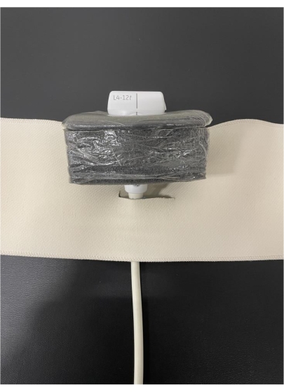
Figure 2: Transducer placed through the elastic belt and foam block. Please click here to view a larger version of this figure.
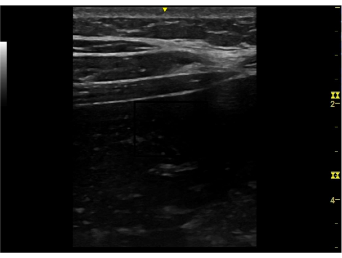
Figure 3: Example of resting image to verify the lateral abdominal wall. Please click here to view a larger version of this figure.
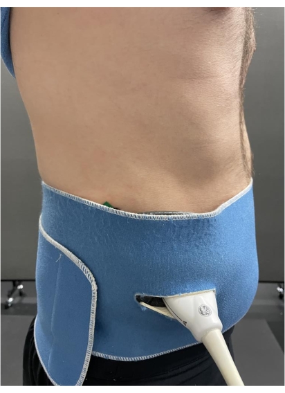
Figure 4: Transducer secured on the lateral abdominal wall by the elastic belt and foam block. Please click here to view a larger version of this figure.
4. Ultrasound static side plank
- On a yoga mat (see Table of Materials), instruct the participant to lay on their right side with their elbow in 90 degrees of flexion, so their trunk and upper extremity are raised off the ground. The participant's feet should be stacked on each other with the knees fully extended. The legs should be parallel touching the yoga mat.
- Press Freeze then Store for each image to capture three static ultrasound images of the lateral abdominal wall.
NOTE: Saved images will be located below the active image screen.
5. sEMG static side plank
NOTE: Simultaneously, the researcher will also obtain the sEMG output during the static positioning described in step 4.1.
- In the sEMG menu in the top left screen of the EMG recording application, select Settings, which is a gear icon.
- Once in the sensor settings page, select the bicep icon. This will open the page for normalization measurements.
- Press Click Play to Begin; there will be a 5 s countdown. During this period, instruct the patient to assume the testing position. The recording will take an additional 5 s.
- Record the "MVC = .XXXXmV", as this will be used for normalization calculation.
NOTE: The EMG mobile suite automatically band pass filters between 20 -450 Hz. EMG (RMS) 333.3, 1125 ms window width.
6. Side plank
- Next, instruct the participant to complete the side plank for 60 s maintaining the correct form22. Press the M on the ultrasound machine to turn on M-mode.
- Press Plot, press the red button and press the Save button again in the EMG application. The participant will be given a 3 s countdown. Once that countdown commences, press Store on the ultrasound to begin the ultrasound recording.
NOTE: The participant will hold the correct form until 60 s, or when the researcher determines that the correct form has been disrupted. - To save the recorded M-mode video, press Store once the exercise has stopped and the Stop button on the sEMG application.
- Click Save file as and type in a filename to save the output, which will appear on the screen when the recording is stopped.
7. Ultrasound static dead bug
- On the yoga mat, instruct the participant to lay supine with their legs in a hook-lying position.
- Press B to enter brightness mode. Press Freeze and then Store for each image to capture three static images of the lateral abdominal wall via ultrasound. Saved images will be located below the active image screen.
8. sEMG static dead bug
- In the menu of the EMG application (top left), select Settings, which is a gear icon.
- Once in the sensor settings page, select the bicep icon. This will open the page for normalization measurements.
- Press Click Play to Begin. During the 5 s countdown, instruct the patient to assume the testing position. The recording will take an additional 5 s.
- Record the "MVC = .XXXXmV", as this will be used for normalization calculation.
9. Dead bug
- Next, instruct the participant to complete the dead bug for 60 s, maintaining the correct form. Press the M on the ultrasound machine to turn on M-mode.
- Instruct the participant to maximally extend their right shoulder while maximally extending their left hip and knee, while also maintaining the starting position of the contralateral extremities.
- Instruct the participant to then flex their shoulder, hip, and knee to return to the starting position. The contralateral extremities will then perform the same motion.
- Ask the participant to perform the exercise to a metronome set to 45 beats per min. This results in 22 repetitions of the dead bug in 60 s.
NOTE: The participant should hold the correct form until 60 s, or until the researcher determines that the correct form has been disrupted or the metronome rhythm has been interrupted. - Press Plot, press the red button and then press Play. Press the Save button again in the EMG application. The participant will be given a 3 s countdown. Once that countdown commences, press Store on the ultrasound to begin the ultrasound recording.
10. Ultrasound static measurement
- Click Enter when the first static image you want to measure is selected.
- Press Measure to open the measurement tool. Measure the maximal muscle thickness during the static positions in centimeters from the superior inferior fascial border to the inferior superior fascial border (Figure 5 [side plank] and Figure 6 [dead bug]).
- Click Enter at the superior inferior border and Enter again at the inferior superior border.
- Repeat steps 10.1-10.3 for side plank and dead bug static measurements. Average the measurements of the three static images.
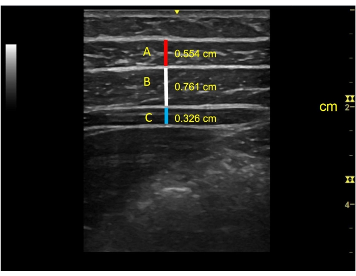
Figure 5: Example of lateral abdominal wall during the side plank static, exercise starting position and measurements of muscles. A = external oblique (0.554 cm), B = internal oblique (0.761 cm), and C = transverse abdominis (0.326 cm). Please click here to view a larger version of this figure.
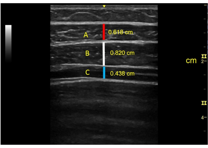
Figure 6: Example of lateral abdominal wall in the dead bug static, exercise starting position and measurements of muscles. A = external oblique (0.618 cm), B = internal oblique (0.820 cm), and C = transverse abdominis (0.438 cm). Please click here to view a larger version of this figure.
11. Ultrasound dynamic measurements
- Measure the maximal thickness of the external oblique, internal oblique, and transverse abdominis thickness during the first 5 s and last 5 s of the exercise. Additionally, record the maximal thickness over the entire 60 s.
NOTE: The side plank is commonly performed for durations of 5 s. Taking sets and repetition guidelines from previous authors, a longer duration of 60 s was chosen for comparison in this protocol. The first 5 s and last 5 s of the task were compared to evaluate both the strength and endurance aspects of the muscle group23,24. - Using the Scroll button, find the first 5 s and last 5 s of each exercise. Additionally, visually inspect for the greatest thickness of each muscle throughout the 60 s exercise.
- Press Measure to open the measurement tool. Measure the maximal muscle thickness during the static positions in centimeters from the superior inferior fascial border to the inferior superior fascial border (Figure 7 [side plank] and Figure 8 [dead bug]).
- Divide each of the three thickness measurements obtained during the exercises by the averaged static position to obtain an activation ratio 25.
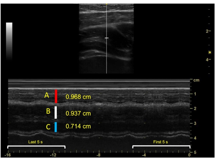
Figure 7: Example of lateral abdominal wall during the side plank exercise and measurements of muscles in M-mode. A = external oblique (0.968 cm), B = internal oblique (0.937 cm), and C = transverse abdominis (0.714 cm). Please click here to view a larger version of this figure.
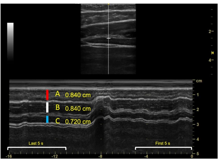
Figure 8: Example of lateral abdominal wall during the dead bug exercise and measurements of muscles in M-mode. A = external oblique (0.840 cm), B = internal oblique (0.840 cm), and C = transverse abdominis (0.720 cm). Please click here to view a larger version of this figure.
12. sEMG Measurement
- Press the Home icon on the EMG data recording page. Select the folder icon in the top right corner of the screen. The saved output from the static and exercise trials will be saved here. Convert each file to an .xlsx file. Export the .xlsx file.
- In a spreadsheet, obtain the maximum values during the first and last 5 s, and the overall maximum value.
- Divide the static sEMG output obtained in steps 5 (static side plank sEMG) and 8 (dead bug sEMG) by the output during the exercises, respectively.
