The present protocol describes a modified and simplified technique with a minimally invasive transverse aortic constriction (TAC) procedure using a self-made retractor. This procedure can be conducted without a ventilator or microscope and introduces pressure overload, eventually leading to cardiac hypertrophy or heart failure.
Transverse aortic constriction (TAC) is a frequently used surgery in research regarding heart failure and cardiac hypertrophy based on the formation of pressure overload in mouse models. The main challenge of this procedure is to clearly visualize the transverse aortic arch and precisely band the target vessel. Classical approaches perform a partial thoracotomy to expose the transverse aortic arch. However, it is an open-chest model that causes a rather large surgical trauma and requires a ventilator during the surgery. To prevent unnecessary trauma and simplify the operating procedure, the aortic arch is approached via the proximal proportion of the sternum, reaching and binding the target vessel using a small self-made retractor that contains a snare. This procedure can be conducted without entering the pleura cavity and does not need a ventilator or microsurgical operation, which leaves the mice with physiological breathing patterns, simplifies the procedure, and significantly reduces operation time. Due to the less invasive approach and less operation time, mice can undergo fewer stress reactions and recover rapidly.
Heart failure is a complex clinical symptom that results from impaired structure and function of ventricular filling or ejecting blood1. The disease stage is mainly defined via the New York Heart Association function classification based on the severity of symptoms and physical activity2. For those patients with an ejection fraction of over 50%, structural and/or functional abnormalities raised natriuretic peptides to support the diagnosis of heart failure with preserved ejection fraction (HFpEF)2. Ischemic heart disease is a leading cause among multiple etiologies of heart failure. Thus, the myocardial infarction model (such as permanent coronary ligation) is often used to study pathophysiology after cardiac hypoperfusion or ischemia-reperfusion injury3,4. Besides acute myocardial injury, other risk factors such as hypertension, diabetes, obesity, and a family history of cardiomyopathy also contribute to the development of heart failure. After patients pass Stage A (at risk for heart failure) and enter Stage B (pre-heart failure), structural modification occurs1. For example, hypertensive patients first go through adaptive left ventricle hypertrophy, and then gradually develop into maladaptive cardiac hypertrophy and transit to heart failure through pathological remodeling5.
As the terminal stage of various cardiovascular diseases, chronic heart failure has been studied for decades6. Multiple mouse models have been widely used in heart failure research, including drug infusion (angiotensin II), metabolic disorders (diabetes or high caloric diet), and aortic constriction7. Among these models, angiotensin II perfusion is accompanied by various organ side effects, such as kidney7. Inducing metabolic disorders usually require a rather long period of time. Ascending aortic constriction has been considered to have limited relevance to human disease7.
TAC is a reliable model that increases afterload and induces cardiac hypertrophy as well as heart failure8. Open-chest TAC model was first described by Rockman et al. and was used in numerous laboratories around the world9. However, this classical TAC procedure causes a rather large trauma to mice and changes their normal behavior, which may take a long recovery time and disturb further treatment10. Other modified closed-chest TAC procedures did reduce some invasive steps but required microsurgical skills or mechanical ventilation10,11.
The present protocol details a step-by-step method with a minimally invasive approach to the aortic arch using a self-made retractor via a 3 mm midline incision of the upper edge of the sternum. This model does not need microsurgical skill, mechanical ventilation, or cutting through the ribs, thereby providing a rapid, surgical trauma-limited, uncomplicated, inexpensive way to perform TAC surgery.
The current protocol is approved by the ethics committee of Tongji Hospital, Tongji Medical College, Huazhong University of Science and Technology, Wuhan, China. This procedure is performed on male adult C57/BL6 mice (>10 weeks of age). All surgical instruments were sterilized by autoclaving before the operation.
1. Preparation of surgical instrument
2. Animal preparation
3. Constriction of transverse aorta
4. Postoperative care
5. Ultrasound imaging
After successful TAC surgery, pressure overload was detected using an ultrasound imaging system. Four weeks after surgery, mice develop decreased heart function. In the present study, the efficacy of TAC surgery was validated via ejection fraction (EF), fractional shortening (FS), left ventricular mass (LV mass) and left ventricular internal diameter (LVID) of mice who underwent TAC surgery after 4 weeks. EF was significantly reduced in TAC mice after 4 weeks compared to sham mice (47% ± 10% vs. 78% ± 4%, p < 0.0001) (Figure 4A). LV mass was significantly elevated in TAC mice (158.1 ± 50.5 vs. 91.8 ± 21.7 mg, p = 0.0226) (Figure 4B). FS was significantly reduced in TAC mice (23 ± 5 vs. 46% ± 3%, p < 0.0001) (Figure 4C). LVID was significantly elevated in TAC mice (2.88 ± 0.39 vs. 1.81 ± 0.52 mm, p = 0.0044) (Figure 4D). Data represents six mice each for TAC and Sham groups. Due to the small invasive procedures, the survival rate is rather high and mainly dependent on bleeding, which can be reduced to less than 5% for a skilled performer. When fully mastered, the general survival rate presented in C57BL/6J mice after 4 weeks is over 95%. An unpaired t-test was performed to compare the sham and TAC groups. All data are presented as mean ± SEM (error bars).
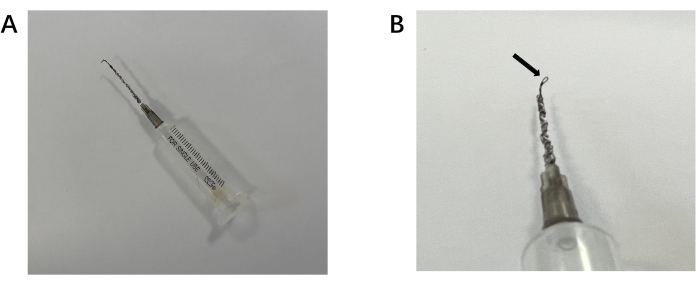
Figure 1: Self-made snare containing retractor for passing silk suture around the aortic arch. (A) Overall view of the retractor.(B) Detail of the retractor. Arrow indicates the snare for the silk to pass through. Please click here to view a larger version of this figure.
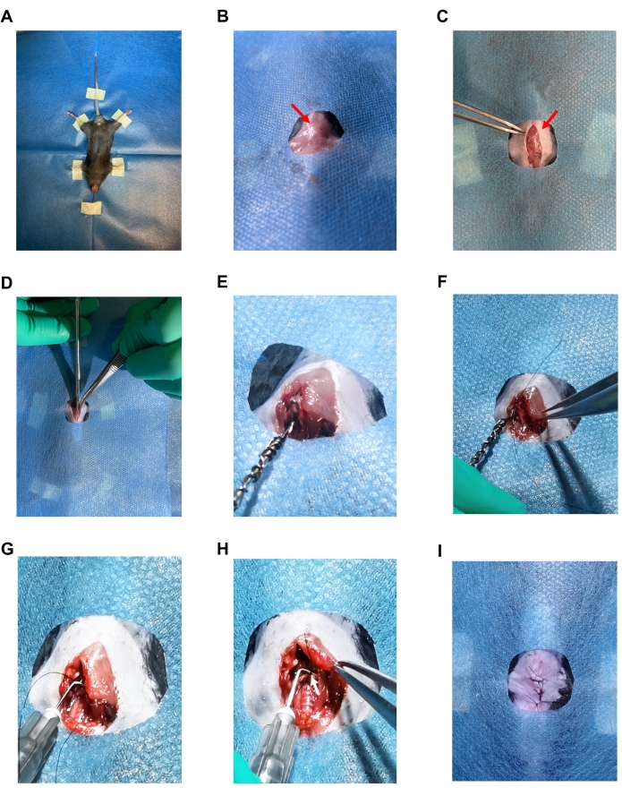
Figure 2: Images of the TAC surgery. (A) Supine position mouse fixed with tape and suture. (B) Sterile drape showing only the surgical area. (C) 1.5 cm vertical skin incision. The red arrow indicates a sternal angel. (D) Longitudinal midline incision that was made to the sternum. (E) Image showing the snare of the self-made retractor passing under the aorta. (F) Image showing the 7-0 silk suture passing through the snare loop. (G) A 27 G needle that was placed parallel to the aorta. (H) Ligation of the aorta with the 27 G needle. The white arrow indicates a ligation knot. (I) Sutured skin with 4-0 silk suture. Please click here to view a larger version of this figure.
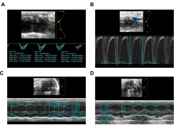
Figure 3: Representative image from ultrasound imaging system of sham and TAC mice after 4 weeks. (A) Pulsed-wave Doppler imaging of sham aortic arch. (B) Pulsed-wave Doppler imaging of aortic arch after TAC. (C) M-mode image of sham mouse calculating EF, LV Mass, wall thickness, and LVID. (D) M-mode image of TAC mouse calculating EF, LV Mass, wall thickness, and LVID. Please click here to view a larger version of this figure.
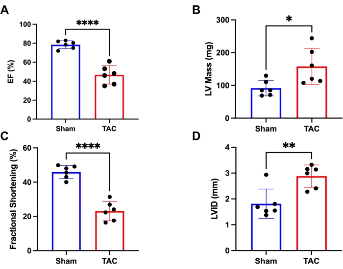
Figure 4: Heart function measured via Ultrasound Imaging System. (A) Ejection fraction (EF) of the mice in two groups. (B) The left ventricular mass (LV mass) of the mice was in two groups. (C) Fractional shortening (FS) of the mice in two groups. (D) The left ventricular internal diameter (LVID) of the mice in two groups. *p < 0.05, **p < 0.005, ***p < 0.0005. Data represents six mice per group. Please click here to view a larger version of this figure.
The induction of sustained pressure overload can gradually cause cardiac hypertrophy and heart failure. This model has been used in numerous laboratories around the world14,15,16. The protocol provided an improved TAC method that does not need microsurgical skills or mechanical ventilation.
The most important step in this protocol is passing silk suture under the aortic arch. When the snare has hooked the aortic arch, all moves must be gentle to reduce unnecessary traction to the artery. Also, the suture around the aorta must not be too tight in case of difficulty when pulling the spacer out. After the operation, abundant food and water are also important for the mouse to recover rapidly.
Previous manuscripts have provided other methods for TAC. Eichhorn et al. published a closed chest method that ligates the transverse aorta10. The whole procedure allows the ribs to remain intact, thus causing very small trauma. Zaw et al. provided a TAC method without entering the pleural cavity17. Tavakoli et al. presented a minimally invasive transverse aortic constriction that does not need intubating and ventilation11. All the above techniques require microsurgical skills. In addition, Lao et al. provided a method to produce TAC models with absorbable sutures18. The protocol in this study offers an alternative way to rapidly (within 10 min) conduct TAC surgery that does not need to operate under a microscope. Minimizing surgical trauma benefits the mice and reduces confounding factors during the experiment. Unlike the open-chest model, this model is minimally invasive and does not affect the normal breathing dynamics of the mouse. When fully mastered, the survival rate of this technique is over 95%. Also, it does not need mechanical ventilation and microscopic surgery skills; a self-made reusable retractor will do all the trick, avoiding systemic inflammatory effects induced by ventilation19. All these together significantly simplify the operating procedure.
There are some limitations of this technique. The acute increase of afterload does not fully reflect the gradual progression of arterial hypertension. The discrepancies in pathophysiology between multifactorial heart failure mouse models and clinical heart failure patients have raised concerns among researchers20. The pathophysiology presented in mice cannot be completely applied to humans.
In conclusion, this protocol provides an alternative procedure to conduct TAC, which can facilitate investigators when inducing heart failure or cardiac hypertrophy in mice.
The authors have nothing to disclose.
This work is funded by the National Natural Science Foundation of China (NSFC 81822002). We thank all the members who took part in this work.
4-0 nonabsorbable suture | Jinhuan | HM403 | Used for suturing the skin |
5 mL syringe | Haifuda Technology Co., Ltd. | BD-309628 | Used for making snare containing retractor |
7-0 nonabsorbable suture | Jinhuan | HM701 | Used for aorta ligation |
Animal temperature monitor | Kaerwen | FT3400 | Used for monitoring body temperature |
Buprenorphine | Sigma | B-044 | Used for post-surgical pain treatment |
Depilatory cream | Veet | N/A | Used for remove body hair from the surgical area |
Heating Pad | Xiaochuangxin | N/A | Used for maintaining body temperature |
Ibuprofen | MCE | HY-78131 | Used for post-surgical pain treatment |
Iron wire (0.5 mm) | Qing Yuan | Iron wire #26 | Used for making snare containing retractor |
Microscopic tweezers | RWD | F12006-10 | Used for penetrating and separating the tissue to open operation space |
Needle holder | RWD | F12005-10 | Used for pinching off the tip of gauge needle and blunting it |
Ophthalmic forceps | RWD | F14012-10 | Used for holding skin and other tissues |
Ophthalmic scissors | RWD | S11001-08 | Used for making sking incision of mouse |
Pentobarbital sodium | Sigma | P3761 | Used for mouse anesthesia |
Sterile operating mat | Hale & hearty | 211002 | Used for placing animal during surgery |
Ultra-sound imaging system | Fujifilm visualsonics | vevo1100 | Used for measure the blood flow velocity, left ventricular wall thickness and ejection fraction, https://www.visualsonics.com/product/imaging-systems/vevo-1100 |