An animal model of acquired hypoparathyroidism (HypoPT) is crucial to understanding how HypoPT affects mineral ion homeostasis and to verify the effectiveness of novel treatments. Here, a technique is presented to generate an acquired hypoparathyroidism (AHypoPT) rat model by parathyroidectomy (PTX) using carbon nanoparticles.
Hypoparathyroidism (HypoPT) is a rare disease involving the parathyroid glands that is characterized by a reduced secretion or potency of the parathyroid hormone (PTH), which leads to high serum phosphorus levels and low serum calcium levels. HypoPT most commonly results from accidental damage to the glands or their removal during thyroid or other anterior neck surgery. Parathyroid/thyroid surgery has become more common in recent years, with a corresponding rise in the occurrence of HypoPT as a postoperative complication. There is a critical need for a HypoPT animal model to better understand the mechanisms underlying the effects of HypoPT on mineral ion homeostasis and to verify the therapeutic effectiveness of novel treatments. Here, a technique is reported to create acquired HypoPT in male rats by performing parathyroidectomy (PTX) using carbon nanoparticles. The rat model shows great promise over the mouse models of hypoparathyroidism. Importantly, the human PTH receptor binding region has an 84.2% sequence similarity with that of the rat, which is higher than the 73.7% similarity shared with mice. Moreover, the effects of estrogen, which can affect the PTH/PTHrP receptor signaling pathway, have not been fully investigated in male rats. Carbon nanoparticles are lymphatic tracers that stain the thyroid lymph nodes black without affecting their function, but they do not stain the parathyroid glands, which makes them easy to identify and remove. In this study, serum PTH levels were undetectable after PTX, and this resulted in significant hypocalcemia and hyperphosphatemia. Thus, the clinical state of postoperative HypoPT can be remarkably represented in the rat model. Carbon-nanoparticle-assisted PTX can, therefore, serve as an extraordinarily effective and readily implementable model for studying the pathogenesis, treatment, and prognosis of HypoPT.
Parathyroid hormone (PTH) is secreted by the parathyroid glands. It is a major modulator of the calcium balance, maintains phosphate metabolism, and participates in bone turnover1,2. Hypoparathyroidism (HypoPT) manifests as a decreased secretion or functional loss of PTH. It is a rare endocrine disorder, with a prevalence of approximately 9-37 per 100,000 person-years3,4,5. HypoPT is characterized by decreased serum PTH and calcium levels accompanied by increased serum phosphorus6,7. HypoPT is classified based on its cause: acquired hypoparathyroidism (AHypoPT) or idiopathic hypoparathyroidism (IHypoPT)8. AHypoPT is more commonly encountered in clinical practice; about 75% of AHypoPT cases are caused by resection or accidental injury of the parathyroid glands during thyroid surgery or other head and neck surgeries. Other causes include radiotherapy and chemotherapy for head and neck tumors and drug toxicity1,8. Upgraded diagnostic methods and an increase in screening for thyroid gland-associated diseases have increased the number of thyroid gland surgical operations. This has led to a corresponding increase in the related parathyroid gland complications9,10.
Easily established animal models with stable characteristics are needed to better investigate AHypoPT and verify the therapeutic effectiveness of novel treatments. Parathyroidectomy (PTX) performed on rats and mice has been reported in previous studies6,11; however, due to the extremely small size of the parathyroid glands and the variability in their anatomical distribution, the success rate is relatively low in practice. Thus, thyro-parathyroidectomy (TPTX) (i.e., the total removal of the thyroid and parathyroid glands) is usually performed to ensure the resection of the parathyroid glands12. However, the resulting low thyroxine levels may complicate studies with this animal model13. HypoPT models established by other methods, such as drug stimulation and gene editing, cannot properly represent the most common AHypoPT pathogenesis. Our group previously used knockout mouse models to label the parathyroid glands and allow the removal of the parathyroid glands without damaging the thyroid glands and surrounding anatomical structures14,15. However, this method utilizes transgenic mouse models, which require a longer development time due to the mating and breeding requirements.
Therefore, we aimed to establish an easily generated model of AHypoPT. This study describes a rat model for PTX using carbon nanoparticle labeling. A carbon nanoparticle suspension of 50 mg/mL, which is commonly used in thyroid surgery, evenly distributes in the thyroid glands after local injection16. The thyroid glands turn black, but the parathyroid glands are left unstained17, thus clearly distinguishing the parathyroid glands from the thyroid glands and allowing the PTX to be performed without affecting the thyroid glands. This method is suitable for rats of different ages. The injection of the carbon nanoparticle suspension is safe and has a negligible effect on thyroid function18. The carbon nanoparticle-labeled PTX rat model generated in this study showed significant hypocalcemia and hyperphosphatemia phenotypes during the 4 week observation period. Thus, this AHypoPT model is easy to establish and has a reproducible phenotype.
This study was approved by the Institutional Animal Care and Use Committee at the State Key Laboratory of Oral Diseases, Sichuan University. Permission was obtained from relevant local agencies before the experiment. Eight 8-10 week old male Sprague-Dawley (SD) rats, with an average weight of 200-250 g, were used for the present study. The animals were obtained from a commercial source (see Table of Materials). Food and water were provided ad libitum throughout the experimental period.
1. Preoperative preparation for the generation of carbon-nanoparticle-assisted PTX rats
2. Parathyroidectomy (PTX)
3. Postoperative recovery and observation
The locations and number of parathyroid glands were initially observed in rats under a dissection microscope. Before the carbon nanoparticle injection, the thyroid glands were a translucent red color, and the parathyroid glands were hardly distinguishable under the microscope (Figure 1A). After the nanoparticle injection, the thyroid glands were stained black, while the parathyroid glands remained unstained (Figure 1B). The careful dissection of the light-colored parathyroid glands left the thyroid glands untouched (Figure 1C). Generally, the parathyroid glands were distributed over the lateral or posterior edges of the thyroid glands.
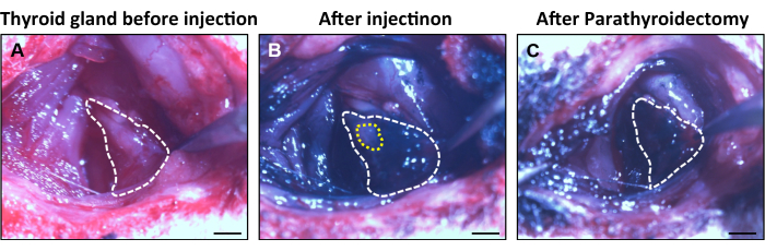
Figure 1: The appearance of the thyroid and parathyroid glands during the surgical procedures. (A) The thyroid glands (white dotted line) are located lateral to the trachea. (B,C) The thyroid glands showed black staining (white dotted line) after the injection of the carbon nanoparticles, while the parathyroid glands (yellow dotted line) exhibited a light color. Scale bars = 2 mm. Please click here to view a larger version of this figure.
The operation time from preoperative preparation to PTX completion was approximately 20 min. The 4 week survival rate of the postoperative rats was 90.9% (60/66). The PTX rats were observed to be hunch-backed 1 week after surgery. A sham-operated control group was simultaneously established by conducting all the steps in the protocol except for step 2.6. All the surviving carbon nanoparticle-labeled PTX rats had a lower mean ionized Ca2+ level, which was 2 SD lower than that of the sham-operated group. The hypoparathyroidism phenotype in the carbon nanoparticle-labeled PTX rats, evidenced by reduced serum calcium, elevated serum phosphate, and undetected PTH, remained steady during the 4 week monitoring period.
At 7 days after surgery, the serum Ca2+ and PTH levels were significantly reduced in the PTX rats compared to the sham group (Ca2+ = 4.97 mmol/L ± 0.99 mmol/L vs. 8.98 mmol/L ± 0.58 mmol/L, p < 0.05; PTH = 13.13 pg/mL ± 6.58 v pg/mL s. 313.06 pg/mL ± 75.24 pg/mL, p < 0.05). Serum Pi was significantly increased after the PTX surgery (Pi = 13.90 mmol/L ± 1.77 mmol/L vs. 7.46 mmol/L ± 1.28 mmol/L). The serum levels of urea and creatinine were comparable between the sham and PTX groups 7 days after the PTX surgery (urea = 8.71 mmol/L ± 0.81 mmol/L vs. 8.84 mmol/L ± 0.89 mmol/L, p > 0.05; creatinine = 49.03 µmol/L ± 13.14 µmol/L vs. 53.15 µmol/L ± 18.28 µmol/L, p > 0.05). At 14 days after the PTX surgery, the urinary Ca2+ and Pi levels were significantly reduced (Ca2+ = 2.33 mmol/L ± 0.53 mmol/L vs. 7.18 mmol/L ± 4.27 mmol/L, p < 0.05; Pi = 2.40 mmol/L ± 1.90 mmol/L vs. 5.29 mmol/L ± 1.52 mmol/L, p < 0.05) (Figure 2).
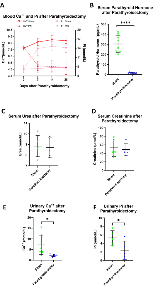
Figure 2: Serum Ca2+, Pi, PTH, urea, and creatinine levels and urinary Ca2+ and Pi levels after carbon-nanoparticle-assisted parathyroidectomy. (A) The PTX rats exhibited stable hypocalcemia and hyperphosphatemia over the 4 week observation period (N = 4). (B) Serum PTH was undetectable in the PTX rats 7 days after the operation (N = 8). (C,D) The serum levels of urea and creatinine were comparable between the sham and PTX groups 7 days after the surgery (N = 5). (E,F) The urinary Ca2+ and Pi levels were significantly reduced 14 days after PTX surgery (N = 8). The error bars indicate the standard deviation. Abbreviations: PTX = parathyroidectomy; Ca++ = ionized calcium in serum; PTH = parathyroid hormone; Pi = ionized phosphorous in serum. Please click here to view a larger version of this figure.
There were no significant differences in body weight between the PTX and sham groups on postoperative day 7 (POD7), POD14, and POD28 (body weight on POD0 = 256.40 g ± 4.76 g vs. 252.56 g ± 6.69 g, p > 0.05; body weight on POD7 = 266.00 g ± 6.93 g vs. 257.44 g ± 30.56 g, p > 0.05; body weight on POD14 = 294.80 g ± 25.90 g vs. 288.22 g ± 37.35 g, p > 0.05; body weight on POD28 = 327.75 g ± 24.82 g vs. 324.17 g ± 57.97 g, p > 0.05). Moreover, serum C-telopeptide of type I collagen (CTX-1) was statistically decreased on POD28 (CTX-1 = 82.03 pg/mL ± 8.98 pg/mL vs. 100.33 pg/mL ± 6.36 pg/mL, p < 0.05). Serum osteocalcin showed no significant difference on POD28 (osteocalcin = 913.66 pg/mL ± 378.03 pg/mL vs. 1066.17 pg/mL ± 549.80 pg/mL, p > 0.05) (Figure 3).
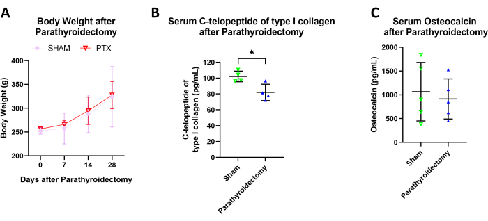
Figure 3: Body weight, blood C-telopeptide of type I collagen, and osteocalcin levels after carbon-nanoparticle-assisted parathyroidectomy. (A) There were no significant differences in body weight between the PTX and sham groups on POD7, POD14, and POD28 (N = 14).(B) The PTX rats exhibited a statistical decrease in serum C-telopeptide of type I collagen (N = 4). (C) There were no significant differences in the serum osteocalcin levels (N = 5). The error bars indicate the standard deviation. Please click here to view a larger version of this figure.
Epidemiological reports indicate that the detection of thyroid diseases has increased significantly, and the number of related surgeries performed has increased accordingly19,20. The incidence rate of postsurgical hypoparathyroidism is approximately 7.6%8,21, while the increased morbidity of acquired hypoparathyroidism has caused this rare disease to gain greater research attention. It is, therefore, particularly important to establish a suitable animal model to investigate the pathogenesis of the disease, as well as to test the outcomes of novel therapeutic treatments. However, at present, there are limited animal models available. Moreover, the success rate, survival rate, and difficulty of surgical procedures in producing such models remain problematic. Our group has previously reported two HypoPT models in mice. In PTHcre+/Rosa-mTmG mice, the parathyroid glands were fluorescently labeled to help accurately dissect the parathyroid glands, and this method was also helpful for finding parathyroid glands with abnormal anatomical distribution to improve the success rate of surgery14. Another modeling approach used transgenic mice, in which parathyroid gland cells could be targeted by diphtheria toxin. The parathyroid glands could then be destroyed by the systemic administration of the diphtheria toxin without requiring surgery14,15. However, the abovementioned methods require extensive crossbreeding of transgenic mice, resulting in relatively high time and cost requirements. Moreover, the systemic administration of diphtheria toxin may have widespread side effects. Currently, thyro-parathyroidectomy (TPTX) is the usual procedure performed to ensure the resection of the parathyroid glands12. Although the technique is easily performed and has a high success rate, the damage to the thyroid glands cannot be ignored. The potential impact of injury to or destruction of the thyroid glands on the experimental results might be significant, meaning this is a major limitation of all studies in this field21,22.
In the current study, a carbon nanoparticle suspension, commonly used to visualize the thyroid glands in clinical practice, was injected to enhance the PTX surgery. This method is safe, fast, and highly feasible. It can effectively label the thyroid glands with a black stain and leave the parathyroid glands unstained, which enables the precise identification and dissection of the parathyroid glands while avoiding injury to the thyroid glands. This labeling method has the same effect as that achieved using the fluorescent labeling of transgenic mice but is not limited by the genotype. Furthermore, the surgery time of carbon-nanoparticle-assisted PTX is around 20 min, which saves time compared with the 2 h surgery required for 5-ALA fluorescence identification23. In addition, due to the biosecurity of the carbon nanoparticles24, this modeling method can be used on rats as young as 7 days old. One critical step to be noted during the surgery is that the dosage of the carbon nanoparticle suspension can be adjusted according to the weight of the rats. The volume of carbon nanoparticle suspension used in this study (1 µL) is enough for surgery on adult rats, even if some amount is lost in the syringe. The distribution of all the parathyroid glands is difficult for beginners to identify, and plenty of practice is recommended.
The current study has some limitations. For instance, it is impossible to identify remote parathyroid glands unattached to the thyroids using carbon nanoparticles. If the serum parameters remain unchanged after surgery, it may indicate that some remote parathyroid glands were present and not removed. The staining period required for the optimal differentiation and identification of the parathyroid glands was not measured; however, the thyroid glands were stained properly within 5 min of nanoparticle administration and retained the stain during the entire surgical procedure. The functioning of the thyroid glands during the follow-up period was not recorded in this study. However, in our previous study, which involved utilizing a transgenic mouse model to identify and remove the parathyroid glands, the thyroid gland function was shown to be preserved15. The tolerance of the rats to the carbon nanoparticles was also not tested in this study; however, these nanoparticles have been commercially used as pharmaceuticals in clinical surgeries16. Generally, this method allows researchers to choose an animal with a desired genotype and operation time point. Ultimately, this approach is expected to provide useful rat models for acquired hypoparathyroidism.
The authors have nothing to disclose.
This work was supported by NSFC grant 81800928, Research Funding from the West China School/Hospital of Stomatology Sichuan University (No. RCDWJS2021-1), and the State Key Laboratory of Oral Diseases Open Funding grant SKLOD-R013.
0.9% Sodium Chloride Solution | Kelun Co. Sichuan, China | ||
10 µL 30G NanoFil Syringe | WPI | ||
6-0 polyglactin 910 suture with needle | Ethicon, Inc | J510G | |
Calcium LiquiColor test | EKF | 0155-225 | For Ca2+ analysis |
Carbon Nanoparticles Suspension Injection | Lummy, Chongqing, China | H20073246 | 1 mL : 50 mg |
Creatinine (Cr) Assay kit ( sarcosine oxidase ) | Jiancheng, Nanjing, China | C011-2-1 | For creatinine analysis |
Disposable Scalpel | Shinva, China | ||
Dumstar Biology forceps | Shinva, China | ||
Micro Dissecting Spring Scissors | Shinva, China | ||
MicroVue Rat intact PTH ELISA | Immunotopics | 30-2531 | For the measurement of PTH in rat serum |
Needle Holder | Shinva, China | ||
Phosphorus Liqui-UV test | EKF | 0830-125 | For Pi analysis |
Ply gauze | Weian Co. Henan, China | ||
Povidone-Iodine | Yongan pharmaceutical Co.Ltd. Chengdu, China | ||
Prism 9.0 (statistics and graphing software) | GraphPad Software, Inc., San Diego, CA, USA | https://www.graphpad.com/scientific-software/prism/ | |
Rat C-telopeptide of type I collagen (CTX-I) ELISA Kit | CUSABIO, Wuhan, China | CSB-E12776r | For CTX-I analysis |
Rat Osteocalcin/Bone Gla Protein (OT/BGP) ELISA Kit | CUSABIO, Wuhan, China | CSB-E05129r | For osteocalcin analysis |
Safety Single Edge Razor Blades | American Safety Razor Company | 66-0089 | |
Sprague-Dawley Rats | 8 to 10 weeks old | ||
Surgical Incise Drapes | Liangyou Co. Sichuan, China | ||
Urea Assay Kit | Jiancheng, Nanjing, China | C013-2-1 | For urea analysis |