Continuous arterial blood pressure recording allows the investigation of impacts of various hemodynamic parameters. This report demonstrates the application of continuous arterial blood pressure monitoring in a large animal model of ischemic stroke for determination of stroke pathophysiology, impact of different hemodynamic factors, and the assessment of novel treatment approaches.
Control of blood pressure, in terms of both absolute values and its variability, affects outcomes in ischemic stroke patients. However, it remains challenging to identify the mechanisms that lead to poor outcomes or evaluate measures by which these effects can be mitigated because of the prohibitive limitations inherent to human data. In such cases, animal models can be utilized to conduct rigorous and reproducible evaluations of diseases. Here we report refinement of a previously described model of ischemic stroke in rabbits that is augmented with continuous blood pressure recording to assess the impacts of modulation on blood pressure. Under general anesthesia, femoral arteries are exposed through surgical cutdowns to place arterial sheaths bilaterally. Under fluoroscopic visualization and roadmap guidance, a microcatheter is advanced into an artery of the posterior circulation of the brain. An angiogram is performed by injecting the contralateral vertebral artery to confirm occlusion of the target artery. With the occlusive catheter remaining in position for a fixed duration, blood pressure is continuously recorded to allow for tight titration of blood pressure manipulations, whether through mechanical or pharmacological means. At the completion of the occlusion interval, the microcatheter is removed, and the animal is maintained under general anesthesia for a prescribed length of reperfusion. For acute studies, the animal is then euthanized and decapitated. The brain is harvested and processed to measure the infarct volume under light microscopy and further assessed with various histopathological stains or spatial transcriptomic analysis. This protocol provides a reproducible model that can be utilized for more thorough preclinical studies on the effects of blood pressure parameters during ischemic stroke. It also facilitates effective preclinical evaluation of novel neuroprotective interventions that might improve care for ischemic stroke patients.
Ischemic stroke (IS) is a leading cause of death and long-term disability worldwide, and its prevalence is projected to increase as society ages1. While substantial advances have been made in acute interventions and secondary prevention strategies, adjunctive neuroprotective treatments have not followed apace2,3,4,5,6,7. Further research is needed into stroke pathobiology because mechanisms by which therapies may or may not prove effective are poorly understood. This is largely due to the heterogeneous nature of the stroke patient population, many of whom have numerous comorbidities that confound analysis1. One driver of limitations in research is the absence of tissue-level data-the gold standard in biomedical research-due to the prohibitive morbidity of sampling tissue from the human central nervous system. Specifically, vascular tissue harvesting in a living human would cause a stroke, so vascular tissue is typically only obtained at autopsy, which is under-representative of the general population and skews toward more advanced disease in elderly patients with concomitant diagnoses.
In such cases, when sufficient human data cannot be utilized, animal models can bridge the data gaps. Large animal models of stroke are limited as most large animals used in research are ungulates having a rete mirabile that prevents direct endovascular access to the cerebral arteries8,9,10,11,12,13,14,15,16,17. Rabbits have a long history of use for the investigation of cardiovascular disease, including intracranial pathologies8,9,10,11,12,13,14,15,16,17. Rabbits present an ideal model for cerebrovascular diseases because they are large enough for endovascular catheterization and lack the rete mirabile that precludes intracranial access in other large mammals9,15,16,17. They have been previously utilized specifically for the investigation of IS through precise and well-controlled occlusion of an intracranial artery with a microcatheter18.
Blood pressure (BP) control, both through modulation of absolute BP or BP variability (BPV), the degree to which arterial BP fluctuates around a mean BP, is an emerging potential therapeutic target for IS patients after reports of worse outcomes in those with poorly controlled BP or BPV19,20,21,22. Mechanistic investigation into how changes lead to poor outcomes in IS patients is lacking. This is partly due to the difficulty in obtaining tissue-level data and performing well-controlled analyses in humans. To test interventions that modulate BP or BPV, animal models must be utilized to overcome these limitations. This report describes the successful pairing of a previously validated rabbit model of IS using controlled occlusion of the posterior cerebral artery in conjunction with continuous intra-arterial measurement of BP18. The method presented here improves on the previous approaches to stroke pathophysiology by applying a validated and reproducible stroke model to a system in which precise measurement and control of BP can be achieved. In this refined model, infarct burden can be assessed with post-procedural histopathologic staining of the harvested brain, which is also amenable to various stains and more advanced analyses such as spatial transcriptomics. Additionally, the occluded posterior circulation artery can also be chosen to be evaluated for morbidity analysis following survival procedures.
This protocol is approved by the Institutional Animal Care and Use Committee (University of Utah IACUC Protocol Number 21-09021). Mature New Zealand White rabbits are obtained from commercial vendors.
1. Animal acquisition
2. Anesthesia and monitoring
3. Surgical preparation
4. Arterial access
5. Cervicocerebral angiography and intracranial access
6. Blood pressure measurement and modulation
7. Euthanasia and tissue harvesting
In the initial experiments with this model, our group successfully achieved the desired outcome of a posterior cerebral or superior cerebellar artery occlusion in 12 out of 14 animals (85.7%). For the experiment, seven males and seven females were studied. The mean animal weight was 3.6 kg (± 0.46 kg). In the two animals in which success was not achieved, profound catheter-induced vasospasm precluded safe access to the intracranial circulation. In one rabbit, intracranial access could not be obtained due to occlusive vasospasm, and in the other animal, intracranial arterial perforation occurred during attempted catheterization, which was likely due to attempting to position the microcatheter too far distally in the posterior cerebral artery.
In all the animals, the brain was successfully harvested and subjected to histopathological analysis with either hematoxylin and eosin (H&E) staining or 2% triphenyltetrazolium chloride (TTC). In keeping with previously published results of the occlusion model, larger infarct volumes occurred with longer occlusion durations, which have been successfully performed from 60 to 240 min18. H&E stain images after 90 min of occlusion with 120 min of reperfusion are provided in Figure 3.
Baseline arterial BPs below normotension (40-60 mmHg systolic BP) were noted in all the animals after induction of anesthesia without use of vasopressors or inflation of an intra-aortic balloon. Partial inflation of the balloon has demonstrated immediate increase in systolic BP, with a sample BP tracing provided in Figure 4. This figure includes tracing of a short duration to visualize both the near-instantaneous change following inflation of the intra-aortic balloon as well as the changes throughout each cardiac cycle.
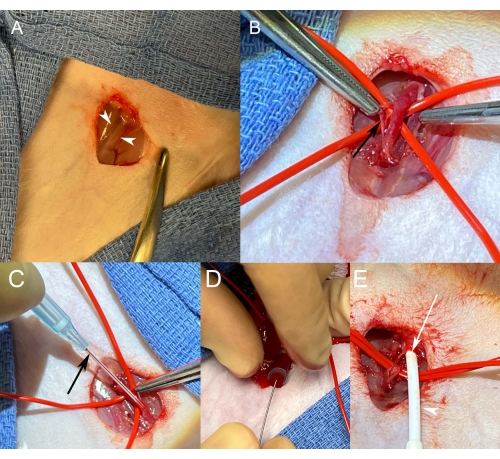
Figure 1: Femoral artery access. (A) Surgical exposure of the right femoral neurovascular bundle before blunt dissection. White arrowheads indicate the medial and lateral borders of the bundle to be exposed with dissection. (B) After isolation, the artery becomes engorged when dripping with lidocaine solution and applying gentle traction to the downstream vessel loop. The vessel can be cleaned by gentle dissection of tissue (black arrow) off the adventitia. (C) Maintaining gentle tension on the vessel, a 22 G angiocatheter is advanced into the vessel. After seeing blood flash in the angiocatheter (black arrow) and its chamber, the angiocatheter is gently advanced into the artery. (D) With the angiocatheter advanced in the artery to its hub, a Cope wire is advanced into the artery through the angiocatheter. (E) After removing the angiocatheter over a Cope microwire, a vascular sheath (white arrowhead) is advanced along with its inner introducer over the wire. The sheath is seen entering the artery, the wall of which can be seen at the arteriotomy site (white arrow). Please click here to view a larger version of this figure.
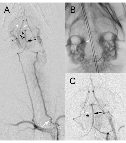
Figure 2: Angiographic images. (A) Low magnification view of digital subtraction angiography during injection of the proximal left vertebral artery (white arrow) demonstrates filling of the basilar artery (black arrow). Note the reflux back down the right vertebral artery into the subclavian artery, which can be used as a roadmap to guide catheterization. Black arrowheads delineate the course of the right superior cerebral artery that will be targeted for occlusion. White arrowheads identify the posterior cerebellar artery, which can also be targeted. (B) High magnification spot fluoroscopic image demonstrates the microcatheter in the right superior cerebellar artery from a right vertebral approach. The white arrowhead indicates the radiopaque marker at the microcatheter tip. (C) High magnification digital subtraction angiography during injection of the left vertebral artery demonstrates persistent filling of the basilar artery (black arrow) while the microcatheter runs through it. No filling is noted beyond the mid-right superior cerebellar artery, where the tip of the microcatheter is indicated by the white arrowhead. The black asterisk identifies non-perfused territory downstream to the occlusion in the superior cerebellar artery. Please click here to view a larger version of this figure.
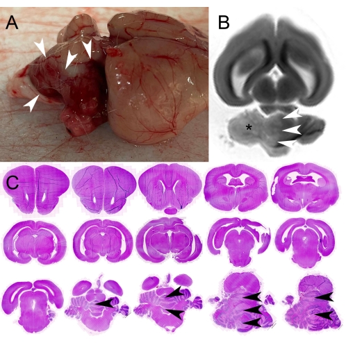
Figure 3: Pathology images. (A) Photograph of intact harvested brain showing the surface of the brain from the animal's right. Note the darkened appearance of the superior cerebellum indicating petechial hemorrhage in the acutely infarcted tissue. White arrowheads demarcate the margin of the infarction. (B) Long-axis T2-weighted magnetic resonance image of the intact brain in formalin. Note the increased signal in the right cerebellum (asterisk), consistent with the infarct, the border of which is delineated by white arrowheads. (C) Bright field images of 1.5 mm thick serial coronal sections following hematoxylin and eosin (H&E) staining demonstrates infarction in the right cerebellum, the margin of which is indicated by black arrowheads on multiple slices. These sections were sliced from blocks of a harvested rabbit brain cut in the coronal plane with a cutting matrix. Please click here to view a larger version of this figure.
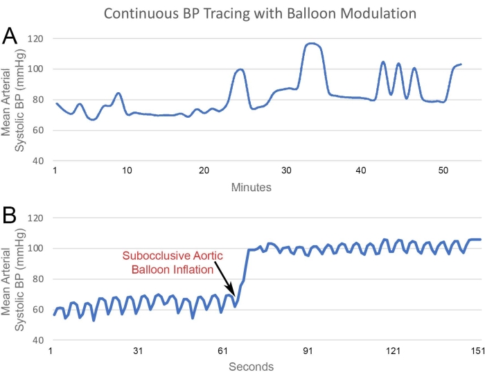
Figure 4: BP Monitoring. BP pressure tracing from a Fogarty balloon catheter positioned in the infrarenal aorta. (A) Data from approximately 1 h of BP monitoring demonstrates real-time arterial pressure changes with changes in balloon inflation. (B) Short-term tracing demonstrates the pressure changes throughout the cardiac cycle. Additionally, small, rapid changes are noted from respiratory variability, which is physiologically normal. An immediate near-doubling of measured BP is noted following the inflation of the balloon. Please click here to view a larger version of this figure.
Substantial progress has been made in the management of IS, particularly considering advances in acute intervention and secondary prevention strategies. However, more work can be done to improve care for IS patients. Limited progress in other aspects of IS treatment, particularly in the realm of neuroprotection, likely results from the limitations in pathophysiological understanding of mechanistic processes at the tissue and molecular level. Impactful data from humans is unrealistic and likely impossible to acquire. In such circumstances, tissue-level data from animal models can bridge knowledge gaps and affect meaningful change.
As detailed above, rabbits provide an optimal combination of size, physiology, and anatomy for the investigation of cerebrovascular pathologies18. Lacking a rete mirabile, there are no structural barriers to the intracranial arteries. Additionally, the intracranial vessels are large enough to accommodate endovascular devices, which is not similarly feasible in rodent models. Data from the intracranial tissues can be analyzed in multiple ways, whether through established histopathological and immunohistochemical stains or cutting-edge methods such as endovascular biopsy samples analyzed with single-cell RNA sequencing or spatial transcriptomics of intact tissues9,15,16,17,18. This reported protocol improves upon previous reports of the rabbit occlusion model given its application of multiple posterior circulation arteries and emphasis on the practical steps to mitigate vasospasm or arterial injury18. This protocol is also an improvement on the existing reports given the feasible and reproducible methods for continuous BP monitoring.
While rabbits present immense potential for advances in the pathobiological understanding of cerebrovascular diseases, they also present technical challenges. According to anecdotal reports from veterinary collaborators, rabbits have a well-earned reputation for being hemodynamically unstable. Hypotension during anesthesia induction is inevitable. To mitigate effects, prompt intubation after sedation is needed. Efficient exposure and prompt access of a femoral artery allow early hemodynamic monitoring by virtue of BP measurement. However, this must be balanced with meticulous techniques to limit blood loss during access. Limiting blood loss must also be a priority throughout all the steps of the endovascular procedure, which can be achieved with concerted observation during device exchanges and using rotating hemostatic valves on all catheters. As the entire protocol occurs over several hours, replacement intravenous fluids are also needed to counteract blood loss and insensible losses. Finally, rabbit arteries are profoundly sensitive and prone to vasospasm, which can be prepared for with topical nitroglycerine, as described above. Minimal instrumentation can limit vasospasm, and this is best achieved by concerted planning to minimize arterial exposure to mechanical stressors. Lidocaine dripped on the artery can counteract this reaction, and verapamil (1 mg/mL) can be similarly dripped on the vessel or infused into the artery through a catheter. Finally, pausing for few minutes can allow vasospasm to resolve.
Despite the challenges, the similarity of rabbit anatomy and physiology to humans can be useful in modeling human diseases and the ability to minimize these challenges make them suitable for experimentation. Coupled with cutting-edge sequencing and imaging, rabbits offer a remarkable opportunity for investigating cerebrovascular disease. In particular, the methods described above allow the well-controlled study of IS and the effects of various hemodynamic parameters on its pathophysiology, diagnosis, and management.
The authors have nothing to disclose.
The research reported in this publication was supported by the National Center for Advancing Translational Sciences of the National Institutes of Health under Award Numbers UL1TR002538 and KL2TR002539 and by Transformational Grant 19TPA34910194 from the American Heart Association.
3-0 Silk Suture | Ethicon | A184H | |
Buprenorphine | Sigma-Aldrich | B9275 | |
Catheter | Terumo | CG415 | 4F glide catheter |
Endovascular Pressure Sensor | Millar | SPR-524 | |
Euthasol | Virbac | PVS111 | |
Guidewire | Terumo | GR1804 | |
Iohexol | ThermoFisher | 466651000 | Iodinated Contrast |
Ketamine | Biorbyt | orb61131 | |
LabChart Software | ADInstruments | ||
Lidocaine | Spectrum | LI102 | |
Microcatheter | Medtronic | EV3 105-5056 | Marathon Microcatheter |
Microwire | Medtronic | EV3 103-0608 | Mirage Microwire |
PowerLab | ADInstruments | ||
Rabbit Brain 2mm Coronal Cutting Matrix | Ted Pella | 15026 | |
Saline | FisherScientific | 23-535435 | |
Sheath | Merit Medical | PSI-5F-11 | |
Xylazine | ThermoFisher | J61430.14 |