Laparoscopic radical antegrade modular pancreatosplenectomy (L-RAMPS) is currently considered technically safe and feasible. However, due to technical challenges and a lack of supporting evidence for widespread clinical applications, only a limited number of institutions are currently conducting L-RAMPS. This article aims to provide detailed techniques for laparoscopic posterior radical antegrade modular pancreatosplenectomy.
Distal pancreatic carcinoma is a highly malignant tumor with strong invasiveness, often growing to the edge of the pancreas and penetrating the pancreatic capsule to infiltrate surrounding tissues. In conventional distal pancreatosplenectomy (DPS), tumor cells are prone to spread along the direction of blood and lymphatic reflux due to surgical compression. Additionally, inflammation makes it challenging to achieve R0 resection, leading to a lower patient survival rate. To address these limitations, radical antegrade modular pancreatosplenectomy (RAMPS) was developed, emphasizing deeper excision, including the left anterior renal fascia, the left anterior renal adipose sac, and even the left adrenal gland, to improve the R0 resection rate. With the advancement of minimally invasive surgical techniques, laparoscopic RAMPS (L-RAMPS) is being considered technically safe and feasible in oncology. However, due to technical difficulties and a lack of supporting evidence for clinical application, only a few institutions are currently conducting L-RAMPS. In this context, this article presents detailed techniques for laparoscopic posterior radical antegrade modular pancreatosplenectomy (L-pRAMPS), offering promise for future clinical applications.
Conventional distal pancreatosplenectomy (DPS) has traditionally been the standard surgical procedure for distal pancreatic carcinoma1,2. Distal pancreatic carcinoma is a highly invasive tumor that easily infiltrates into retroperitoneal tissues. Simultaneously, pancreatic cancer is often accompanied by chronic pancreatitis, making the boundary between the pancreas and adjacent tissues unclear. Consequently, during conventional DPS, there is a risk of tumor cells spreading along the direction of blood circulation and lymphatic return. In addition to the inherent inflammation associated with pancreatic cancer, achieving R0 resection is challenging, leading to a relatively low postoperative survival rate for patients3.
With the advancement of surgical techniques and a deeper understanding of the pancreatic lymphatic system, the high positive rate of the surgical margin and incomplete lymph node dissection in conventional DPS have garnered more attention. In response, radical antegrade modular pancreatosplenectomy (RAMPS) has emerged to address these challenges3,4. RAMPS involves the complete removal of tissues around the body and tail of the pancreas, including the tumor, to ensure a negative incisional margin and thorough dissection of lymph nodes in N1 nodes, the superior mesenteric artery (SMA), and the anterior and left of the celiac trunk3,4.
In 2003, Strasberg first reported RAMPS3. With the development of minimally invasive surgical techniques, there have been sporadic reports of laparoscopic RAMPS (L-RAMPS) in recent years. L-RAMPS is preliminarily considered technically safe and feasible in oncology5,6,7,8,9. However, due to technical difficulties and a lack of supporting evidence for clinical application, only a few institutions are currently conducting L-RAMPS. In light of this situation, this article presents the techniques of laparoscopic posterior radical antegrade modular pancreatosplenectomy (L-pRAMPS) in detail, holding great promise for future clinical application.
The surgical procedure was approved by the institutional review board at The First Affiliated Hospital of Jinan University. The patient, a 58-year-old female, was admitted with a chief complaint of "abdominal pain for 2 months." A computed tomography (CT) scan conducted at an external hospital revealed a space-occupying lesion in the distal pancreas, raising concerns about the possibility of pancreatic carcinoma. Before the surgery, the patient provided written informed consent. The surgical tools and equipment used for this procedure are listed in the Table of Materials.
1. Preoperative preparation
2. Surgical technique
3. Postoperative procedures
The surgery proceeded smoothly, and the intraoperative rapid frozen pathological examination indicated the absence of cancer at the incisal margin of the pancreas. Throughout the surgery, the patient's vital signs remained stable, and the anesthesia was effective. The operation duration was 150 min, with an intraoperative blood loss of 80 mL. Anal exhaust occurred 32 h postoperatively. There were no complications such as abdominal hemorrhage, pancreatic leakage, abdominal infection, or incision infection. The postoperative hospital stay was 6 days.
The histopathological analysis of postoperative paraffin sections revealed moderately differentiated pancreatic ductal adenocarcinoma (Figure 10). No tumor tissues were identified at the incisal margin of the pancreas. Fourteen lymph nodes were removed, and one exhibited metastasis. The tumor stage was determined to be pT3N1M0, IIB (Table 1). A postoperative CT scan demonstrated the successful removal of the tumor without significant recurrence or metastasis (Figure 11).
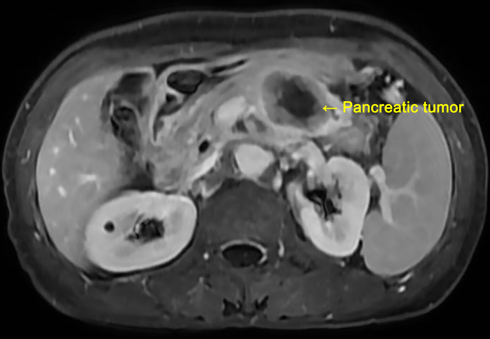
Figure 1: Presurgical abdominal MRI image of the patient. The abdominal MRI indicates that the size of the pancreatic body and tail mass is approximately 3.5 cm × 3.0 cm × 3.0 cm, suggesting an epithelial-derived malignant tumor. Please click here to view a larger version of this figure.
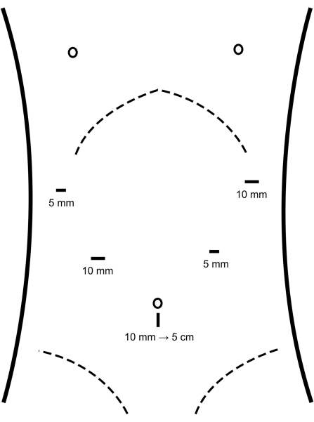
Figure 2: Location of the trocars. Trocars are placed into the abdominal cavity at the locations depicted in the figure. Please click here to view a larger version of this figure.
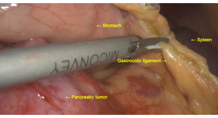
Figure 3: Exploration of the pancreatic tumor. The gastrocolic ligament is cut to facilitate the exploration of the pancreatic tumor. Please click here to view a larger version of this figure.
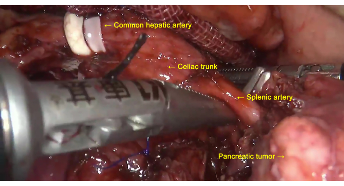
Figure 4: Lymph node dissection. Lymph nodes around the common hepatic artery, celiac trunk, and splenic artery are dissected. Please click here to view a larger version of this figure.
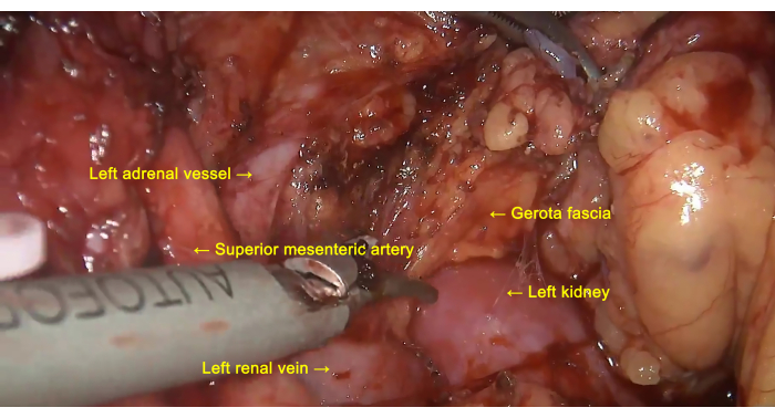
Figure 5: Removal of the anterior layer of Gerota fascia. Lymph nodes around the superior mesenteric artery (SMA) are dissected, and the anterior layer of Gerota fascia is peeled from the surface of the left kidney along the left renal vein. Please click here to view a larger version of this figure.
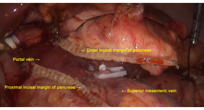
Figure 6: Pancreatic transection. The pancreas is severed at the neck using a linear stapling device. Please click here to view a larger version of this figure.
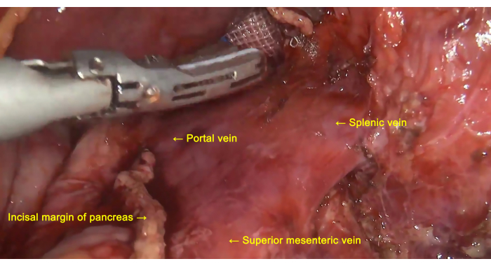
Figure 7: Treatment of the splenic vein. The splenic vein is dissected, exposed, ligated, and severed at the root. Please click here to view a larger version of this figure.
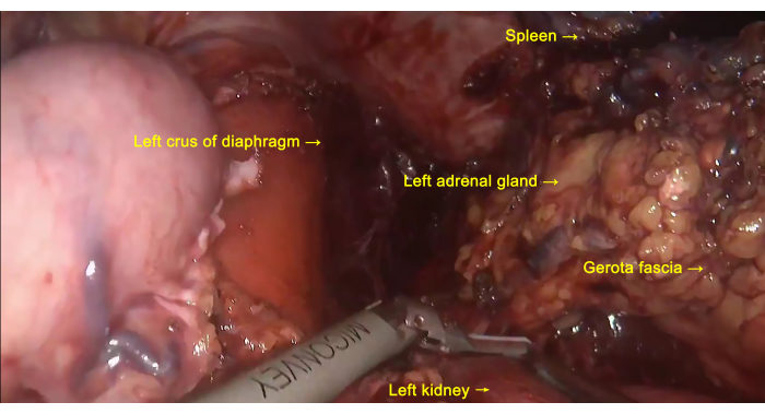
Figure 8: En Bloc resection. The distal pancreas, the anterior layer of Gerota fascia, the left adrenal gland, and the spleen are removed as a single entity. Please click here to view a larger version of this figure.
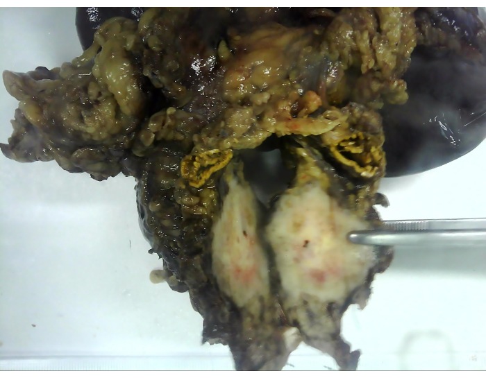
Figure 9: The excised specimen. The excised specimen includes the distal pancreas, the anterior layer of Gerota fascia, the left adrenal gland, and the spleen. Please click here to view a larger version of this figure.
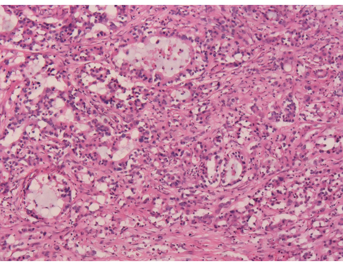
Figure 10: Postoperative histopathological examination. Postoperative paraffin sections reveal moderately differentiated pancreatic ductal adenocarcinoma. Magnification: 40x. Please click here to view a larger version of this figure.
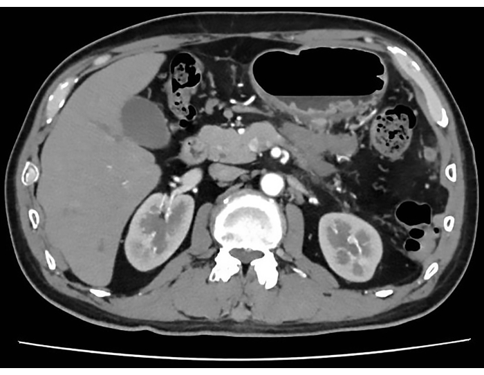
Figure 11: Postoperative CT scan. The postoperative CT scan indicates successful tumor removal without significant recurrence or metastasis. Please click here to view a larger version of this figure.
Operation time (min) | 150 |
Intraoperative blood loss (mL) | 80 |
First flatus (h) | 32 |
First postoperative liquid diet (days) | 1 |
Postoperative hospital stay (days) | 6 |
Postoperative complications (yes/no) | no |
Bleeding (yes/no) | no |
Pancreatic leakage (yes/no) | no |
Abdominal infection (yes/no) | no |
Incision infection (yes/no) | no |
Pathological result | Pancreatic ductal adenocarcinoma |
Differentiation | Moderate |
TNM stage | pT3N1M0 |
AJCC stage | IIB |
Table 1: The surgical outcomes of the patient.
DPS has been widely used as the standard radical surgery for distal pancreatic carcinoma1,2. However, due to the highly invasive nature of pancreatic cancer, it is easy for the tumor to grow to the edge of the pancreas and even break through the surface of the pancreas. Meanwhile, accompanying chronic pancreatitis makes the boundary between the pancreas and the surrounding tissues unclear. Therefore, it is difficult to achieve R0 resection during conventional surgical procedures3,4. In conventional DPS, due to the incomplete understanding of the pancreatic lymphatic system and limited surgical techniques, a surgical mode has been developed that neither completely removes the lymphatic reflux loop nor cleans up any lymph nodes around the celiac trunk or SMA. Therefore, the survival rate of patients is low3,4.
With the development of surgical techniques and the deepening understanding of the pancreatic lymphatic system, the high positive rate of surgical margin and incomplete lymph node dissection in DPS have received attention. RAMPS is a new technique that differs from DPS, and its concept stems from the more commonly mentioned radical resection of pancreatic head carcinoma. In 2003, Strasberg applied the concept of Whipple surgery to radical resection of pancreatic body and tail carcinoma and proposed RAMPS, which pioneered the whole resection of pancreatic body and tail combined with the spleen from right to left3. RAMPS aims at the complete resection of tissues around pancreatic body and tail, including the tumor, to ensure a negative incisal margin and thorough dissection of lymph nodes. In 2007, Strasberg's team reviewed the clinical data from 23 cases of RAMPS and found that the negative margin rate was as high as 91%, with an average survival time of 21 months and a 5-year overall survival rate of 26%, which was very similar to the results of Whipple surgery4. Reviewing the literature since RAMPS was proposed, it has been found that RAMPS can achieve a significantly improved R0 resection rate, N1 lymph nodes dissection rate, and postoperative survival rate4.
The basic procedure for RAMPS is to first cut off the neck of the pancreas, ligate and detach the splenic artery and vein from the root, remove the lymph nodes surrounding the celiac trunk and the SMA, and then completely remove the distal pancreas, the spleen, and the anterior layer of the Gerota fascia13,14. The Gerota fascia, also known as the renal fascia, is derived from extraperitoneal tissue and has a relatively tough texture15. The Gerota fascia is divided into two layers, including the prerenal fascia and the retrorenal fascia. Two layers of fascia wrap around the kidney and adrenal gland. At the superior and lateral borders, the two layers of fascia fuse with each other and are continuous with the subphrenic fascia and the fascia transversalis, respectively16,17,18. RAMPS accords with the concept of surgical oncology. RAMPS first cuts off the pancreatic neck and disconnects the splenic blood vessels, then follows the lymphatic reflux direction of the distal pancreas and resects it as a whole (en bloc). By using this operation to pre-block the tumor cell proliferation pathway, which is in line with the "no-touch" concept of surgical oncology. Because of the theoretical rationality and perioperative safety, RAMPS helps to improve the R0 resection rate of distal pancreatic carcinoma and has become increasingly widespread in recent years19,20,21.
There are two surgical approaches for RAMPS, including the anterior radical antegrade modular pancreatosplenectomy (aRAMPS) and the pRAMPS. If the preoperative CT scan indicates that the tumor has not penetrated the posterior pancreatic capsule, aRAMPS is feasible, and the resection boundary is located behind the prerenal fascia. On the contrary, the posterior pathway (pRAMPS) can be used to remove the left adrenal gland and prerenal fascia. The aRAMPS enters the lesser omentous sac by cutting off the gastrocolic ligament, dissociates pancreatic tissues adjacent to SMV to establish a tunnel behind the pancreatic neck, dissects relevant lymph nodes, and severs the splenic artery and vein after ligation. The posterior boundary of aRAMPS is the abdominal aorta and the upper margin of the left renal vein. During this procedure, the prerenal fascia is removed, but the left adrenal gland is preserved. The posterior dissection of pRAMPS is deeper and needs to sweep to the diaphragm and retroperitoneal muscles, exposing the left renal vein throughout. During this procedure, the left adrenal gland is excised along with the prerenal fascia22. There is currently no clear definition of the choice between aRAMPS and pRAMPS. In general, pRAMPS may be more appropriate for patients with periadrenal adipose tissue invasion, and the removal of a unilateral adrenal gland will not cause complications such as adrenal insufficiency23. A retrospective study found no statistically significant difference between aRAMPS and pRAMPS in the incidence of pancreatic leakage, mean length of stay, and 90-day mortality24. For this surgical patient, the possibility of periadrenal adipose tissue invasion could not be ruled out through preoperative imaging examination. In order to achieve more thorough lymph node dissection, we performed pRAMPS for the patient. A thorough dissection of the surrounding lymph nodes, complete resection of the distal pancreas, spleen, prerenal fascia, and adrenal gland as a whole (en bloc), are critical steps within the protocol.
With the development and progress of minimally invasive surgical techniques, various surgical fields have seen the application of minimally invasive methods, including pancreatic surgery. In 2007, the first case of L-RAMPS was reported by Fernández-Cruz25. In 2011, Choi reported a case of modified L-aRAMPS26. In 2014, Lee proposed the Yonsei criteria for minimally invasive RAMPS, specifying that the tumor should be confined to the pancreas and located more than 1 to 2 cm from the abdominal axis, and the fascia layer between the distal pancreas and the left adrenal gland and kidney should remain intact27. This study demonstrated the feasibility and safety of minimally invasive RAMPS in both techniques and oncology outcomes for patients selected by Yonsei criteria. In 2014, Han reported a case of robot-assisted aRAMPS that met Yonsei criteria, and the patient survived for 5 years without recurrence28. L-RAMPS is preliminarily considered technically safe and feasible in oncology.
However, due to technical difficulties and a lack of supporting evidence for clinical applications, only a few institutions are currently conducting L-RAMPS. Moreover, in the early stages of developing this technique, it may require a longer operative time and a longer learning curve, which are the limitations of this technique.
According to our experience, L-RAMPS can be preliminarily considered feasible and safe in both techniques and oncology outcomes. However, due to the widespread technical difficulties and the lack of supporting evidence for clinical application in the academic community, further evaluation remains to be validated by high-quality multi-center, large-sample clinical studies. We recommend that laparoscopic or robotic RAMPS should be performed selectively by experienced surgeons. In general, L-pRAMPS is technically challenging to treat distal pancreatic carcinoma that invades the retroperitoneal space, and L-aRAMPS may be a priority for surgeons who are in the early stages of the learning curve.
The authors have nothing to disclose.
This work was supported by grants from the Basic and Applied Basic Research Project of Guangzhou Basic Research Program (No. 2023A04J1917), the Fundamental Research Funds for the Central Universities (No. 21622312), the Special Foundation for Scientific Research Development of the Affiliated Shunde Hospital of Jinan University (No. 202101004), and Guangdong Basic and Applied Research Foundation (No. 2022A1515012581).
10-mm trocar | Xiamen Surgaid Medical Device Co., LTD | NGCS 100-1-10 | Sterile, ethylene oxide sterilized, disposable |
12-mm trocar | Xiamen Surgaid Medical Device Co., LTD | NGCS 100-1-12 | Sterile, ethylene oxide sterilized, disposable |
5-mm trocar | Xiamen Surgaid Medical Device Co., LTD | NGCS 100-1-5 | Sterile, ethylene oxide sterilized, disposable |
Hem-o-lok | America Teleflex Medical Technology Co., LTD | 544240 | Sterile, ethylene oxide sterilized, disposable |
Linear stapling device | America Ethicon Medical Technology Co., LTD | PSEE60A | Sterile, ethylene oxide sterilized, disposable |
Pneumoperitoneum needle | Xiamen Surgaid Medical Device Co., LTD | NGCS 100-1 | Sterile, ethylene oxide sterilized, disposable |
Suction and irrigation tube | Tonglu Hengfeng Medical Device Co., LTD | HF6518.035 | Sterile,dry heat sterilized, reusable |
Ultrasounic-harmonic scalpel | Chongqing Maikewei Medical Technology Co., LTD | QUHS36S | Sterile, ethylene oxide sterilized, disposable |