1. Cell lysate buffer and media preparation
2. Cell lysate preparation (4 day protocol)
3. Preparation of lyophilized hydrogels (Method A)
4. Preparation of the 14x energy solution stock
5. Preparation of the 4x amino acid stock
6. Cell-free buffer calibration
NOTE: The CFPS buffer used for the hydrogel reactions was calibrated for optimal DTT, Mg-glutamate, and K-glutamate concentrations following the protocol in Banks et al.34 modified from Sun et al.35. The calibration reaction compositions were selected using the design of experiments (DOE) method, with seven factor levels being selected for K-glutamate (200 mM, 400 mM, 600 mM, 1,000 mM, 1,200 mM, 1,400 mM), Mg-glutamate (0 mM, 10 mM, 20 mM, 30 mM, 40 mM, 50 mM, 60 mM), and DTT (0 mM, 5, 10 mM, 15 mM, 20 mM, 25 mM, 30 mM) stocks. JMP Pro 15 was used to generate a custom DOE design (Table 2) and conduct the analysis to determine the optimal factor concentrations.
7. CFPS in rehydrated lyophilized hydrogels (Method A)
NOTE: The plasmids used in the CFPS reactions were created using components from the EcoFlex modular toolkit for E. coli36 and described in Whitfield et al.11 The mCherry and eGFP were under the control of the constitutive JM23100 promoter. These components are available from Addgene.
8. Cell-free protein synthesis in deployable hydrogels (Method B)
This protocol details two methods for embedding CFPS reactions into hydrogel matrices, with Figure 1 presenting a schematic overview of the two approaches. Both methods are amenable to freeze-drying and long-term storage. Method A is the most utilized methodology for two reasons. First, it has been shown to be the most applicable method for working with a range of hydrogel materials11. Second, Method A allows for the parallel testing of genetic constructs. Method B is more appropriate for the fabrication of an optimized system and field deployment. Both protocols allow many samples to be prepared in one go to aid in experimental reproducibility. This feature is also useful for the long-term development of the technology, as freeze-dried devices may be shipped in a dry state and reconstituted on site when needed.
The approach outlined in the protocol and Figure 1 can be used for the expression of single gene constructs or for the co-expression of multiple genes. The data presented in Figure 2 show the expression of both eGFP and mCherry in a 0.75% agarose gel. Confocal microscopy was used to confirm that protein expression was homogenous throughout the hydrogel, including within the internal planes. Specifically, protein synthesis was not confined to the outer edges of the hydrogel, and internal fluorescence was not the result of protein diffusion. To confirm this, by placing an eGFP-expressing hydrogel in physical contact with an mCherry-expressing hydrogel, it was possible to see protein diffusion from one hydrogel to another. The rate of diffusion between the two was insufficient to explain the extensive localization of either red or green fluorescence inside the material. This experiment also illustrates a key advantage of deploying cell-free devices in hydrogels-the device functionality can be spatially organized in a manner that is not possible in liquid cell-free reactions. In addition, for the creation of gene networks, the simultaneous synthesis of more than a single gene product is needed. The results shown in Figure 2 (bottom row) confirmed the co-expression of both mCherry and eGFP in agarose. In this work, both proteins were expressed, and there was no spatial competition between the proteins. Again, an overlay of the red and green wavelength range demonstrates the even spatial distribution of both proteins within the hydrogel.
Table 1: The 14x energy solution stocks. Please click here to download this Table.
Table 2: Design of an experimental array for the optimization of DTT, Mg-glutamate, and K-glutamate within the cell-free protein synthesis reactions. Please click here to download this Table.
Table 3: The 2x CFPS buffer components. Please click here to download this Table.
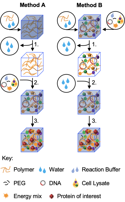
Figure 1: Schematic of the two protocols. In the first method (Method A, demonstrated in this paper) hydrogel materials are prepared first and then freeze-dried (step 1) without cell-free components. These dried hydrogels can be stored and reconstituted when required (step 2) with the correct volume of cell-free reaction prior to incubation for protein production (step 3) The variant method, Method B, incorporates all, or some, of the cell-free reaction components in the initial hydrogel fabrication. Following freeze-drying (step 1), the hydrogels may then be reconstituted in water alone or in buffer containing an analyte of interest (step 2). Protein production (step 3) continues as before. A third method, in which freeze-dried cell-free components are reconstituted with hydrogel polymers, is described in Whitfield et al.11 but has found use with only a limited number of hydrogels to date. Please click here to view a larger version of this figure.
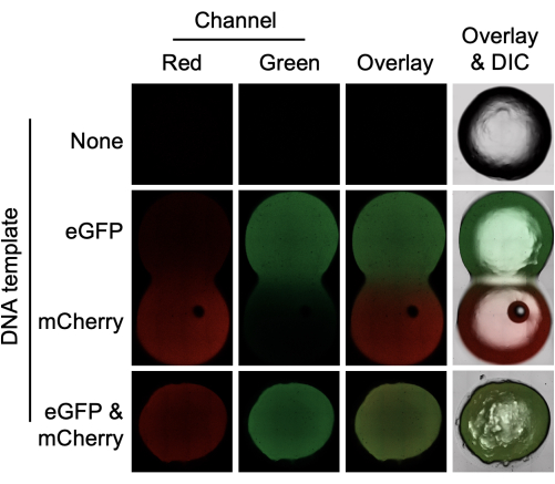
Figure 2: Cell-free protein synthesis of eGFP and mCherry in a hydrogel using E. coli cell lysates. Agarose gels (0.75%) were prepared without DNA template (top) with 4 µg of either eGFP or mCherry template (middle) or with 4 µg of both eGFP and mCherry template (bottom). The hydrogels were incubated for 4 h before confocal microscopy in the red and green channels. An overlay of the two channels is also shown, and the overlay includes the differential interference contrast (DIC) image. Hydrogels containing either eGFP or mCherry template were prepared separately but incubated in physical contact with each other. The gel diameter is 6 mm. Please click here to view a larger version of this figure.
Material | |||
3-PGA | Santa Cruz Biotechnology | sc-214793B | |
Acetic Acid | Sigma-Aldrich | A6283 | |
Agar | Thermo Fisher Scientific | A10752.22 | |
Agarose | Severn Biotech | 30-15-50 | |
Amino Acid Sampler Kit | VWR | BTRABR1401801 | |
ATP | Sigma-Aldrich | A8937-1G | |
cAMP | Sigma-Aldrich | A9501-1G | |
Coenzyme A (CoA) | Sigma-Aldrich | C4282-100MG | |
CTP | Alfa Aesar | J14121.MC | |
DTT | Thermo Fisher Scientific | R0862 | |
Folinic Acid | Sigma-Aldrich | F7878-100MG | |
GTP | Carbosynth | NG01208 | |
HEPES | Sigma-Aldrich | H4034-25G | |
K-glutamate | Sigma-Aldrich | G1149-100G | |
Lysozyme | Sigma-Aldrich | L6876-1G | |
Mg-glutamate | Sigma-Aldrich | 49605-250G | |
NAD | Sigma-Aldrich | N6522-250MG | |
PEG-8000 | Promega | V3011 | |
Potassium Hydroxide (KOH) | Sigma-Aldrich | 757551-5G | |
Potassium Phosphate Dibasic (K2HPO4) | Sigma-Aldrich | P3786-500G | |
Potassium Phosphate Monobasic (KH2PO4) | Sigma-Aldrich | RDD037-500G | |
Protease Inhibitor cocktail | Sigma-Aldrich | P2714-1BTL | |
Qubit Protein concentration kit | Thermo Fisher Scientific | A50668 | |
Rossetta 2 DE 3 E.coli | Sigma-Aldrich | 71397-3 | |
Sodium Chloride (NaCl) | Sigma-Aldrich | S9888-500G | |
Spermidine | Sigma-Aldrich | 85558-1G | |
Tryptone | Thermo Fisher Scientific | 211705 | |
Tris | Sigma-Aldrich | GE17-1321-01 | |
tRNA | Sigma-Aldrich | 10109541001 | |
UTP | Alfa Aesar | J23160.MC | |
Yeast Extract | Sigma-Aldrich | Y1625-1KG | |
Equipment | |||
1.5 mL microcentrifuge tubes | Sigma-Aldrich | HS4323-500EA | |
10K MWCO dialysis cassettes | Thermo Fisher Scientific | 66381 | |
15 mL centrifuge tube | Sarstedt | 62.554.502 | |
50 mL centrifuge bottles | Sarstedt | 62.547.254 | |
500 mL centrifuge bottles | Thermo Fisher Scientific | 3120-9500 | |
Alpha 1-2 LD Plus freeze-dryer | Christ | part no. 101521, 101522, 101527 | |
Benchtop Centrifuge | Thermo Fisher Scientific | H-X3R | |
Black 384 well microtitre plates | Fischer Scientific | 66 | |
Cuvettes | Thermo Fisher Scientific | 222S | |
Elga Purelab Chorus | Elga | ##### | |
Eppendorf Microcentrifuge 5425R | Eppendorf | EP00532 | |
High Speed Centrifuge | Beckman Coulter | B34183 | |
JMP license | SAS Institute | 15 | |
Magnetic Stirrer | Fischer Scientific | 15353518 | |
Parafilm | Amcor | PM-966 | |
Photospectrometer (Biophotometer) | Eppendorf | 16713 | |
Pipettes and tips | Gilson | ##### | |
Precision Balance | Sartorius | 16384738 | |
Qubit 2.0 Fluorometer | Thermo Fisher Scientific | Q32866 | |
Shaking Incubator | Thermo Fisher Scientific | SHKE8000 | |
Sonic Dismembrator (Sonicator) | Thermo Fisher Scientific | 12893543 | |
Static Incubator | Sanyo | MIR-162 | |
Syringe and needles | Thermo Fisher Scientific | 66490 | |
Thermo max Q8000 (Shaking Incubator) | Thermo Fisher Scientific | SHKE8000 | |
Varioskan Lux platereader | Thermo Fisher Scientific | VLBL00GD1 | |
Vortex Genie 2 | Cole-parmer | OU-04724-05 | |
VWR PHenomenal pH 1100 L, ph/mv/°c meter | VWR | 662-1657 |
Synthetic gene networks provide a platform for scientists and engineers to design and build novel systems with functionality encoded at a genetic level. While the dominant paradigm for the deployment of gene networks is within a cellular chassis, synthetic gene networks may also be deployed in cell-free environments. Promising applications of cell-free gene networks include biosensors, as these devices have been demonstrated against biotic (Ebola, Zika, and SARS-CoV-2 viruses) and abiotic (heavy metals, sulfides, pesticides, and other organic contaminants) targets. Cell-free systems are typically deployed in liquid form within a reaction vessel. Being able to embed such reactions in a physical matrix, however, may facilitate their broader application in a wider set of environments. To this end, methods for embedding cell-free protein synthesis (CFPS) reactions in a variety of hydrogel matrices have been developed. One of the key properties of hydrogels conducive to this work is the high-water reconstitution capacity of hydrogel materials. Additionally, hydrogels possess physical and chemical characteristics that are functionally beneficial. Hydrogels can be freeze-dried for storage and rehydrated for use later. Two step-by-step protocols for the inclusion and assay of CFPS reactions in hydrogels are presented. First, a CFPS system can be incorporated into a hydrogel via rehydration with a cell lysate. The system within the hydrogel can then be induced or expressed constitutively for complete protein expression through the hydrogel. Second, cell lysate can be introduced to a hydrogel at the point of polymerization, and the entire system can be freeze-dried and rehydrated at a later point with an aqueous solution containing the inducer for the expression system encoded within the hydrogel. These methods have the potential to allow for cell-free gene networks that confer sensory capabilities to hydrogel materials, with the potential for deployment beyond the laboratory.