Archived blood films and project design were approved by the Institutional Biosafety Committee, the Institutional Animal Care and Use Committee of the Faculty of Veterinary Science, Chulalongkorn University (IBC No. 2031033 and IACUC No. 1931027), and Human Research Ethics Committee of King Mongkut's Institute of Technology Ladkrabang (EC-KMITL_66_014).
1. Preparation of raw images
- The image dataset preparation
- Obtain at least 13 positive slides with blood-parasite infections, including T. brucei, T. cruzi, and T. evansi, confirmed by parasitologist experts. Separate the 13 slides for training (10 slides) and testing (three slides).
- Acquire images of the Giemsa stained-thin blood films described above under an oil-immersion field of a light microscope with a digital camera. Obtain images containing multiple objects of the trypomastigotes of all three parasite species under microscopic examination; look for a slender shape, long tails, an undulating membrane, and a kinetoplast at the anterior end.
NOTE: Creating both thick and thin smears would enhance the detection of acute phase trypanosomiasis31. The blood collection by finger-prick is recommended by WHO32. Nevertheless, thin films are more effective in identifying Trypanosoma cruzi and other species, as these organisms tend to become distorted in thick films33. In light of this, we utilized thin blood film images to maintain the appropriate morphology of the parasites for this study. - Store all images in a parasite-specific folder with the following specifications: 1,600 x 1,200 pixels, 24-bit depth, and JPG file format. Split the images into the training and test sets at a ~6:1 ratio.
NOTE: See https://gitlab.com/parasite3/superior-auto-identification-of-medically-important-trypanosome-parasites-by-using-a-hybrid-deep-learning-model/-/blob/main/JOVEimage.zip; 650 images were split to train (560 images) and test (90 images) model. - Define the region of interest as a rectangular label for two classes: trypanosomes and non-trypanosomes. Use the auto-cropping module to crop all detected images by using the well-trained object detection model. The auto-cropping module is the module developed in the in-house CiRA CORE program (see Table of Materials). Collect a single object per image for training the object classification.
NOTE: For this paper, 1,017 images were split for training (892 images) and testing (126 images). The model training was performed with four labeled classes, including leukocyte, T. brucei, T. cruzi, and T. evansi.
2. Training process with in-house CiRA CORE platform
- Starting a new project
- Open the CiRA CORE application from the computer desktop (see Table of Materials) and create a new project by double-clicking on the program's icon.
- Choose the operation icon on the left vertical toolbar to select the required tools.
- Object detection model training
- Select the training-DL model function for data labeling and training by using the drag-and-drop method. Go to the General toolbar | CiRA AI | Drag DeepTrain | Drop DeepTrain on the screen (right-hand side).
NOTE: For additional options, right-click on the selected tool and perform the appropriate functions: Copy, Cut, or Delete. - Import the images using DeepTrain tool's settings. Click on the Load images button and navigate to the image directory. Label the objects by holding the left-click and naming the selected object. Adjust the rectangle line thickness and font size by clicking on the Display Setting button and Save GT as a .gt file in the same directory.
NOTE: Save as needed to avoid any undesired conditions such as power shortage, automatic program closures, and hanging within the labeling process. - Prior to model training, expand the data to gather sufficient information using the four augmentation techniques: Rotation, Contrast, Noise, and Blur. Click the Gen Setting button to access this feature.
- Initiate model training by clicking the Training button in the DeepTrain tool. The training part has two sub-functions: Generate Training Files and Train. Under the Generate Training Files function, select the desired models, batch size, and subdivisions. Click the Generate button to generate data and save it in the directory. In the Train function, choose the following options: i) use another generated training location for conditions and backup, ii) use prebuilt weights for continued training, or iii) override parameters for current training design. This will design the model configuration and training conditions.
NOTE: The generation process time depends on the image file size, augmentation usage, and available memory space. - Once all necessary configurations are complete, begin the model training by clicking on the Train button. Allow the program to continuously execute, evaluating the training loss and adjusting the weight of the dataset during the training process. If the model achieves optimal loss, save the trained weight file in the specified directory by clicking on the Export button.
- Select the training-DL model function for data labeling and training by using the drag-and-drop method. Go to the General toolbar | CiRA AI | Drag DeepTrain | Drop DeepTrain on the screen (right-hand side).
3. Object detection model evaluation
- Select the object detection model evaluation function for model evaluation using the drag-and-drop method. Go to the Plugin toolbar | Evaluate | Drag EvalDetect | Drop EvalDetect on the screen (right-hand side).
- Click on Setting and wait for three functions: Detection, Evaluate, and Plot. Initiate model evaluation by importing the trained weight file from the directory (step 2.2.5) by clicking on Load Config.
- Under the Detection function, select non-maximum suppression (NMS) value to enhance accuracy by eliminating redundant false positive (FP) detections. NMS removes duplicate model-generated detections for improved reliability.
- Proceed with the following steps under the Evaluation function:
- Import test images from the image file directory by clicking on Browse. Import the GT file from the directory where it was saved in step 2.2.2 by clicking on Load GT.
- Choose the Intersection over Union (IoU) value to assess accuracy on the specific image test dataset.
- Click the Evaluation button to assess the detection model in the specified directory. Once the evaluation is completed, the results will be automatically saved as a CSV file in the same directory, sorted by class name. This CSV file will provide essential parameters such as True Positive (TP), False Positive (FP), False Negative (FN), Recall, and Precision for each class.
- To plot the Precision-Recall (PR) curve, follow these steps under the Plot function: Import the CSV files from the previous section (step 3.4) directory by clicking on Browse. Choose classes from the list and click the Plot button to display the editable PR curve image.
- Finally, to save an image with the AUC values of the PR curve in the required image format at the specified directory, click on the Save button of the image.
4. Image cropping for a single object per image
- Prior to cropping the images, complete the following steps:
- Import the images from the image file directory by accessing the settings of the Image Slide tool.
- Import the trained weight file (saved in step 2.2.8) by accessing the settings of the Deep Detect tool. Click on the Config button | + button, select the backend (CUDA or CPU), provide a name, click OK, choose the weight file directory, and click Choose. Within the Deep Detect tool, select the detection parameters (threshold and non-maxima suppression (nms)); drawing parameters; tracking parameters; and region of interest (ROI) parameters.
- Select the directory where the cropped images will be saved by accessing the settings of the Deep Crop tool. Click Browse | choose the directory to save the cropped images | click Choose | select the image format (jpg or png) | enable the Auto Save option.
- Crop images to obtain a single object per image for image classification and segmentation. To carry out this process, utilize four tools and establish connections between them: go to the General toolbar | General | Button Run. Next, navigate to General toolbar | CiRA AI | DeepDetect; then, go to General toolbar | CiRA AI | DeepCrop. Finally, go to Image toolbar | Acquisition | ImageSlide.
- Once all the necessary settings are in place, initiate the image cropping process by clicking on the Button Run tool.
- Obtain a new image training dataset consisting of single-object images with a size of 608 x 608.
5. Image classification as model training
- Use drag-and-drop to select the image classification model training function for data training. Go to the Image toolbar | DeepClassif | Drag ClassifTrain | Drop ClassifTrain on the screen.
- Import images for model training using the ClassifTrain tool's settings. Click on the Open folder button and navigate to the desired image directory. Before training, expand the data by clicking on the Augmentation button for more information using techniques such as Rotation, Contrast, Flipping (horizontal and/or vertical), Noise, and Blur.
- To commence model training, click on the GenTrain button of the ClassifTrain tool. Under the GenTrain function, select the models, batch size, and subdivisions. Assign a directory to save the generated file. Click the Generate button to proceed with data for training. In the Train function, tick the appropriate options: Continue training with default weight or custom weight.
NOTE: The generation process may take time depending on factors such as image file size, augmentation usage, class balancing, and available memory space. - Once all preparations are complete, initiate the model training by clicking the Start button. Allow the program to execute continuously, evaluating the training loss and adjusting the weight of the dataset during the training process. If the model achieves the desired level of loss, save the trained weight file to the specified directory by clicking on the Export button.
6. Classification model evaluation
- Select the image classification model evaluation function for model evaluation using the drag-and-drop method. Go to the Plugin toolbar | Evaluate | Drag EvaluateClassif | Drop EvaluateClassif on the screen (the right-hand side).
- Click on Setting to access additional functions within the EvaluateClassif tool, namely Evaluate and PlotROC.
- To initiate model evaluation, click on the Evaluate button in the EvaluateClassif tool. Follow these steps under the Evaluate function.
- Import the test images from the image file directory by clicking on the Load folder image. Import the trained weight file from the directory (saved in step 5.4) by clicking on Load Config. Click the Start button to evaluate the classification model.
- Once the evaluation is complete, save the evaluated file as CSV in the specified directory by clicking on the Export to CSV button. For evaluation of data at every threshold, save the CSV file with class names in the specified directory by clicking on Start all threshold. The saved CSV file includes parameters such as Recall (True Positive Rate), False Positive Rate, and Precision for each class.
- To plot the Receiver Operating Characteristics (ROC) curve, click on the PlotROC button within the EvaluateClassif tool. Follow these steps under the PlotROC function.
- Import CSV files from the directory obtained earlier by clicking on Browse. Inspect the imported class list and select each class label to plot the ROC curve.
- Click the Plot button to visualize the ROC curve as an image. Make the desired edits to adjust image properties, including font size, font colors, rounding the decimal, line styles, and line colors.
- Finally, save an image of the ROC curve with the AUC values in the required image format at the specified directory by clicking on the Save button.
7. Testing the process with the CiRA CORE application
- Object detection as model testing
- To perform model testing, utilize four tools and establish connections between them. Go to the General toolbar | General | Button Run. Then, General toolbar | General | Debug. After that, click on General toolbar | CiRA AI | DeepDetect, and finally Image toolbar | Acquisition | ImageSlide.
- Before testing the images, follow these steps:
- Import the test images from the image file directory by clicking on the Setting option in the Image Slide tool.
- Import the saved trained weight file from step 2.2.8 by clicking on the Setting option in the DeepDetect tool. Click on the Config button, then the + button, select the backend (CUDA or CPU), provide a name, click OK, choose the weight file directory, and click Choose. Under the DeepDetect tool, select the detection parameters (Threshold and nms), drawing parameters, tracking parameters, and ROI parameters.
- View the test image results by clicking on the image function in the Debug tool.
- Finally, check the predicted results for each image by clicking on the Run button on the Button Run tool.
- Image classification as model testing
- To perform model testing, utilize four tools and establish connections between them. Go to the General toolbar | General | Button Run; then, General toolbar | Debug. After that, navigate to Image toolbar | Acquisition | ImageSlide, and finally, Image toolbar | DeepClassif | DeepClassif.
- Before testing the images, follow these steps:
- Import the test images from the image file directory by clicking on the Setting option in the Image Slide tool.
- Import the saved trained weight file from section 5.5 by clicking on the Setting option in the DeepClassif tool. Click on the Config button | + button | select the backend (CUDA or CPU) | provide a name | click OK | choose the weight file directory | click Choose. Under the DeepClassif tool, select the classification parameters (Threshold and number of top-class predictions), Guide map parameters (threshold, alpha, beta, and color map), and various parameters in the color map.
- View the test image results by clicking on the image function in the Debug tool.
- Finally, check the predicted results for each image by clicking on the Run button on the Button Run tool.
8. Hybrid (detection and classification) as model testing
- To perform this model testing, utilize four tools and establish connections between them. Go to the General toolbar | General | ButtonRun. Then, General toolbar | General | Debug. After that, Image toolbar | Acquisition | ImageSlide, and finally, Image toolbar | DeepComposite | DeepD->C.
- Before testing the images, follow these steps: Import test images from the image file directory by clicking on the Setting option in the Image Slide tool. Import the two saved trained weight files from section 2.1.5 and section 4.4 by clicking on the Setting option in the DeepD->C tool:
- For the Detect function, click on the Config button |+ button, select the backend (CUDA or CPU) | provide a name | click OK | choose the weight file directory | click Choose. Under the Detect function, select the detection parameters (Threshold and nms), drawing parameters, tracking parameters, and ROI parameters.
- For the Classif function, click on the Config button |+ button, select the backend (CUDA or CPU) | provide a name | click OK | choose the weight file directory | click Choose. Under the Classif function, select the classification parameters (Threshold and number of top-class predictions) and Guide map parameters (threshold, alpha, beta, and color map).
- View the test image results by clicking on the image function in the Debug tool. Finally, check the predicted results for each image by clicking on the Run button on the Button Run tool.
9. Five-fold cross-validation
NOTE: To validate the performance of the proposed model more effectively, K-fold cross-validation is used.
- Divide the dataset into five sections, corresponding to the five folds of cross-validation. During each iteration of model training and testing, use one section as the validation set for testing and the remaining four sections for training. Repeat this process five times, with each fold being used as the validation set once.
- For Folds 1 through 5:
- Repeat section 5 to train the model using the training data from the four folds.
- Repeat section 7.2 to test the model using the remaining fold as the test set.
10. Model evaluation
- Confusion matrix
- Based on the test results, the four conditions will happen as follows:
- True Positive (TP): When the input image is true, and the prediction is also true.
- False Positive (FP): When the input image is false, but the prediction is true.
- False Negative (FN): When the input image is true, but the prediction is false.
- True Negative (TN): When the input image is false, and the prediction is also false.
- Using these four conditions, evaluate the performances with the confusion matrix.
- Based on the test results, the four conditions will happen as follows:
- Performance evaluations
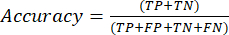
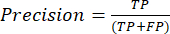
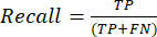
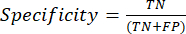
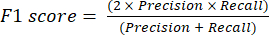
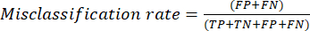
- The most commonly used classification performance metrics are accuracy, precision, recall, specificity, and F1-score values. Calculate all evaluation metrics in equations (1-6) used to evaluate model performance from values from the confusion matrix.
 (1)
(1)
 (2)
(2)
 (3)
(3)
 (4)
(4)
 (5)
(5)
 (6)
(6)
- The most commonly used classification performance metrics are accuracy, precision, recall, specificity, and F1-score values. Calculate all evaluation metrics in equations (1-6) used to evaluate model performance from values from the confusion matrix.
- ROC curve
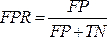
NOTE: The ROC curve is a performance measure for classification problems with different threshold settings. The area under the ROC curve (AUC) represents the degree or measure of separability, while the ROC is a probability curve.- The ROC curve is a two-dimensional graph with the true positive rate (TPR) and false positive rate (FPR) values plotted on the Y and X axes, respectively. Construct the ROC curves using the TPR and TFR values obtained from the confusion matrix. The TPR value is the same as the sensitivity; calculate the FPR value using the equation (7).
 (7)
(7) - After obtaining the TPR and FPR values, plot the ROC curve using the Jupyter Notebook open-source web tool in a Python environment. The AUC is an effective way to assess the performance of the proposed model in ROC curve analysis.
- The ROC curve is a two-dimensional graph with the true positive rate (TPR) and false positive rate (FPR) values plotted on the Y and X axes, respectively. Construct the ROC curves using the TPR and TFR values obtained from the confusion matrix. The TPR value is the same as the sensitivity; calculate the FPR value using the equation (7).
- PR curve
- Use the PR curve to evaluate models by measuring the area under the PR curve. Construct the PR curve by plotting the models' precision and recall using the model's confidence threshold functions. Because the PR curve is also a two-dimensional graph, plot Recall on the x-axis and Precision on the y-axis.
- Plot the PR curve, like the ROC curve, using the open-source Jupyter Notebook web tool in a Python environment. The area under the Precision-Recall curve (AUC) score is also helpful in multilabel classification.
In this study, hybrid deep learning algorithms were proposed to help automatically predict the positivity of a blood sample with a trypanosome parasite infection. Archived, Giemsa-stained blood films were sorted to localize and classify the parasitized versus non-parasitic by using the object detection algorithm based on a darknet backbone neural network. Within any rectangular box prediction result obtained by the previous model, the best-selected classification model was developed to classify all three species of medically and veterinary important trypanosomes including T. brucei, T. cruzi, and T. evansi. The final output of the hybrid models used revealed the robustness of the proposed models against the variation of 100x microscopic images that might affect the prediction result, including the blood-stage morphology of the parasite. In addition, environmental factors may disturb the image quality of staining color change by storing time, intensity from the light sources of the microscope, and blood film preparation skills. Nevertheless, the best-selected model can achieve the goal with high performance.
Localization and classification of multi-class labels
Since the detection of parasitic protozoa from Giemsa staining blood film under oil-immersion microscopy is tedious and lengthens turn-around time, this leads to prone error bias. Well-trained-AI approaches require a large pool of image data with rescaling 416 x 416 pixels and varying feature characteristics of 3-RGB color channels to increase the correct prediction of localization and classification. The number of parameters during training and optimizing models is set up with a learning rate of 0.002, burn-in of 1,000, and ranging steps between 400,000 and 450,000. Low training loss but high training accuracy were considered the optimum level or saturation under momentum of 0.9, hue of 0.1, and decay of 0.0005. In the testing phase with unseen data, correct localization and classification were performed by using the concepts of intersection over union (IOU) and percentage of the probability. The testing interpretation result was performed at a threshold of 50% and a non-maximum suppression (NMS) of 0.4, which gave the correct answer with a % probability.
As with all parasitized blood films studied, discrimination of the trypanosome out of the non-trypanosome has been performed by using a detection neural network model that can function for both localization and classification (Figure 1)22. The prediction result of the detection task proposed revealed an outstanding result with a mean average precision of 93.10% (Table 1). Although the trained detection model can be used to identify the non-trypanosome class more than that used to identify the trypanosome parasite, it brings us greater precision than 91% for both class labels. In addition, the precision versus recall curve showed a highly average AUC value of 0.969, which gave the AUC values for the parasite and non-parasite at 0.976 and 0.961, respectively (Figure 2). This led us to assure ourselves that the trained model could be trustworthy. The rectangular box of the first detection result was cropped by using the image capture module under the in-house CiRA CORE program. The cropped images mentioned above were sorted into three folders that are specific to the trypanosome species. This process was prepared to input data for the training classification model that is illustrated in the next subsection.
Classification model-wise classification
To find an appropriately trained model for classifying the well-known species of the parasite, T. brucei, T. cruzi, and T. evansi were kept in folders that were assigned their relative class names. During AI training, rescaled 256 x 256 pixels of images were fed into three RGB channels, learning rate of 0.1, burn-in of 1000, momentum of 0.9, hue of 0.1, and decay of 0.0005. The training loss and training accuracy were used to find the optimum trained model. The classification prediction was analyzed using the concepts of pixel-wise determination and % probability at a threshold of 50%.
The comparison of three popular classification neural network algorithms was studied to find the best one27,30. These three neural networks have been widely used in classifying multiclass labels in medical and veterinary fields27,34,35. The inference result of the trained model with a % probability that ranked 0 to 1, was justified above the threshold of 50%. Additionally, different pattern recognitions of each parasite were highlighted and specific to the nucleus of the middle portion of T. evansi by the attention map. The largest kinetoplast organelle of the anterior portion of T. cruzi when compared to the other two species was also highlighted. Both nuclease and kinetoplast were emphasized by the attention map found for T. brucei (Figure 3).
Several statistical metrics were used to measure those three models proposed, including accuracy, misclassification rate, recall (true positive rate), specificity (true negative rate), false positive rate, false negative rate, precision, and F1 score. As a result, almost all evaluation metrics using the Densenet201 neural network showed superior values to the others. On average, the metric values of accuracy, recall, specificity, precision, and F1 score were remarkably greater and equal to 98%. However, the model performance importance revealed less than and equal to 1.5% of the misclassification, false positive, and false negative rates (Table 2). Considering the class-wise comparison, the Densenet201 model seems to correctly identify T. evansi without error while doing this with unseen testing data, suggesting the potential trained model is for distinguishing the parasite species.
In Figure 4A–C, the AUC under the ROC curve gave the greatest degree of average accuracy at 0.931 obtained from the best classification model (Figure 4C), which was representative of confirming the best selected model studied. The AUC of T. evansi was 0.817, which is lower than others (0.980-1.00 for T. brucei and 0.955-0.977 for T. cruzi) and a contrast to the statistical metrics above. This may be because these two values are calculated by different formulas. The AUC was obtained from all thresholds but the statistical metrics from only a threshold of 50%, suggesting these two values cannot be compared. Hence, consistent AUC values by class names obtained from all three models indicate the general accuracy of T. brucei > T. cruzi > T. evansi, respectively.
k-fold cross validation
To assess the robustness of the best-selected classification model studied in terms of estimating the true prediction error and tuning the model parameters as described above36, the five-fold cross-validation technique was used. A random split of the data into five folders was done. Assigned trained data by four-folders and tested data for the rest folder were prepared before training with the selected classification algorithm.
As a result, the average statistical metrics; accuracy, recall (true positive rate), specificity (true negative rate), precision, and F1 score, provided similar values of the statistical metrics studied that showed greater than 98% (Table 3). Considering each metric studied, a ranking of 0.992-1.000 in accuracy was found. High specificity values ranging from 0.994 to 1.000 were provided. Both recall and F1 scores ranging from 0.988 to 1.000 were shown, likewise, 0.989-1.000 were studied by precision. Interestingly, low rates of misclassification, false negatives, and false positives were found at less than 1.2%. This quality performance supported the outstanding trained model with varied data folds and represented robustness.
Accompanying the metrics proposed, the average AUC under the ROC curve obtained revealed closed values ranging from 0.937 to 0.944, giving similar values of general accuracy among the five folds of the data (Figure 5). The class-wise comparison provided a varied AUC of 0.831 for T. evansi, 0.982-1.000 for T. cruzi, and 1.000 for T. brucei. Although T. evansi's AUC value was lower than the others, the values may be exposed to the high degree of false positive rate (~33%) belonging to the thresholds 1% to 97% which results in smaller AUC values when compared to those of the other two classes (Figure 6).
The hybrid deep learning a practical screening
In this section, the contribution of the hybrid deep learning approach between object detection and, on the other hand, the classification technique is shown in Figure 7. The parasite and non-parasite features were distinguished and their relative classes identified within the pink bounding box by using the first detection model. Next, the specific species of the parasite were diagnosed in different colors by using the well-trained classification model. The green label was for T. evansi, the pink label for T. brucei, and the orange label for T. cruzi. The second classification label would not be shown if the first detection model failed, suggesting the well-connected functions between these two different neural network backbones in the D-C module of the in-house CIRA CORE platform.
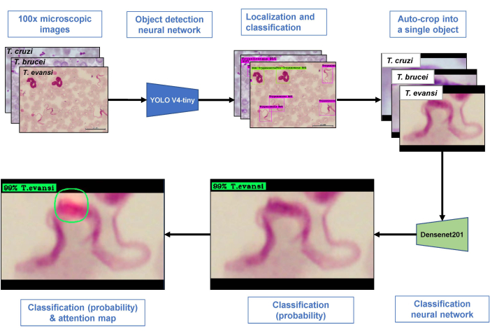
Figure 1: Architecture for a hybrid model. All three parasite species of trypanosomes (including Trypanosoma evansi, T. brucei, and T. cruzi) were used as input. Multi-objects within a 100x microscopic image were detected by using the detection model. A single cropped object from the previous model was then classified according to its relative species by using the best-classification model. An attention map integrated with the best classification model highlighted areas specific to each class label. Please click here to view a larger version of this figure.
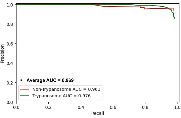
Figure 2: PR curve. The area under the PR curve, or AUC value, in this study, is used to measure the ability to discriminate between non-trypanosome and trypanosome classes. All samples can be detected on both class labels. An AUC of 1 is a perfect prediction, while an AUC of 0.5 is a random prediction. The curve is used to measure the performance of the proposed detection model. This model can detect the trypanosome class at a higher rate (AUC = 0.976) than the non-trypanosome class (AUC = 0.961). The average AUC value of 0.969 was obtained from the binary result of two class labels, the non-trypanosome and the trypanosome. Abbreviations: PR = precision versus recall; AUC = area under the curve. Please click here to view a larger version of this figure.
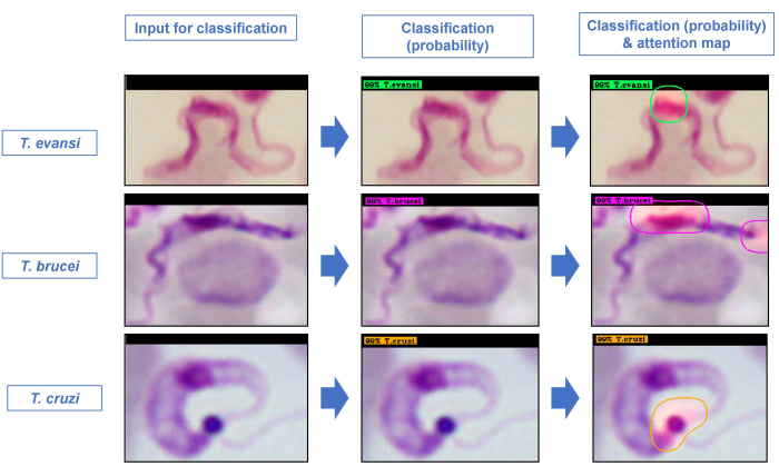
Figure 3: Predictive result of the classification model. All three trypanosome species were used to test the best proposed trained models. Output images of species classification-based probability and attention maps are shown. Specifically, the attention maps highlighted the significant areas within the unseen object that were guiding the discrimination of the parasite species. Please click here to view a larger version of this figure.
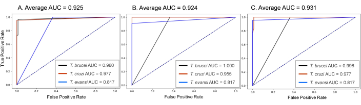
Figure 4: Model-wise comparison-based ROC curves. The AUC under the ROC curve is a graphical plot of the performance of a classification system based on its varied threshold of discrimination. Similar to the AUC-PR curve, the AUC-ROC of 1 is a perfect prediction, while the AUC of 0.5 is a random prediction, which is indicated by dashed lines in each graph. Three classification models were compared, including (A) the 1st classification model with an average AUC of 0.925, (B) the 2nd classification with an average AUC of 0.924, and (C) the best classification with an average AUC of 0.931. Therefore, the higher the AUC, the better the performance. Abbreviations: ROC = receiver operating characteristics; AUC = area under the curve. Please click here to view a larger version of this figure.
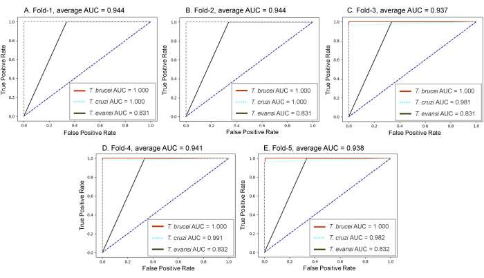
Figure 5: Five-fold cross-validation. All experiments based on the best classification neural network models were compared. Similar AUC values of five-fold data included (A) 0.944, (B) 0.944, (C) 0.937, (D) 0.941, and (E) 0.938, which suggest the robustness of the proposed trained model used against the variation of the biological data. Please click here to view a larger version of this figure.
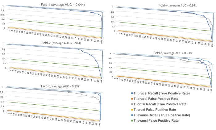
Figure 6: true positive rate and false positive rate per class name. The X-axis is representative of thresholds from 1% to 97%. The Y-axis is representative of the degrees of the statistical metrics. Please click here to view a larger version of this figure.
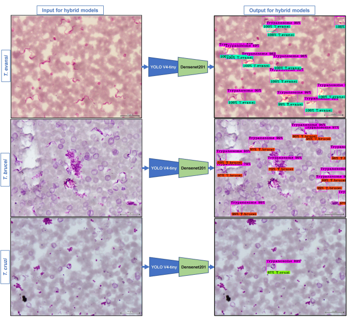
Figure 7: Final output of the hybrid models. The final step of the hybrid model contribution can be applied with input data as a raw microscopic image by 20 µm. The predictive result can be obtained from both the object detection and the classification models. The first predictive result provided whether the unseen testing image contained trypanosome parasites with a rectangle (pink-colored labels). Then the classification results specific to the parasite species will be followed by the first detection with multi-color labels; green for T. evansi, pink for T. brucei, and orange for T. cruzi. Please click here to view a larger version of this figure.
Table 1: Average precision by class and mean Average Precision (mAP) of the detection model. Please click here to download this Table.
Table 2: Classification model-wise comparison. Eight evaluation metrics were used to measure the model's performance, including accuracy, misclassification rate, recall (true positive rate), specificity (true negative rate), false positive rate, false negative rate, precision, and F1-score. The bold value is representative of the greatest value per class label. The italic value is representative of the average value of each evaluation metric. Please click here to download this Table.
Table 3: Five-fold cross-validation. The bold value is representative of the average value per evaluation metric. Please click here to download this Table.
