This protocol demonstrates microscopy-guided isolation and immunofluorescence staining of murine pulmonary veins. We prepare tissue samples containing the left atrium, pulmonary veins, and the corresponding lungs and stain them for cardiac Troponin T and Connexin 43.
Pulmonary veins (PVs) are the major source of ectopic beats in atrial arrhythmias and play a crucial role in the development and progression of atrial fibrillation (AF). PVs contain myocardial sleeves (MS) composed of cardiomyocytes. MS are implicated in the initiation and maintenance of AF, as they preserve similarities to the cardiac working myocardium, including the ability to generate ectopic electrical impulses. Rodents are widely used and may represent excellent animal models to study the pulmonary vein myocardium since cardiomyocytes are widely present all over the vessel wall. However, precise microdissection and preparation of murine PVs is challenging due to the small organ size and intricate anatomy.
We demonstrate a microscopy-guided microdissection protocol for isolating the murine left atrium (LA) together with the PVs. Immunofluorescence staining using cardiac Troponin-T (cTNT) and connexin 43 (Cx43) antibodies is performed to visualize the LA and PVs in full length. Imaging at 10x and 40x magnification provides a comprehensive view of the PV structure as well as detailed insights into the myocardial architecture, particularly highlighting the presence of connexin 43 within the MS.
Atrial fibrillation (AF) is the most common sustained arrhythmia1. The prevalence of AF is increasing even further with an expected number of ~17.9 million patients in Europe in 20601. AF is clinically highly important since it is an essential risk factor for the development of myocardial infarction, heart failure, or stroke, resulting in an enormous individual, social, and socioeconomic burden1. Even though AF has been known for decades, the pathophysiology of AF is still not fully understood2.
Already in the late 1990s, studies demonstrated the great impact of pulmonary veins (PVs) in initiating and maintaining AF, as they are the main source of AF-triggering ectopic beats3. It has been demonstrated that PVs structurally differ from other blood vessels. While typical blood vessels contain smooth muscle cells, the tunica media of PVs also contains cardiomyocytes4. In rodents, this cardiac musculature is ubiquitously present throughout the whole PVs, including intra- and extrapulmonary parts, as well as the orifice region5. In humans, PVs also contain cardiomyocytes, which can be observed within extensions of the left atrial (LA) myocardium-so-called myocardial sleeves (MS)6,7.
MS have morphological similarities to the atrial myocardium8. The shape and size of atrial and PV cardiomyocytes do not vary significantly between each other and show comparable electrophysiological properties8. Electrophysiologic recordings within the PV have proven the electrical activity of MS, and angiographic imaging has revealed contractions synchronized with the heartbeat9,10.
Gap junctions are pore-forming protein complexes composed of six connexin subunits, which allow the passage of ions and small molecules11. Gap junctions exist in the cell-to-cell appositions, interconnect neighboring cardiomyocytes, and enable an intercellular electrical coupling between cardiomyocytes12,13. Several connexin isoforms are expressed in the heart with connexin 43 (Cx43) being the most common isoform expressed in all regions of the heart14. Previous studies provide evidence for the expression of Cx43 in cardiomyocytes of the PVs15,16.
It remains challenging to investigate MS within intact PVs due to their delicate structure, especially in small animal models. Here, we demonstrate how to identify and isolate PVs together with LA and lung lobes in mice using microscopy-guided microdissection. Additionally, we demonstrate immunofluorescence (IF) staining of PVs to visualize cardiomyocytes and their interconnections within the PVs.
Animal care and all experimental procedures were conducted following the guidelines of the Animal Care and Ethics Committee of the Ludwig-Maximilians-University of Munich, and all the procedures using mice were approved by the Regierung von Oberbayern (ROB 55.2-2532. Vet_02-20-215, ROB 55.2-2532. Vet_02-18-46, ROB 55.2-2532. Vet_02-19-86, ROB 55.2-2532. Vet_02-21-178, ROB 55.2-2532. Vet_02-22-170). C57BL6/N mice were commercially obtained.
1. Preparation
2. Organ harvest and tissue preparation
NOTE: An extensive protocol detailing the procedure of mouse anesthesia and harvesting the heart has been previously published17,18. Thus, we present only a brief description of that part. Experiments were performed on 12 to 16 weeks old C57BL6 mice (six male, four female). The male's body weight extended from 26 g to 28 g and the female's body weight from 19 g to 22 g. The following steps were performed without prior systemic heparin injection.
3. Microscopy-guided preparation of LA and PVs
4. Tissue embedding
5. Cutting and collection of frozen tissue sections
NOTE: To cut the tissue blocks, the machine settings were adjusted to a specimen temperature of -18 °C, and blade temperature of -25 °C.
6. Immunofluorescence staining of cryosections from the PVs
7. Imaging of the immunofluorescence staining slices
8. Image editing with ImageJ
We performed the microdissection, staining, and imaging of the PVs in 10 12-16-week-old mice. Following the protocol, we successfully microdissected PVs together with the LA in all experimental mice and obtained sections with a comprehensive view of the PVs in eight mice. Overview images were taken at 10x magnification to identify the PV orifice (PVO) region at the LA-PV junction, the extrapulmonary PVs (PVex) (PVs in between the lung hilum and the LA-PV junction), and the intrapulmonary PVs (PVin) (PVs surrounded by lung tissue) (Figure 2). Zoomed-in images of the mentioned regions were obtained at a 40x magnification objective (Figure 3).
We found specific cTNT signals with a typical muscular striation in the PVO, the PVex, and the PVin (Figure 3). Cx43 was found in all three PV regions and was mostly projected between neighboring cardiomyocytes (Figure 3 and Figure 4A). Cx43-related signals were observed at both the polar side (Figure 3, yellow arrows) and the lateral side of the cardiomyocytes in the MS (Figure 3, red arrows).
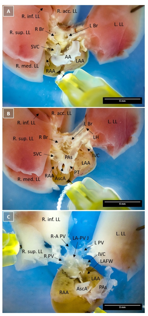
Figure 1: Identification of murine pulmonary veins under the microscope. Brightfield microscopy (10x magnification). The heart is pinned to the dissection dish with four pins at the atrium, the left lung lobe, the right middle lung lobe, and the right inferior lung lobe. (A) Overview of the heart base after separating the ventricular tissue from the atrium with the aortic arch in the middle. The atrium is located at the bottom; the lung lobes are spread out in the upper half of the picture. PVs are concealed by the AA, the main bronchi, and the PAs. (B) Top view image of the heart base after removing trachea, parts of the main bronchi, the aortic arch, as well as connective and fat tissue. The PAs in the middle of the image show their full course from the PT to the LH and cover the PVs. (C) Top view image of the PVs after cutting the PAs from the lung lobes and removing the extrapulmonary bronchial tissue. Scale bars = 5 mm. Abbreviations: AA = aortic arch; AscA = ascending aorta; IVC = inferior vena cava; L Br = left main bronchus; L PV = left pulmonary vein; LA-PV J = left atrium-pulmonary vein junction; LAA = left atrial appendage; LAFW = left atrial free wall; LH = lung hilum; L. LL = left lung lobe; PAs = pulmonary arteries; PT = pulmonary trunk; R Br = right main bronchus; R PV = right pulmonary vein; R-A PV = right ascending pulmonary vein; RAA = right atrial appendage; R. acc. LL = right accessory lung lobe; R. inf. LL = right inferior lung lobe; R. mid. LL = right middle lung lobe; R. sup. LL = right superior lung lobe; SVC = superior vena cava. Please click here to view a larger version of this figure.
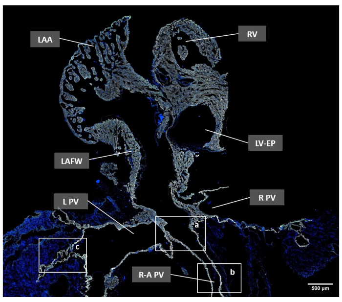
Figure 2: Overview of the adult mouse pulmonary veins and the left atrium in a transversal section image by immunofluorescence microscopy. Cell nuclei were stained with Hoechst-33342 (blue); cardiomyocytes were labeled with anti-cTNT antibody (white); and gap junctions were detected using anti-Cx43 antibody (green). The white rectangles highlight the following regions: a, PV orifice (PVO); b, extrapulmonary PV (PVex); and c, intrapulmonary PV (PVin). Scale bar = 500 µm. Abbreviations: L PV = left pulmonary vein; LAA = left atrial appendage; LAFW = left atrial free wall; LV-EP = left ventricle – ejection path; R PV = right pulmonary vein; R-A PV = right ascending pulmonary vein; RV = Right ventricle. Please click here to view a larger version of this figure.
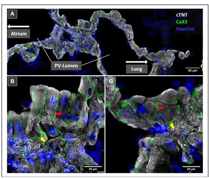
Figure 3: Zoomed-in view of the right PV myocardium using immunofluorescence microscopy. Cell nuclei were stained with Hoechst-33342 (blue); cardiomyocytes were labeled with anti-cTNT antibody (white); and Cx43+ gap junctions were detected by anti-Cx43 antibody (green). (A) EDF tile scans showing a longitudinal section through the right extrapulmonary PV. The LA and PV orifice are not shown in the image and would be located to the left, while intrapulmonary PV and corresponding lung parenchyma (also not shown) would be located to the right. (B,C) EDF single tile scans of the right extrapulmonary PV. Arrows highlight Cx43+ gap junctions at the cellular borders of cardiomyocytes. The yellow arrows indicate gap junctions at the polar sides of cardiomyocytes, colocalized with intercalated discs, while the red arrows indicate gap junctions at the lateral side of cardiomyocytes. Scale bars = 50 µm (A), 20 µm (B,C). Abbreviations: PV = pulmonary vein; EDF = extended depth of field; LA = left atrium. Please click here to view a larger version of this figure.
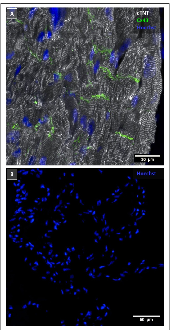
Figure 4: Immunofluorescence staining of control tissue. (A) Positive control staining of the murine LV. Cell nuclei were stained with Hoechst 33342 (blue); cardiomyocytes were labeled with anti-cTNT antibody (white); and Cx43+ gap junctions were detected with anti-Cx43 antibody (green). (B) Negative control staining of the right extrapulmonary PV by using secondary antibodies only. Cell nuclei are stained with Hoechst 33342 (blue). Scale bars = 20 µm (A), 50 µm (B). Abbreviations: LV = left ventricle; PV = pulmonary vein. Please click here to view a larger version of this figure.
Filter-Cube | Excitation Filter | Emission Filter |
DAP | 350 / 50 | 460 / 50 |
L5 | 480 / 40 | 527 / 30 |
TXR | 560 / 40 | 630 / 75 |
Table 1: Excitation and emission filter characteristics of the Leica DM6 B filter cubes.
Compound | Final concentration | g or mL / 100 mL required |
Fixing solution | ||
Paraformaldehyde (PFA) 16% | 4% | 25 mL |
Phosphate Buffered Saline (PBS) 1x concentrated | 75 mL | |
Permeabilization Solution | ||
Triton X-100 | 0.1% | 0.1 mL |
Phosphate Buffered Saline (PBS) 1x concentrated | 99.9 mL | |
Blocking Buffer | ||
Normal goes serum (NGS) | 10% | 10 mL |
Bovine Serum Albumin (BSA) | 0.5% | 0.5 mg |
Tween 20 | 0.1% | 0.1 mL |
Phosphate Buffered Saline (PBS) 1x concentrated | 89.9 mL | |
Washing Buffer | ||
Bovine Serum Albumin (BSA) | 0.5% | 0.5 mg |
Tween 20 | 0.1 mL | |
Phosphate Buffered Saline (PBS) 1x concentrated | 99.9 mL |
Table 2: Buffer Recipes.
With this protocol, we share a method to distinguish and isolate the PVs of the mouse heart and perform immunofluorescence staining on them. After the organ harvest, the heart and lungs were dehydrated in sterilized sucrose solution, followed by separating the ventricles from the atrium and lung lobes under microscopic guidance. Afterwards, the heart base was prepared to visualize the PVs followed by cutting them from the lungs at the hilum. The subsequent immunofluorescence staining was performed using a cryotechnique by embedding the tissue in O.C.T. compound, cutting it with a cryotome, and performing immunofluorescence staining for cTNT and Cx43.
Isolating the PVs can be useful for studying their role in the arrhythmogenesis of AF. However, PV isolation in mice is challenging. In addition to their small size, PVs are hidden behind the main bronchi and the prominent vessels at the heart base, convoluting their detection massively. This protocol provides a simple and transparent way to investigate the murine PVs and permits an economic approach to study their morphological characteristics in detail. The proposed harvesting and preparation approach preserves the anatomical relations between PVs, the corresponding lung lobes, and the LA, facilitating investigations of the entire LA-PV tissue complex.
MS have a heterogeneous arrangement. Significant variability in continuity and fiber orientation has been described in multiple species, including rats and humans, and are mostly found around the PV orifices4,20. Those diversities result in anisotropic electrical conduction and can induce the initiation of reentry excitations and autonomic activity in PVs21. Furthermore, the electrical interconnection between MS and atrial myocardium at the LA-PV junction can lead to the transition of desynchronized electrical impulses from the PVs to the left atrium, resulting in AF2. Preserving the morphological proportions at the LA-PV junction is, therefore, crucial for studying the interaction between the atrial myocardium and PVs. To achieve this, the accurate identification of the main bronchi and PAs is essential. We identified the landmarks according to their anatomic positions and relations using a microscope with 10x magnification and by systematically following their course from the heart base to the LH.
We performed IF staining of the murine PVs. Staining approaches based on immunoreactive dyes are widely established in different species and allow the direct detection of target antigens, providing high-quality visual contrast in microscopy. This approach enables both qualitative and quantitative characterization of intact tissue slices on a protein level. We use the cardiac isotype of TNT to distinguish between myocardium and non-cardiac musculature. The presence of cardiomyocytes in PVs, as well as the SVC and IVC has been described previously7. However, other tubular structures of the lungs, including the PAs, bronchi, and private lung vessels, do not contain heart muscle cells, which makes them easy to distinguish by IF staining7. By labeling structure proteins such as cTNT, cardiomyocytes show their characteristic cross striation depending on their orientation towards the beam path. The direction of the striation can be mapped in ImageJ with plugins such as OrientationJ or Directionality Plugin. This aids in quantifying and characterizing the directionality of cardiomyocytes and might be useful for identifying regions with anisotropic electrical conduction. Furthermore, this study supports previous findings regarding the abundant presence of Cx43 in the murine PV myocardium, which is organized in a similar manner as in the LV (Figure 4A)15,16. Remarkably, we observed gap junctions at the lateral membrane of cardiomyocytes in murine PVs. This condition, known as lateralization, has been demonstrated as a pathophysiologic mechanism in AF, especially in male mice22. Thus, this protocol highlights the potential for similar studies in mouse MS cardiomyocytes.
PV isolation is an important technique for arrhythmia research since it allows a number of investigations, including IF-staining, flow cytometry, fluorescence-activated cell sorting, optical mapping, and patch clamp. However, since the current protocol was established for IF imaging of fixed and embedded tissue, direct proof that cardiomyocytes can be isolated from PV preparations in sufficient quality to allow experiments such as patch clamp is currently missing. The success of the microdissection depends on the experience of the researcher due to the small size and fragility of the murine cardiac tissue. The quality of IF imaging is also largely dependent on the embedding step. Horizontal arrangements are difficult to achieve, the tissue integrity of the sections can suffer, and PVs can rupture if they are not filled with O.C.T. compound. Furthermore, the validity of the protocol is limited to 2D imaging, as the method does not allow the 3D reconstruction of the PVs.
The authors have nothing to disclose.
This work was supported by the German Centre for Cardiovascular Research (DZHK; 81X3600221to H.V., 81X2600255 to S.C.), the China Scholarship Council (CSC201808130158 to R.X.), the German Research Foundation (DFG; Clinician Scientist Program in Vascular Medicine (PRIME), MA 2186/14-1 to P. T.), and the Corona Foundation (S199/10079/2019 to S. C.).
Adhesion slides | Epredia | 10149870 | |
AF568-secondary antibody | Invitrogen | A11036 | Host: Goat, Reactivity: Rabbit |
Agarose | Biozym LE | 840104 | |
Alexa Fluor 488-secondary antibody | Cell Signaling Technology | 4408S | Host: Goat, Reactivity: Mouse |
Anti-Connexin 43 /GJA1 antibody | Abcam | ab11370 | Polyclonal Antibody, Clone: GJA1, Host: Rabbit |
Anti-cTNT antibody | Invitrogen | MA5-12960 | Monoclonal Antibody, Clone: 13-11, Host: Mouse |
Bovine serum albumin | Sigma-Aldrich | A2153 | |
Brush | Lukas | 5486 | size 6 |
Cover slips | Epredia | 24 mm x 50 mm | |
Cryotome Cryo Star NX70 | Epredia | Settings: Specimen temperature: -18 °C, Blade Temperature: -25 °C | |
DFC365FX camera | Leica | ||
DM6 B fluorescence microscope | Leica | ||
Dry ice | |||
Dubecco's phosphate-buffered saline (DPBS) 1x conc. | Gibco | 14040133 | 500 mL |
Dumont #5FS Forceps | F.S.T. | 91150-20 | 2 pieces needed |
Fine Scissors | F.S.T. | 14090-09 | |
Fluorescence mounting medium | DAKO | S3023 | |
Graefe Forceps | F.S.T. | 11052-10 | |
Hoechst 33342 | Invitrogen | H3570 | Cell nuclei counterstaining |
ImageJ | FIJI | analysis and processing software | |
LAS X | Leica | Imaging software for Leica DM6 B | |
Microtome blades S35 | Feather | 207500000 | |
Microwave | |||
Normal goat serum | Sigma-Aldrich | S26-M | |
O.C.T. compound | Tissue-Tek | 4583 | |
Paraformaldehyde 16% | Pierce | 28908 | methanol-free |
Pasteur pipettes | VWR | 612-1681 | |
Petri dish | TPP | 93100 | 100 mm diameter |
Rocker 3D digital | IKA Schüttler | 00040010000 | |
Slide staining jars | EasyDip | M900-12 | |
Specimen Molds | Tissue-Tek Cryomold | 4557 | 25 mm x 20 mm x 5 mm |
StainTray M920 staining system | StainTray | 631-1923 | Staining system for 20 slides |
Sterican Needle | Braun | 4657705 | G 27 – used for injection (step 2) and pinning (step 3 and 4) in the protocol |
Student Vannas Spring Scissors | F.S.T. | 91500-09 | |
Super PAP Pen Liquid Blocker | Super PAP Pen | N71310-N | |
Syringes | Braun | 4606108V | 10 mL |
Tris base | Roche | TRIS-RO | component for 1x Tris-Buffered Saline (TBS) |
Triton X-100 | Sigma-Aldrich | T8787 | |
Tween 20 | Sigma-Aldrich | P2287 |