1. HRM-PCR Protocol
- Thaw the DNA samples. Dilute the DNA samples in 1.5 mL tubes with water to adjust them to a concentration of 10 ng/µL
NOTE: The total volume of diluted DNA should be between 2 µL and 10 µL. - Thaw the primer solutions. Dilute the primer solutions in 1.5-mL tubes with water to adjust them to the same concentration of 6 µM.
NOTE: The primer sequences and reaction conditions are provided in Table 1. - Thaw the HRM-PCR kit solutions and mix carefully by vortexing to ensure the recovery of all contents. Briefly spin the three vials containing the enzymatic mixture with DNA binding dye, MgCl2, and water in a microcentrifuge before opening them. Store them at room temperature.
- In a 1.5 mL tube at room temperature, prepare the PCR mix for one 20 µL reaction by adding the following components in the order listed below:
- 10 µL of enzymatic mixture with DNA binding dye;
- 2 µL of 25 mM MgCl2;
- 1 µL of primer 1, 6 µM (final concentration: 300 nM);
- 1 µL of primer 2, 6 µM (final concentration: 300 nM); and
- 5 µL of water.
NOTE: To prepare the PCR mix for more than one reaction, multiply the volumes above by the number of reactions to be run, plus one additional reaction.
- Mix carefully by vortexing.
- Pipette 19 µL of PCR mix, prepared above, into each well of a white multiwell plate.
- Add 1 µL of concentration-adjusted DNA template.
NOTE: For control reactions, always run a negative control with the samples. To prepare a negative control, replace the template DNA with water. - Seal the white multiwell plate with sealing foil.
- Place the white multiwell plate in the centrifuge and balance it with a suitable counterweight (i.e., another multiwell plate). Centrifuge for 1 min at 1,500 x g in a standard swing-bucket centrifuge containing a rotor for multiwell plates with suitable adaptors.
- Load the white multiwell plate into the HRM-PCR instrument.
- Start the HRM-PCR program with the following PCR conditions:
Denaturation: 95 °C for 10 min; 1 cycle.
Amplification: 95 °C for 10 s, 62 °C for 15s, and 72 °C for 20s; 47 cycles.
Melting curve: 95 °C; ramp rate: 0.02 °C/s; 25 acquisitions per °C; 1 cycle.
Cooling: 40 °C for 30 s; ramp rate 2.2 °C/s; 1 cycle.
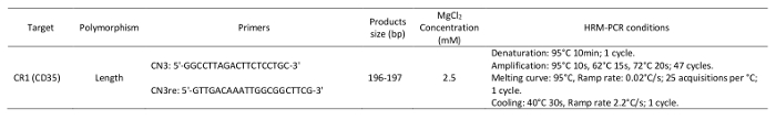
Table 1: Primers and parameters used in the high-resolution melting analysis.
2. HRM Analysis to Determine the CR1 Length Polymorphism
NOTE: The methodology described (Figure 1) is specific to our software (See the Table of Materials), although other software packages may be used.
- Open a gene scanning software to perform the CR1 length polymorphism scanning analysis.
- Open the experiment containing the amplification program and the melting curve program.
- Click Sample editor in the Module bar and then select the Scanning workflow.
- Define the properties of the samples (i.e., name; unknown or negative control).
- Click Analysis in the Module bar.
- In the Create New Analysis list, select Gene Scanning.
- Click the Normalization tab to normalize the melting curves.
- Click the Temperature shift tab to reset the temperature axis (x-axis) of the melting curves.
NOTE: The lower graph shows melting curves that are both normalized and temperature-shifted. - Click the Calculate button to analyze the results and determine the grouping.
- Click the Difference plot tab in the charts area to view the Normalized and Shifted Melting Curves and the Normalized and Temperature Shifted Difference Plot.
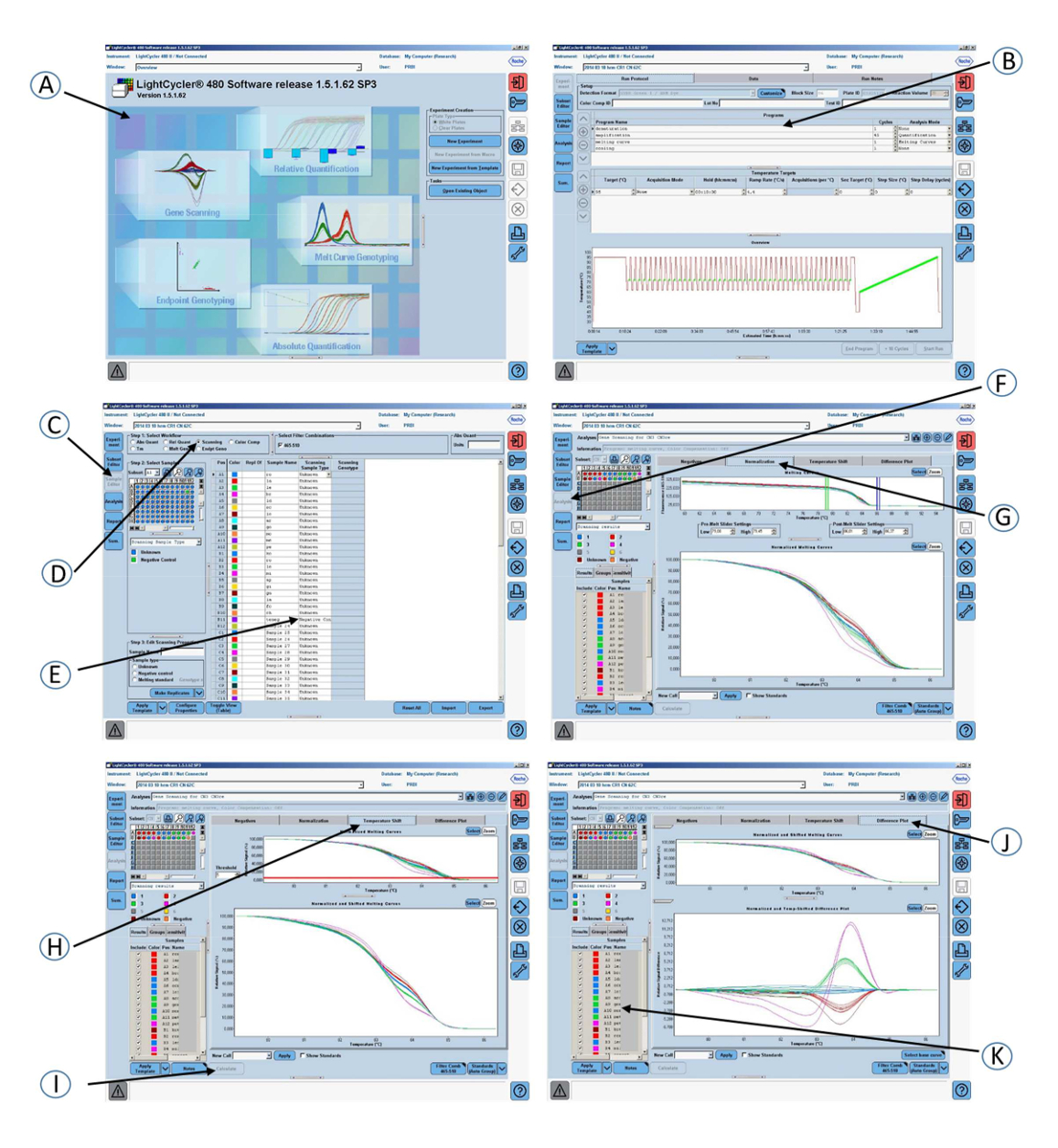
Figure 1: Screenshots of the graphical interface of the software used in step 2 of the protocol. (A) Open the gene scanning software. (B) Amplification program and melting curve program. (C) Click on Sample editor in the Module bar. (D) Select Scanning. (E) Define the properties of the samples. (F) Click on Analysis in the Module bar. (G) Click on the Normalization tab to normalize the melting curves. (H) Click on the Temperature shift tab to show the melting curves that are both normalized and temperature-shifted. (I) Click on the Calculate button to analyze the results and determine the grouping. (J) Click on the Difference plot tab to view the Normalized and Shifted Melting Curves and the Normalized and Temperature Shifted Difference Plot. (K) Colored grouping of the samples according to the CR1 length genotypes.
