A subscription to JoVE is required to view this content. Sign in or start your free trial.

Method Article

An Immunofluorescence Staining Technique to Detect Adult Hippocampal Neurogenesis

1.3K views

⸱

July 8th, 2025

In This Article

Abstract

Source: Ansorg, A., et al. Immunohistochemistry and Multiple Labeling with Antibodies from the Same Host Species to Study Adult Hippocampal Neurogenesis. J. Vis. Exp. (2015)

The video illustrates an immunofluorescence staining method used to examine neurogenesis in the adult hippocampus. Initially, a mouse brain section undergoes treatment with primary antibodies directed at marker proteins associated with neurogenesis in neuronal progenitor cells. Next, it is labeled with secondary antibodies tagged with fluorophores. The brain section is then observed under a confocal microscope to distinguish various subtypes of progenitor cells, based on the expression patterns of distinct combinations of marker proteins.
Protocol

All procedures involving animal models have been reviewed by the local institutional animal care committee and the JoVE veterinary review board.

1. Tissue Preparation

  1. Prepare 4% formaldehyde in 0.1 M phosphate buffer pH 7.4 on the day before perfusion. Store at 4 °C.
  2. Transcardially perfuse the deeply anesthetized mice (3.5% isoflurane) via the left ventricle with 10 ml ice-cold phosphate-buffered saline (PBS), then with 40 ml ice-cold formaldehyde (flow rate 5 ml/min). Dissect the brain and post-fix in the same fixative for 24 hr at 4°C.
  3. Transfer the brains consecutively into 10% (2....

Access restricted. Please log in or start a trial to view this content.

Results

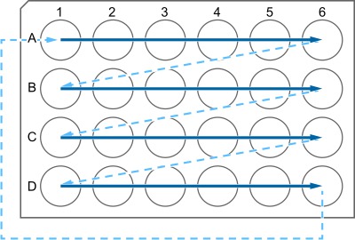
Microtiter plate sample mixing process, diagram showing horizontal sample flow pattern for analysis.
Figure 1. Schematic illustration of transferring microtome slices into a 24-well plate. Start at A1 and place subsequent slices into row A; after A6, go to the next row B, and so forth. When reaching D6, go back to A1 and continue. This arrangement of s.......

Access restricted. Please log in or start a trial to view this content.

Disclosures

No conflicts of interest declared.

Materials

List of materials used in this article
NameCompanyCatalog NumberComments
Tissue preparation
Isoflurane-Actavis Piramal Healthcare 700211
Paraformaldehyde powder (PFA) Riedel-De Häen 16005toxic, flammable
Perfusion pump PD5206 Heidolph Instruments 523-52060-00
Masterflex Tygon lab tubing, Ø 0.8 mmThermo Fischer Scientific 06409-13
Feeding needle, straight, 21 G, 1.75 mm olive tip, 40 mmAgnthos 1036
Freezing microtome Microm HM 400Thermo Fischer Scientific
24-well Cell culture multiwell plates Greiner Bio-One 662160
Immunohistochemistry
Tefal vitacuisine steamer Tefal VS 4001
Netwell 24 mm polyester mesh membrane insertsCorning 3479pre-loaded in 6-well culture plates
Netwell 15 mm polyester mesh membrane insertsCorning 3477pre-loaded in 12-well culture plates
Netwell plastic 6-well carrier kit Corning 3521for 24 mm polyester mesh membrane inserts
Netwell plastic 12-well carrier kit Corning 3520for 15 mm polyester mesh membrane inserts
Vectastain Elite ABC kit Vector Laboratories PK-6100
DAB (3,3′-Diaminobenzidine tetrahydrochloride hydrate)Sigma-Aldrich D-5637 carcinogenic, light sensitive
Fluoromount-G SouthernBiotech 0100-01
Primary antibodies
rabbit IgG1 α-Ki67
rabbit α-GFAP, AS-3-GFNovocastra/ Leica BiosystemsNCL-L-Ki67MM1DAB 1:400/IF 1:100; requires epitope retrieval
goat IgG (H+L) α-GFPSynaptic Systems173 00209:20
mouse IgG1 α-nestinAcris AntibodiesR1091P06:00
guinea pig IgG (H+L) α-DoublecortinAbcamab61421:200; requires epitope retrieval
rat IgG2a α-BrdU (ascites)Merck MilliporeAB225309:20
rat IgG2a α-BrdU (purified)AbD Serotec/ Bio-RadOBT0030CXfor detection of BrdU; DAB 1:500/IF 1:400
mouse IgG1κ α-BrdUAbD Serotec/ Bio-RadOBT0030for detection of CldU; DAB 1:500/IF 1:250-400
mouse IgG1 α-NeuNBD Biosciences347580for detection of IdU; DAB 1:500/IF 1:350
Secondary antibodiesMerck MilliporeMAB37709:20
donkey α-guinea pig IgG (H+L)-Biotin
donkey α-rat IgG (H+L)-BiotinDianova711-065-15209:20
donkey α-mouse IgG (H+L)-BiotinDianova712-065-15009:20
goat α-rat IgG (H+L)-Alexa Fluor 488Dianova715-065-15109:20
donkey α-goat IgG (H+L)-Alexa Fluor 488Molecular ProbesA1100605:10
donkey α-mouse IgG (H+L)-FITC, Fab-FragmentMolecular ProbesA1105505:10
donkey α-mouse IgG (H+L)-Alexa Fluor 647Dianova715-097-00302:40
donkey α-guinea pig IgG (H+L)-Alexa Fluor 647Dianova715-605-15105:10
donkey α-rat IgG (H+L)-Rhodamine Red-XDianova706-605-14805:10
donkey α-rabbit IgG (H+L)-Rhodamine Red-XDianova712-295-15005:10
donkey α-guinea pig IgG (H+L)-Rhodamine Red-XDianova711-295-15205:10
Streptavidin-Rhodamine Red-XDianova706-296-14805:10
goat α-rabbit IgG (H+L)-AMCADianova016-290-08409:20
Hoechst 33342Dianova111-155-1441:250, works only with rabbit α-GFAP
DAPIMolecular ProbesH357017:40
BlockingMolecular ProbesD130617:40
Fab-fragment donkey α-mouse IgG (H+L)
Fab-fragment donkey α-rabbit IgG (H+L)Dianova715-007-00301:20
Normal donkey serumDianova711-007-00301:20
Normal rabbit serumMerck MilliporeS30
Normal goat serumDianova011-000-010
Bovine Serum AlbumineDianova005-000-001
HistologySigma-AldrichA7906
Cresyl violet
Neo-ClearSigma-AldrichC5042
Neo-MountMerck Millipore109843non-toxic xylene substitute
MicroscopyMerck Millipore109016permanent mounting medium
Axioskop 2
LSM 710Carl Zeiss Microscopy
Carl Zeiss Microscopy

Explore More Articles

Confocal MicroscopyPrimary Antibody CocktailFluorophore-Labeled Secondary AntibodiesHost Species SerumMonovalent Fab FragmentsNeuronal Progenitor SubtypesMarker Protein ExpressionFluorescence Microscopy