Summary

Orthotopic Mouse Model of Colorectal Cancer

Published: December 04, 2007
doi:

Summary

Two techniques can be used to establish this model: injection of a cancer cell suspension into the cecal wall or transplantation of a piece of subcutaneous tumor onto the cecum. This model is useful for studying the natural progression of colorectal cancer and testing new therapeutic agents against colorectal cancer.

Abstract

The traditional subcutaneous tumor model is less than ideal for studying colorectal cancer. Orthotopic mouse models of colorectal cancer, which feature cancer cells growing in their natural location, replicate human disease with high fidelity. Two techniques can be used to establish this model. Both techniques are similar and require mouse anesthesia and laparotomy for exposure of the cecum. One technique involves injection of a colorectal cancer cell suspension into the cecal wall. Cancer cells are first grown in culture, harvested when subconfluent and prepared as a single cell suspension. A small volume of cells is injected slowly to avoid leakage. The other technique involves transplantation of a piece of subcutaneous tumor onto the cecum. A mouse with a previously established subcutaneous colorectal tumor is euthanized and the tumor is removed using sterile technique. The tumor piece is divided into small pieces for transplantation to another mouse. Prior to transplantation, the cecal wall is lightly damaged to facilitate tumor cell infiltration. The time to developing primary tumors and liver metastases will vary depending on the technique, cell line, and mouse species used. This orthotopic mouse model is useful for studying the natural progression of colorectal cancer and testing new therapeutic agents against colorectal cancer.

Protocol

I. Cell Preparation

  1. Colorectal cancer cells are grown in culture and harvested when subconfluent.
  2. A single cell suspension is prepared in phosphate buffered saline and kept on ice.

II. Tumor Preparation

  1. A mouse with a previously established subcutaneous colorectal tumor is euthanized.
  2. The subcutaneous tumor is removed using sterile technique and divided into 2-3 mm pieces
  3. The tumor pieces are kept in phosphate buffered saline on ice.

III. Mouse Preparation

Note: In our laboratory we use inhaled isoflurane to anesthetize the mouse; alternatively, one can use injectable anesthetics to achieve the same effect

  1. The depth of anesthesia is assessed using toe pinch. There should be no withdraw reflex with toe pinch.
  2. Antibiotics may be given at this point.
  3. The anesthetized mouse, which was previously shaved, is properly positioned.
  4. The abdomen is prepped with a betadine solution.
  5. The abdomen and surgical site are draped in a sterile fashion.

IV. Laparotomy

  1. A small nick is made in the skin
  2. The abdominal wall musculature is grasped and lifted up
  3. The abdominal cavity is entered and a single blade of the scissors is used to push the intra-abdominal contents away
  4. The incision is extended to 2-3 cm

V. Exposure of the Cecum

  1. The cecum with its blind ending pouch is identified and exteriorized
  2. The cecum is isolated from the rest of the mouse using a pre-cut, sterile gauze
  3. Warm saline is used to keep the cecum moist

VI. Injection of Cells into the Cecal Wall

  1. A 27 G or finer needle is used to inject a 50 µL volume of cells into the cecal wall
  2. The needle is removed
  3. The injection site is inspected to ensure no leakage
  4. The cecum is returned to the abdominal cavity

VII. Transplantation of Tumor onto the Cecum

Note: In addition to injecting cells into the cecal wall, an alternative approach is to transplant tumor onto the cecum

  1. A figure of 8 stitch is placed onto the cecum using 6-0 or 7-0 sized suture
  2. The cecal wall is lightly damaged
  3. Then, the tumor piece is positioned
  4. The stitch is tied down
  5. The cecum is returned to the abdominal cavity

VIII. Mouse Abdominal Wall Closure and Recovery

  1. The mouse abdominal wall is closed using three interrupted stitches using 3-0 or 4-0 sized suture
  2. Alternatively, one can use a simple running stitch
  3. Post-operative analgesics and a fluid bolus may be given at this point
  4. The mouse is allowed to recover from anesthesia.

Note: With inhaled anesthetics this typically takes 30 seconds

IX. Results – Primary Tumor and Liver Metastasis


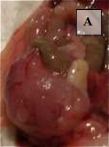
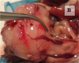
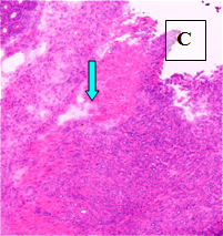
Primary Tumor – shown in situ (A) and with evidence of neovascularization (B); on H&E staining, tumors are locally invasive (C)


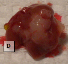
Liver Metastases – shown ex vivo (D)

Discussion

Although mouse subcutaneous tumor models are easy to establish and monitor, it is clear that this model cannot replicate the original anatomic site of colorectal cancer. Due to the difference in microenvironment, colorectal cancer cells growing under the skin have been shown to change their phenotype and almost always fail to progress and metastasize 1,2. In fact, tumor response to therapy can vary dramatically depending on whether cancer cells are implanted in an ectopic (subcutaneous) versus orthotopic location 3. Orthotopic mouse models of colorectal cancer, which feature cancer cells growing in their natural location, replicate human disease with high fidelity. The two techniques to establish this model have unique advantages and disadvantages. Injection of a cell suspension into the cecal wall introduces colorectal cancer cells which have been previously growing in an in vitro environment and are arguably homogeneous. Cells may have reduced invasive or metastatic potential after several passages in culture. Transplantation of a piece of subcutaneous tumor introduces a more heterogeneous population of cancer cells that have been established in vivo. However, tumors contain stromal cells and frequently have necrotic portions which may affect the consistency of this model (personal communication, I. Fidler, MD Anderson). From a technical standpoint, our laboratory has found that injection of a cell suspension into the cecal wall is more difficult and carries the risks of intraluminal injection and leakage post-injection. Interestingly, an orthotopic mouse model of rectal cancer has been described that does not require mouse laparotomy 4. Mice are anesthetized and the rectal mucosa is prolapsed with digital pressure. A small volume of colorectal cancer cells in single cell suspension is injected submucosally. Primary, invasive rectal cancers develop in mice as early as one week after injection; however, none of the mice develop liver metastases and the authors did not comment on the frequency of lung metasases.

Acknowledgements

Department of Surgery, University of California, San Francisco
Raymond Shaheen, M.D.

Materials

Material Name Tipo Company Catalogue Number Comment
Cell culture media and components       will vary depending on cell line used
Phosphate Buffered Saline   BioWhitaker 17-512F also sold by a number of other vendors
15 mL centrifuge tubes, polypropylene   Corning 430790  
6 well plates with lid   Becton Dickinson Labware 353046  
Iris forceps, straight, serrated tips   World Precision Instruments 15914 best to have a pair of these instruments
Iris dissecting scissors, straight, sharp   World Precision Instruments 14393  
Needle Holder (regular)   World Precision Instruments 14109  
Needle Holder (Castroviejo)   World Precision Instruments 14137  
Cidex Plus   World Precision Instruments 7364 instruments should be autoclaved periodically, but may also be sterilized by soaking in Cidex
6-0 or 7-0 suture       for cecum; courtesy of Department of Surgery, UCSF
3-0 or 4-0 suture       for abd wall; courtesy of Department of Surgery, UCSF
Anesthetic machine / Isoflurane   J.A. Baulch and Associates 3206 also sold by a number of other vendors
Heating Pad, disposable, 6″ x 6″   Prism Technologies 20419  
Tape, orange, 13 mm x 13 m   Fisher Scientific 15901F for restraining anesthetized mouse
Pro-Cord/Cordless Trimmer   Oster 78997-010 for shaving mouse fur
Betadine   Fisher Scientific 19-027132 may also be purchased at any medical supply store
2 x 2 gauze, 3-ply   Johnson and Johnson 7635 may also be purchased at any medical supply store
Sterile Field, Barrier, 16″ x 29″   Johnson and Johnson 0905 may also be purchased at any medical supply store
Cotton tipped applicators, 6″   Fisher Scientific 14-960-3Q may also be purchased at any medical supply store
Syringe, 10 cc, Luer lock tip   BD 309604  
Syringe, 1 cc, tuberculin, slip tip   BD 309602  
27 1/2 G needle   BD 305109  
30 G needle   BD 305106  

Riferimenti

  1. Heijstek, M. W., Kraneburg, O., Rinkes Borel, I. H. Mouse models of colorectal cancer and liver metastases. Dig Surg. 22, 16-25 (2005).
  2. Kobaek-Larsen, M., Thorup, I., Diederichsen, A., Fenger, C., Hoitinga, M. R. Review of colorectal cancer and its metastases in rodent models: comparative aspects with those in humans. Comp Med. 50, 16-26 (2000).
  3. Wilmanns, C., Fan, D., O'Brian, C. A., Bucana, C. D., Fidler, I. J. Orthotopic and ectopic organ environments differentially influence the sensitivity of murine colon carcinoma cells to doxorubicin and 5-fluorouracil. Int J Cancer. 52, 98-104 (1992).
  4. Kashtan, H., Rabau, M., Mullen, J. B., Wong, A. H. C., Roder, J. C., Shiptz, B., Stern, H. S., Gallinger, S. Intra-rectal injection of tumor cells: a novel animal model of rectal cancer. Surg Oncol. 1, 251-256 (1992).
check_url/it/484?article_type=t

Play Video

Citazione di questo articolo
Tseng, W., Leong, X., Engleman, E. Orthotopic Mouse Model of Colorectal Cancer. J. Vis. Exp. (10), e484, doi:10.3791/484 (2007).

View Video