A method to rapidly and completely remove cellular components from an intact porcine heart through retrograde perfusion is described. This method yields a site specific cardiac extracellular matrix scaffold which has the potential for use in multiple clinical applications.
Perfusion-based whole organ decellularization has recently gained interest in the field of tissue engineering as a means to create site-specific extracellular matrix scaffolds, while largely preserving the native architecture of the scaffold. To date, this approach has been utilized in a variety of organ systems, including the heart, lung, and liver 1-5. Previous decellularization methods for tissues without an easily accessible vascular network have relied upon prolonged exposure of tissue to solutions of detergents, acids, or enzymatic treatments as a means to remove the cellular and nuclear components from the surrounding extracellular environment6-8. However, the effectiveness of these methods hinged upon the ability of the solutions to permeate the tissue via diffusion. In contrast, perfusion of organs through the natural vascular system effectively reduced the diffusion distance and facilitated transport of decellularization agents into the tissue and cellular components out of the tissue. Herein, we describe a method to fully decellularize an intact porcine heart through coronary retrograde perfusion. The protocol yielded a fully decellularized cardiac extracellular matrix (c-ECM) scaffold with the three-dimensional structure of the heart intact. Our method used a series of enzymes, detergents, and acids coupled with hypertonic and hypotonic rinses to aid in the lysis and removal of cells. The protocol used a Trypsin solution to detach cells from the matrix followed by Triton X-100 and sodium deoxycholate solutions to aid in removal of cellular material. The described protocol also uses perfusion speeds of greater than 2 L/min for extended periods of time. The high flow rate, coupled with solution changes allowed transport of agents to the tissue without contamination of cellular debris and ensured effective rinsing of the tissue. The described method removed all nuclear material from native porcine cardiac tissue, creating a site-specific cardiac ECM scaffold that can be used for a variety of applications.
1. Tissue Preparation and Experiment Setup
2. Tissue Rinses
3. Decellularization and Solution Perfusion
4. Disinfection and Final Processing
The effect of decellularization on whole porcine hearts naturally varies due to differences in size, pressures, and vessel arrangement. Therefore, the exact composition of the derived extracellular matrix scaffolds will not be the same from heart to heart. The completion of the described protocol will yield a heart that appears white or translucent, indicating the loss of cellular material. However, it is widely accepted that a tissue can be considered “decellularized” based on the combination of a few more quantitative parameters 8. A successful decellularization protocol will produce a matrix with less than 50 ng of double stranded DNA per mg of tissue (Figure 6). In order to avoid a host immune response upon implantation of the matrix, the remaining DNA should also contain less than 200 base pairs (Figure 7). To confirm these findings, Hematoxylin and Eosin staining should reveal the absence of nuclear staining in representative sections of the ventricles and ventricular septum (Figure 8). Masson’s Trichrome further confirms the loss of cardiac muscle bundles and retention of collagen networks (Figure 9).
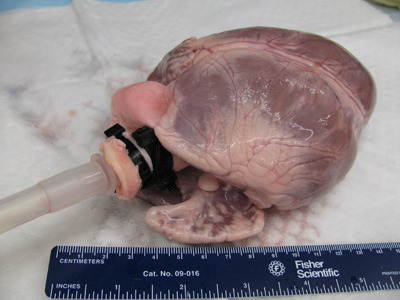
Figure 1. The barbed end of the tubing is inserted into the aorta of the native heart. The tubing must be secured with hose clamps or zip ties above the aortic valve to ensure perfusion through the coronary arteries.
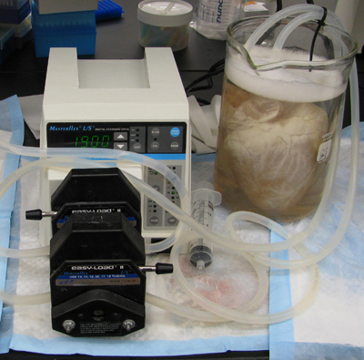
Figure 2. The heart is submerged in water in a 4L beaker and air bubbles must be removed from the tubing. If bubbles are observed emerging from the aorta near the tubing, additional ties must be used to secure the tubing to the aorta in order to maintain adequate pressure in the tissue.
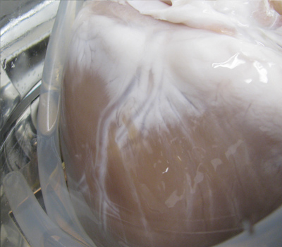
Figure 3. As solutions are perfused through the coronary arteries, the heart will lose its native color, progressing from the atria to the apex of the heart and localized around the coronaries.
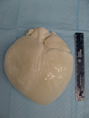
Figure 4. After completion of the disinfection and rinse steps of the protocol, the tubing is removed and the heart is placed on an absorbent pad to allow the excess water to drain out of the heart. This ensures an accurate measurement when weighing the tissue and also allows the tissue to relax before sectioning.
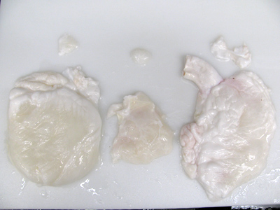
Figure 5. The left ventricle (LV), right ventricle (RV), and ventricular septum are all removed from the decellularized heart for histologic processing, freezing and lyophilization, and DNA quantification.
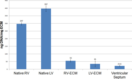
Figure 6. Quantitative analysis of DNA content using a Pico Green assay. The ventricles from cECM hearts show a significant decrease in DNA content when compared to native ventricles. The DNA values observed from this protocol are observed at or below the 50 ng/mg standard for decellularized tissues.
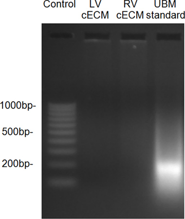
Figure 7. DNA fragment size, as determined by ethidium bromide gel, showed little residual DNA in the decellularized ventricles when compared to a urinary bladder matrix (UBM) standard.
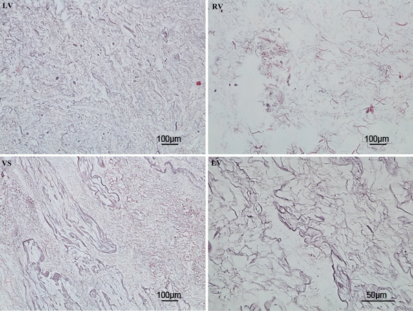
Figure 8. Hematoxylin and Eosin staining showed complete removal of nuclear material from the ventricles following completion of the decellularization protocol.
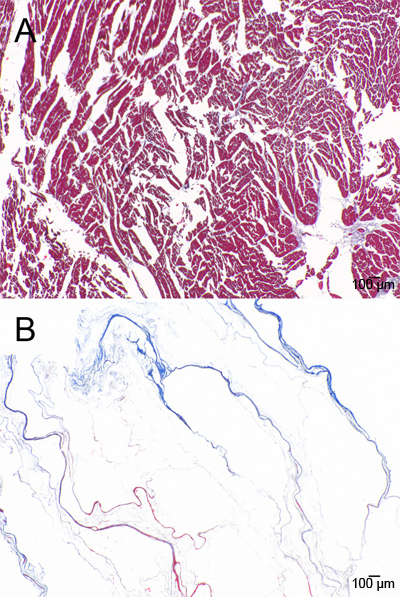
Figure 9. Masson’s Trichrome staining of A) native and B) decellularized ventricle.
The current study described methodology for consistent and efficient decellularization of a porcine heart. The protocol was a modification to a previously published report 1, and included longer exposure to flow and increased pressure, which provided more repeatable results. The resulting decellularized tissue met all of the published criteria for successful decellularization of tissue 2. Frequent solution changes were performed to limit the reintroduction of cellular material to the tissue, and the duration of exposure to each decellularization agent was minimized to reduce adverse effects on the ECM. During the beginning stages of the protocol, the perfusion rate was gradually increased to condition the tissue and allow for higher flow rates during the later stages of the protocol. Without conditioning the tissue in the early stages, the vasculature of the heart can rupture, making perfusion of the heart impossible. The protocol was used due to its efficiency, and no claims are made to its superiority over other protocols. The precise composition of decellularization agents and rates of perfusion may conceivably be varied to yield a protocol with better mechanical or biologic characteristics, but the general principles for delivery of the agents to the heart are applicable.
The preservation of the native three-dimensional structure of the heart was attributed to several procedures performed throughout the decellularization protocol. First, the tissue was trimmed and frozen upon arrival. Freezing promoted cell lysis and was important for pre-conditioning the tissue for the perfusion cycles. The tissue was thoroughly inspected for cuts and 2 cm intact intact aorta superior to the aortic valve. If any pericardium or epicardium was cut, the organ was discarded because the perfusate did not reach downstream regions of the heart, and the heart was not fully decellularized. Next, the tissue was fully thawed in type I water before use. The water allowed the tissue to relax as it thawed and also aided the removal of residual blood clots within the heart. Finally, as the tubing was inserted, care was taken to ensure that the aortic valve remained intact so that it formed a water-tight seal around the tubing, so that a proper pressure was maintained and that the solution entered the coronary arteries.
After each decellularization protocol was completed, a series of quality control measures were completed to ensure complete removal of cellular material. The current study verified that the protocol eliminated histologic staining for cell nuclei, showed that less than 50 ng of DNA was present per mg of dry weight of the tissue, and that any DNA was less than 200 bp in size 6. Previously published methods for cardiac decellularization showed similar levels of decellularization in DNA staining and quantification 2,5,9,10. Complete decellularization was accomplished in these studies using similar treatments of enzymes and detergents. However, in the present study, the length of exposure to each chemical was increased, there were more solution changes, and the flow rates were increased. The present protocol also increased the length of chemical rinses, potentially leading to more efficient removal of chemical residues from the extracellular matrix.
Recellularization of decellularized rat hearts with cardiac specific cells has yielded promising results in vitro 2,5. Whole organ perfusion decellularization allowed for maintenance of the native vasculature, which is critical in recellularization of the tissue. The inherent growth factors, matrix proteins, and three-dimensional fiber architecture also promoted proper cell attachment, migration, and signaling to reconstitute contractile myocardial tissue. Porcine cardiac extracellular matrix will be more difficult to recellularize due to the size of the scaffold and the number of cells necessary for proper cell communication and nutrient transport. However, patches cut from the cardiac matrix may be useful for in vivo applications. Multiple studies have shown the potential advantage of using a site-specific matrix for reconstruction of damaged tissue in animal models 11-13. Thus, an extracellular matrix scaffold derived from cardiac tissue is desirable for myocardial reconstruction applications. The inherent architecture of the cardiac tissue may present advantages over an ECM scaffold derived from another organ or an artificial biomaterial. A site-specific scaffold may support host cell infiltration and promote a constructive remodeling response, as opposed to scar tissue formation. To date, cardiac ECM patches have been investigated in vivo to reconstruct a defect created in the myocardial wall 14. Future studies will be performed in vitro to examine the ability of the scaffold to support cardiac cells seeded and cultured on the matrix. The methods described herein may also be applicable to decellularization of human hearts.
In conclusion, porcine heart decellularization is possible and the methods are straight-forward. Continued investigation of this material will provide insight into its potential for clinical use.
The authors have nothing to disclose.
The authors would like to acknowledge Brogan Guest, Michelle Weaver, and Kristen Lippert. Funding for this study was provided by NIH Grant R03EB009237, as well as NIH Training Grants T32EB001026-06 from the National Institute of Biomedical Imaging And Bioengineering and T32HL076124-05.
Name of Reagent/Material | Company | Catalogue Number | Comments |
Trypsin | Gibco | 15090 | |
EDTA | Fisher | BP120-500 | |
NaN3 | Sigma | S2002-500G | |
Triton X-100 | Sigma | X100-1L | |
10X PBS | Fisher | BP399-20 | |
Sodium Deoxycholate | Sigma | D6750-500G | |
Peracetic Acid | Pfaltz and Bauer | P05020 | 35% CAS# 79-21-0 |
Ethanol | Pharmco | 111000200 | |
Masterflex Pump Drive | Cole Parmer | SI-07524-50 | |
Masterflex Tubing | Cole Parmer | 96400-18 | Size 18 |
Barbed Reducer | Cole Parmer | EW-30612-20 | |
4L Beaker | Fisher Scientific | 02-540T |