A breast cancer brain metastasis mouse model is established with ultrasound imaging-guided intracardiac injection of MDA-MB231/Br-GFP cells. Development of multifocal intracranial metastases has been monitored longitudinally using high-resolution 9.4 T MRI.
Breast cancer brain metastasis, occurring in 30% of breast cancer patients at stage IV, is associated with high mortality. The median survival is only 6 months. It is critical to have suitable animal models to mimic the hemodynamic spread of the metastatic cells in the clinical scenario. Here, we are introducing the use of small animal ultrasound imaging to guide an accurate injection of brain tropical breast cancer cells into the left ventricle of athymic nude mice. Longitudinal MRI is used to assessing intracranial initiation and growth of brain metastases. Ultrasound-guided intracardiac injection ensures not only an accurate injection and hereby a higher successful rate but also significantly decreased mortality rate, as compared to our previous manual procedure. In vivo high resolution MRI allows the visualization of hyperintense multifocal lesions, as small as 310 µm in diameter on T2-weighted images at 3 weeks post injection. Follow-up MRI reveals intracranial tumor growth and increased number of metastases that distribute throughout the whole brain.
Brain metastasis is the most common intracranial malignancy in adults. The prognosis is extremely poor, with a median survival of 4-6 months even with aggressive treatment. Breast cancer is one of the three major primary cancers with a high morbidity of brain metastasis1-3. Several brain-tropic breast cancer lines are capable of developing brain metastases after intracardiac or intracarotid injection4. Direct injection of tumor cells into the left ventricle can bypass the lung capillary bed and thus increase the incidence of forming brain metastases while minimizing visceral metastases. The MDA-MB231Br line is one of the most widely used human breast cancer lines to develop brain metastasis in rodent models5,6.
Like many other studies4,7, we have performed a manual procedure of intracardiac injection in our previous studies. However, only 50% successful rate was obtained with the manual injection and a fraction of mice died from the repeated invasive procedures if prior trials failed. Here, we are introducing the use of an imaging-guided procedure to secure the injection of brain-seeking breast cancer cells into the left ventricle of athymic mice. Longitudinal high resolution MRI is applied to follow intracranial development of brain metastases.
All animal procedures were approved by the Institutional Animal Care and Use Committee of University of Texas Southwestern Medical Center.
1. Preparation of the MDA-MB231/Br-GFP Cells
2. Ultrasound Imaging-guided Intracardiac Injection
3. MRI Monitoring of Intracranial Tumor Development
4. H&E Staining Confirming the Metastases
With the high spatial resolution of MRI (78 µm in plane resolution), hyperintense lesions can be identified as small as 310 µm in diameter (Figure 2). Since the metastases in this study are very small and development of necrosis and edema is minimal, the hyperintense lesion on T2-weighted images truly represented the tumor mass.
Longitudinal MRI studies allow in vivo noninvasive evaluation of tumor growth. As shown in Figure 3, the high resolution MRI was able to detect several small lesions 3 weeks after intracardiac injection (Figure 3A). On week 4, the lesions that were seen in the previous scan all became larger; more new lesions appeared on T2-weighted images (Figure 3B).
H&E staining revealed either diffuse or cluster type metastatic lesions (Figure 4). Enlarged vessels were often seen around the tumor, indicating nonsprouting angiogenesis (Figure 4D).
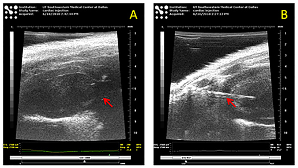
Figure 1. Ultrasound-guided intracardiac injection. (A) Identification of the ascending aorta (arrow) as the landmark of left ventricle of the mouse heart. (B) A needle (arrow) insertion into the left ventricle to inject the tumor cells. Click here to view larger image.
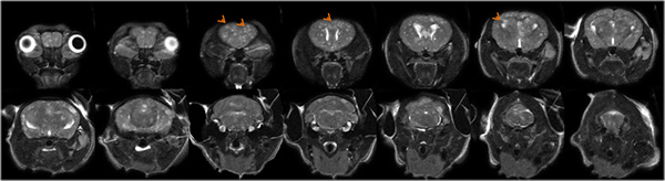
Figure 2. High resolution T2-weighted images of breast cancer brain metastases. Fourteen consecutive MRI slices of a representative mouse brain, acquired four weeks after intracardiac injection, clearly revealed the multifocal metastases distributing through the whole mouse brain, from olfactory bulb to pontine and medulla. Click here to view larger image.
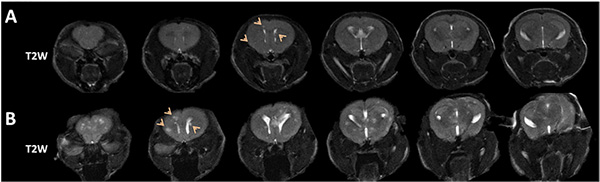
Figure 3. Longitudinal MRI of development of brain metastases. A. Six consecutive coronal MRI sections at week 3 identified multiple lesions with hyper-intensity on T2-weighted images. B. An increased number of lesions appeared on the images at week 4, and those lesions seen on week 3 became larger. Click here to view larger image.
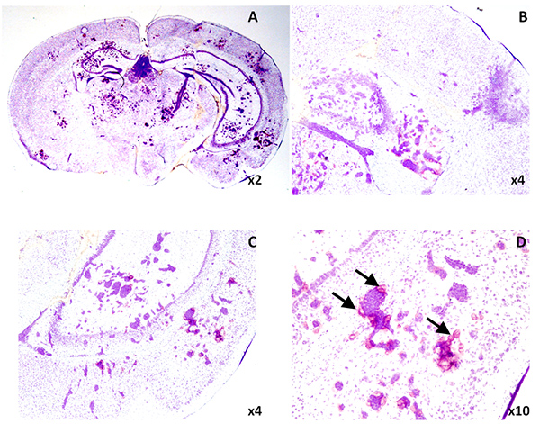
Figure 4. Microscopic lesions were observed on H&E staining. A. A whole mount coronal section depicted multiple lesions. B-D. Higher magnification images showed either diffuse or cluster type lesions (B and C). Enlarged vessels were seen around the tumor. Click here to view larger image.
In the present study, we have demonstrated that ultrasound imaging-guided left ventricular injection ensures the accuracy so that every animal in this study developed brain metastases and no animal death was observed. The beauty of image-guided injection is that the path of needle penetration of skin and finally into the left ventricle can be monitored and adjusted under image, which is distinct from the manual procedure requiring strict anatomic landmarks to follow.
Current understandings of intracranial development of brain metastases are largely based on histological studies on animal models4,10. However, histological studies normally require a large number of mice that are killed at different time points after tumor implantation. More importantly, information about temporal development in individual lesions is lacking from histological studies.
In vivo imaging promises greater efficiency since each animal serves as its own control and multiple time points can be examined sequentially7,11. High resolution T2-weighted images enable the detection of multifocal tumor initiation at very early stage (lesions with diameter as small as 310 µm are visible). Longitudinal MRI allows individual brain metastases to be examined over time. Moreover, in vivo noninvasive MRI will be particularly valuable in longitudinal study of therapeutic response, e.g. whole brain radiation.
Formation of multifocal brain metastases is the characteristics of the MDA-MB231Br model. Besides the MDA-MB231/Br-GFP cell line we showed in the present study, the parental clone MDA-MB231/Br and MDA-MB231/Br-luc also exhibit similar metastatic lesions. In contrast, several other brain-seeking lines such as MCF-7Br and 4T1 have been shown to develop a solitary metastasis after intracardiac injection12. Both of the models, mirroring clinical counterparts, are useful animal models of brain metastasis. However, in addition to brain metastases, visceral metastases, such as lung and bone metastases are often observed in this model. Establishment of animal models that develop brain metastases exclusively will be critical, in particular, for evaluation of treatment response.
In conclusion, we have demonstrated the usefulness of imaging-guided intracardiac injection to establish a brain metastasis mouse model and the high resolution MRI to assess intracranial development of multifocal metastases in a mouse model.
The authors have nothing to disclose.
We are grateful to Drs. Diane Palmieri and Patricia Steeg of NCI for providing us MDA-MB231Br cells. We thank Dr. Ralph Mason, Mr. Jason Reneau and Ms. Ramona Lopes for technical and collegial support. This work was supported in part by the DOD IDEA Awards W81XWH-08-1-0583 and W81XWH-12-1-0317. MRI experiments were performed in the Advanced Imaging Research Center, an NIH BTRP # P41-RR02584 facility, and ultrasound-guided intracardiac injection was performed with VisualSonics Vevo 770 under 1S10RR02564801.
DMEM | HyClone | SH30022.01 | |
Trypsin | Mediatech | 25-051-C1 | |
Automatic cell counter | Biorad | TC10 | |
Isoflurane | Baxter International Inc. | 1001936060 | |
VisualSonics Vevo 770 High-Resolution Imaging System | Visual Sonics Inc. | ||
9.4T horizontal bore magnet with a Varian INOVA Unity system | Agilent | ||
O.C.T Compound | Sakura Finetek USA | 4583 |