Molecular Profiling of the Invasive Tumor Microenvironment in a 3-Dimensional Model of Colorectal Cancer Cells and Ex vivo Fibroblasts
Summary
Molecular profiling of laser microdissected cells and stroma from synthetic, tissue-engineered colorectal cancer models represents a novel and manipulatable approach to characterize and dissect the distinctive biology at the interface between tumor cells at the invasive front and cancer associated stromal cells.
Abstract
Invading colorectal cancer (CRC) cells have acquired the capacity to break free from their sister cells, infiltrate the stroma, and remodel the extracellular matrix (ECM). Characterizing the biology of this phenotypically distinct group of cells could substantially improve our understanding of early events during the metastatic cascade.
Tumor invasion is a dynamic process facilitated by bidirectional interactions between malignant epithelium and the cancer associated stroma. In order to examine cell-specific responses at the tumor stroma-interface we have combined organotypic co-culture and laser micro-dissection techniques.
Organotypic models, in which key stromal constituents such as fibroblasts are 3-dimentioanally co-cultured with cancer epithelial cells, are highly manipulatable experimental tools which enable invasion and cancer-stroma interactions to be studied in near-physiological conditions.
Laser microdissection (LMD) is a technique which entails the surgical dissection and extraction of the various strata within tumor tissue, with micron level precision.
By combining these techniques with genomic, transcriptomic and epigenetic profiling we aim to develop a deeper understanding of the molecular characteristics of invading tumor cells and surrounding stromal tissue, and in doing so potentially reveal novel biomarkers and opportunities for drug development in CRC.
Introduction
Organotypic co-cultures are tissue-engineered models which aid in reconstructing the in vivo tumor microenvironment in 3-dimensions by juxtaposing malignant epithelial cells and stromal cells in a collagen gel containing essential extracellular matrix components1-3. Principally conceived as a method of measuring tumor invasion, organotypics reduce reliance on in vivo animal models, and avoid the shortcomings of other in vitro techniques such as Transwell assays, in which invading cells are forced artificially into a mono-dispersed state2-4. The inclusion of stromal cells in these models, such as fibroblasts, reflects the essential role of the tumor microenvironment in regulating malignant invasion and metastasis3-4, however the phenotypic heterogeneity of the stroma is such that in order to maximize the physiological relevance of organotypic models, organ specific, anatomically accurate ex vivo fibroblasts should be included wherever possible3,6.
Organotypics are a versatile platform to study interactions between tumor and stromal cells and are increasingly used in novel ways to examine the impact on tumor invasion of chemical inhibitors and targeted gene alterations7,8. Studying the invasive tumor margin is a particularly exciting prospect. In organotypic models, established colorectal cancer (CRC) cell lines typically produce a well stratified epithelial layer, and in cross section, cells that have acquired the capacity to invade the extracellular matrix (ECM) are easily discernible. In light of the established micro-topographic heterogeneity of tumor and tumor associated stromal cells in vivo, extracting these cells using Laser microdissection and studying them in isolation, may reveal important biological insights regarding the origins of colorectal cancer metastasis and how it may be more efficiently targeted. In the following methodology, all patients who donated tissue samples provided written informed consent, and the study was approved by the institutions regional research ethics committee.
Protocol
1. Establishment of Primary Fibroblast Cultures from Colonic Explants
- Obtain samples of normal human colon mucosa tissue directly from the surgical operating theatre, and suspend in 7-10 ml of PBS supplemented with 100 units/ml penicillin, 100 μg/ml streptomycin, and 0.25 μg/ml Fungizone.
- In the laboratory, place the specimen at the center of a 10-cm tissue culture dish and wash 3 times with PBS/Pen-strep/Fungizone. Do not aspirate after the final wash.
- With sterile forceps and scalpel, cut the tissue into small pieces (approximately 2mm) and place each piece at the center of a cross drawn with the scalpel blade in a 12-well plate. Rock the scalpel gently over the tissue to allow it to adhere.
- Culture the specimens in 750 μl primary fibroblast growth media (DMEM supplemented with 20% fetal calf serum (FCS), 100 U/ml penicillin, 100 μg/ml streptomycin and 292 μg/ml L-Glutamine), which should submerge the tissue without enabling it to float. Avoid disturbing the plate over the next 5 days except to re-feed every 2 days.
- Over the next 5 days fibroblasts will slowly start to grow out from the edge of the tissue. At approximately 7 days, this will be more visible. After approximately 3-4 weeks, and if there are sufficient fibroblasts growing out, add 750 μl trypsin to each well. After cells have detached (which may take 5-10 min), pool and transfer to a T-25 tissue culture flask.
2. Organotypic Preparation
Preparation of the fibroblast impregnated organotypic gel:
- Prepare a fibroblast cell suspension containing 5 x 10⁵ cells per organotypic in DMEM supplemented with 10% FCS and 292 μg/ml L-Glutamine.
- Make up the organotypic gels on ice, in the following ratios:
- 7 volumes of Rat-Tail Collagen: Matrigel mix (1:1)
- 1 volume of filtered 10x DMEM
- 1 volume of FCS
- 1 volume of fibroblast cell suspension containing 5 x 10⁵ fibroblasts
For 9 gels (1 ml per gel) prepare 10 ml and mix gently to avoid bubbles.
- Add 1 ml to each well of a 24-well plate and incubate for 1 hour at 37 °C in a humidified atmosphere to allow gels to set. After 1 hr add 1 ml DMEM (10% FCS)/Glut on top of the gels and return to the incubator overnight.
- The following day, aspirate medium from the top of the gels and plate 1ml of media containing 5 x 10⁵ CRC epithelial cells.
Preparation of gel coated nylon sheets:
- Prepare sterile, autoclaved nylon sheets measuring 1.5 cm x 1.5 cm, in advance.
- Using sterile forceps, place the required number of nylon sheets in a 10 cm culture dish (usually 4 nylon sheets can be accommodated per dish).
- Make up gel on ice in the following order (250 μl total volume is required per nylon sheet):
- 7 volumes of Rat-Tail Collagen
- 1 volume of filtered 10x DMEM
- 1 volume of FCS
- 1 volume of DMEM 10% FCS/Glut
- If the solution is yellow, neutralize by adding 0.1 M NAOH in 50 μl aliquots until the solution turns pink.
- Add 250 μl of this solution to each nylon sheet and incubate at 37 °C in a humidified atmosphere of 5% CO2 for 30 min to allow gel to set.
- Make up 1% glutaraldehyde solution in PBS. Add 10 ml to each 10 cm culture dish once the gel has set and incubate at 4 °C for 1 hr.
- Wash nylon sheets 3x in PBS and 1x in DMEM (10% FCS)/Glu and incubate overnight in DMEM/Glu at 4 °C.
Raising organotypic gels onto steel grids for invasion:
- Pre-prepare scaffolding grids by folding stainless steel sheets into a tripod formation. Autoclave prior to use.
- Use forceps sterilized in ethanol to place a steel grid into each well of a six well plate.
- Place a gel-coated nylon sheet with the collagen side uppermost, onto each grid.
- Carefully transfer the organotypic gels from the 24-well plate to the raised nylon sheet using a spatula sterilized in ethanol.
- Fill the well with DMEM 10% FCS/Glut supplemented with 10% FCS until the nylon sheet is in contact with the medium but not submerged. Make sure the medium does not touch the organotypic gel.
- Incubate for 14 days at 37 °C in a humidified atmosphere of 5% CO2, replacing media every 2 days.
3. Organotypic Fixation
- Remove whole organotypic including nylon sheet from well and place on cling film.
- Bisect organotypic and nylon sheet using a clean disposable scalpel and fix both halves in formaldehyde for 24 hr at room temperature.
- Replace formaldehyde with 70% ethanol after 24 hr and leave overnight prior to embedding in paraffin, sectioning, and staining.
4. Laser Microdissection of the Invasive Margin
- Section to 10 μm-thickness onto membrane mounted slides.
- Deparaffinize sections by applying Xylene for 1 min, then remove Xylene and fix in 75% ethanol for a further 1 min.
- Remove ethanol and stain with 0.125% Cresyl Violet solution for 1 min. Cresyl Violet highlights epithelial cells and permits easy discrimination from the stroma.
- Remove Cresyl Violet and apply 100% ethanol for a further 1 min before rinsing with 100% ethanol. Allow slides to air dry for 30 min.
- Conduct laser microdissection using chosen platform (e.g. the Leica AS system).
- Place the glass slide with the stained section to be microdissected face down on the microscope stage.
- Mount a 0.5 ml microcentrifuge tube into the collection cassette and add 50 μl of cell lysis buffer (e.g. DNA, RNA or protein lysis buffer) to the cap into which microdissected tissue will collect.
- Under direct vision, use the joystick to identify tissue of interest, and using the software interface, annotate the stained organic section, highlighting cells to be microdissected at the tumor invasion front.
- Instruct the laser to fire, which should both cut out the highlighted section and propel microdissected tissue into the cap of the microcentrifuge tube.
- Once sufficient material has been collected, eject the collection cassette, close the microcentrifuge tube and spin gently to draw the lysis buffer to the bottom of the tube.
- Place samples on ice until microdissection is complete. Process samples by an appropriate method to extract the analyte of interest.
Representative Results
We have applied the above method to multiple combinations of CRC cell lines and stromal cells. One example presented here is SW480 epithelial CRC cell lines with primary human ex vivo colonic fibroblasts. 3-dimensional co-cultures are constructed (Figure 1), subjected to microscopy and laser capture microdissection (Figure 2), and analyzed by microRNA (miRNA) profiling (Figure 3) for comparison of differentially expressed miRNAs between cells at the invasive tumor front and those in the stratified (non-invading) tumor zones.
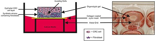
Figure 1. Schematic and photographic representations of the organotypic model. CRC cells are seeded onto a synthetic stromal layer containing fibroblasts and essential extracellular matrix components. Cells are co-cultured at an air-liquid interface created by elevating the organotypic gel onto a sterile stainless-steel grid. Click here to view larger image.
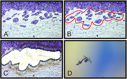
Figure 2. LMD of SW480 CRC cells invading the organotypic stroma. Cresyl Violet is used to highlight CRC epithelial cells (A); invasive tumor islands are then defined (B); before laser dissection occurs (C) and the cut piece is transported to a collection device (D). Non-invading cells and stromal cells are identified and isolated using the same methodology. Click here to view larger image.

Figure 3. MicroRNA microarray data. Arrays were scanned using a GenePix Pro 3.0.5 scanner to detect Cy5 at a wavelength of 635 nm. Mean fluorescent intensities were normalized and expression ratios calculated between laser microdissected invasive margin CRC cells and non-invasive margin CRC cells in the stratified epithelial layers. Data was sorted by fold change and data values +/- 2 fold change from a representative experiment are presented showing differential miRNA gene expression between the invasive margin cells and non-invasive margin cells. Click here to view larger image.
Discussion
Here we describe a method to specifically isolate and characterize tumor cells which have acquired the capacity to invade the cancer associated stroma during the earliest stages of metastatic progression in a 3-dimensional co-culture construct of epithelial and stromal cells.
Co-culture models comprised of CRC epithelial cells juxtaposed with a synthetic stroma containing ex vivo human colonic fibroblasts were used to study CRC invasion in 3-dimensions. This physiologically relevant system which mimics in vivo conditions can be adapted for other cancer scenarios by substituting cells in the epithelium and constructing the stroma using ex vivo fibroblasts of various anatomical origins. One limitation of this approach is the oversimplification of the stromal compartment using fibroblast cells only, as the in vivo colonic stroma is a complex and dynamic tissue with varying cell constituents. One example is the immune-derived cells, which are present in significant quantities in primary tumor material but absent from this model. Despite this, the reliable reproducibility of this model coupled with the ease with which the cell constituents can be experimentally manipulated (e.g. by over expression or knockdown of individual genes in the epithelial and stromal compartments), make this a valuable and cost-effective model compared to conventional in vivo experimentation.
In this manuscript we also demonstrated that CRC cells at the tumor invasive front in this model system express different miRNA expression profiles to identical cells not at the invasive front and situated in stratified epithelial layers. This represents a proof of concept that laser microdissected tissue from organotypic co-culture models is a valid approach to study early processes in metastasis. The focus here was exclusively on miRNA expression, since miRNAs are strongly implicated in the pathogenesis of numerous malignancies and deregulated expression both in epithelial and cancer associated stromal cells have important consequences in CRC2,3,5. Moreover miRNAs are stable to extraction and analysis from conventional archival formalin-fixed paraffin-embedded tissue, making them powerful biomarkers for diagnosis and prognostication on human tissue subjected to conventional processing in routine histopathological laboratories9,10. However, our methodology could very easily be adapted for genomic and proteomic analysis, further emphasizing the versatility of this approach.
Divulgazioni
The authors have nothing to disclose.
Acknowledgements
MB is supported by grant funding from an MRC fellowship. KP and AHM are supported by grant funding from Wessex Medical Research and Cancer Research UK/RCS (England) (C28503/A10013). We are thankful for the support of the University of Southampton Histochemistry Research Unit.
Materials
| Colorectal cancer cell lines – example shown SW480 | ATCC | ATCC CCL-228 | |
| Collagen | BD Biosciences | 354265 | |
| Matrigel | BD Biosciences | 354234 | |
| Nylon membrane | Merck Millipore | VVLP01300 | |
| Metal grid | The Mesh Company | WSS20-A4 | themeshcompany.com |
| Laser microdissection platform | Leica Microsystems | Leica AS LMD | |
| Membrane mounted slides | Molecular devices | ||
| Cresyl Violet | Merck Millipore | 1052350025 |
Riferimenti
- Nystrom, M. L., Thomas, G. J., Stone, M., Mackenzie, I. C., Hart, I. R., Marshall, J. F. Development of a quantitative method to analyse tumour cell invasion in organotypic culture. J Pathol. 205 (4), 468-475 (2005).
- Zhang, L., et al. miR-153 supports colorectal cancer progression via pleiotropic effects that enhance invasion and chemotherapeutic resistance. Cancer Res. , (2013).
- Bullock, M. D., et al. Pleiotropic actions of miR-21 highlight the critical role of deregulated stromal microRNAs during colorectal cancer progression. Cell Death Dis. 20 (4), (2013).
- Jenei, V., Nystrom, M. L., Thomas, G. J. Measuring invasion in an organotypic model. Methods Mol Biol. 769, 223-232 (2011).
- De Wever, O., Demetter, P., Mareel, M., Bracke, M. Stromal myofibroblasts are drivers of invasive cancer growth. Int J Cancer. 123 (10), 2229-2238 (2008).
- Fries, K. M., et al. Evidence of fibroblast heterogeneity and the role of fibroblast subpopulations in fibrosis. Clin Immunol Immunopathol. 72 (3), 283-292 (1994).
- Moutasim, K. A., et al. Betel-derived alkaloid up-regulates keratinocyte alphavbeta6 integrin expression and promotes oral submucous fibrosis. J Pathol. 223 (3), 366-377 (2011).
- Winter, J., Jung, S., Keller, S., Gregory, R. I., Diederichs, S. Many roads to maturity: microRNA biogenesis pathways and their regulation. Nat Cell Biol. 11 (3), 228-234 (2009).
- Li, J., et al. Comparison of miRNA expression patterns using total RNA extracted from matched samples of formalin-fixed paraffin-embedded (FFPE) cells and snap frozen cells. BMC Biotechnol. 7 (36), (2007).
- Szafranska, A. E., et al. Accurate molecular characterization of formalin-fixed, paraffin-embedded tissues by microRNA expression profiling. J Mol Diagn. 10 (5), 415-423 (2008).
