Protocols for the study of biofilm formation in a microfluidic device that mimics porous media are discussed. The microfluidic device consists of an array of micro-pillars and biofilm formation by Pseudomonas fluorescens in this device is investigated.
Several bacterial species possess the ability to attach to surfaces and colonize them in the form of thin films called biofilms. Biofilms that grow in porous media are relevant to several industrial and environmental processes such as wastewater treatment and CO2 sequestration. We used Pseudomonas fluorescens, a Gram-negative aerobic bacterium, to investigate biofilm formation in a microfluidic device that mimics porous media. The microfluidic device consists of an array of micro-posts, which were fabricated using soft-lithography. Subsequently, biofilm formation in these devices with flow was investigated and we demonstrate the formation of filamentous biofilms known as streamers in our device. The detailed protocols for fabrication and assembly of microfluidic device are provided here along with the bacterial culture protocols. Detailed procedures for experimentation with the microfluidic device are also presented along with representative results.
Recently, we demonstrated bacterial biofilm formation dynamics in a microfluidic device that mimics porous media1. Bacterial biofilms are essentially colonies of surface aggregated bacteria that are encased by extracellular polymeric substances (EPS)2-4. These thin films of bacteria can form in almost every conceivable niche ranging from smooth surfaces to the much more complex habitat of porous media. Valiei et al.1 used a microfluidic device with an array of micro-pillars to simulate a porous media structure and studied biofilm formation in this device as a function of fluid flow rate. They found that in a certain flow regime, filamentous biofilms known as streamers began to emerge between different pillars. Streamers can be tethered at one or both ends to solid surfaces, but the rest of the structure is suspended in liquid. Streamer formation typically starts after an initial layer of biofilm has formed and its formation can dictate the long-term evolution of biofilm in such complex habitats. Recently, several researchers have investigated the dynamics of streamer formation. Yazdi et al.5 showed that the streamers can form in vortical flows originating from an oscillating bubble. In another experiment, Rusconi et al.6 investigated the effect of channel curvature and channel geometry on the formation of streamers. They found that the streamers can form in curved sections of microchannels, and streamer morphology is related to motility. Recent research has demonstrated that streamers can have wide repercussions in various natural and artificial scenarios as they can act as precursors to the formation of mature structures in porous interfaces, lead to rapid and catastrophic biofilm proliferation in a biomedical systems, and also cause substantial flow-structure interactions, etc1,7-9.
Biofilm streamers often form in complex habitats such as porous media. Understanding biofilm growth in porous media environment is relevant to several environmental and industrial processes such as biological wastewater treatment10, maintaining well-bore integrity in situations such as CO2 capture11 and plugging of pores in soil12. Observing biofilm formation in such complex habitats can often be challenging due to the opacity of porous media. In such situations, microfluidics based porous media platforms can prove extremely advantageous as they allow real-time and in situ monitoring. Another advantage of microfluidics is the ability to build multiple bioreactors on a single bio-microfluidic platform and simultaneously allow for online monitoring and/or incorporation of sensors. The flexibility to implement multiple laboratory experiments in one device and the ability to collect significant pertinent data for accurate statistical analysis is an important advantage of microfluidic systems13,14.
In the context of the above discussion, understanding streamer formation dynamics in a porous media environment would be beneficial to several applications. In this study, we develop the protocol for investigating streamer formation in a device that mimics porous media. Fabrication of the microfluidic platform, necessary steps for cell culture and experimentation are described. In our experiments, the wild type bacterial strain of Pseudomonas fluorescens was employed. P. fluorescens, found naturally in soil, plays a key role in maintaining soil ecology15. The bacterial strain employed had been genetically engineered to express green fluorescent protein (GFP) constitutively.
Perform the experimental protocols here in the order described below. Microfabrication protocols for creating the microfluidic platform are discussed in Step 1. Step 2 describes the bacterial culture protocol (Figure 2), and Step 3 pertains to assembly of the experimental setup (Figure 3). Finally, the actual experimental step is described in Step 4.
1. Chip Fabrication Procedure
NOTE: Proper safety procedures must be followed for the processes described below. Consult the institutional safety officer for details.
2. Bacterial Culture
NOTE: Proper biosafety protocols must be followed for Steps 2-4. Consult the institutional safety officer for details.
3. Prepare the Experimental Setup
4. Run the Experiment
Using the above mentioned microfabrication protocol, a PDMS based microfluidic device was constructed. Figure 1 shows the scanning electron microscope (SEM) images of the PDMS device. Figure 1a shows the entrance section of the device. A fork-like entrance is created to equalize pressure head across the device. Further SEM imaging also showed that the pillar walls are almost vertical (Figure 1b). The cultured bacterial solution (Figure 2) was diluted and its optical density was adjusted to a value of 0.1. We examined biofilm formation in the microfluidic device as a function of input flow rate. When P. fluorescens was injected into the device at a low flow-rate of 0.8 μl/hr, bacterial attachment and biofilm formation occurred at the walls of the device. Even after a prolonged period of time (>20 hr), no other bacterial structures other than surface-hugging biofilms were observed. Next, the same experiment was repeated at a flow rate of 8 μl/hr. In this case, biofilm formation again started after a few minutes of infusion of the diluted bacterial culture. However, after a few hours, appearance of filamentous structures extending between micro-pillars was observed near the mid-section of the device (Figure 4). These filamentous structures could be visualized through the presence of immobile bacteria. These structures are known as streamers and they are filamentous biofilms that are only tethered at one or both ends to surfaces. The rest of the structure is often suspended in the liquid medium (as in this case). Figure 4 shows the time-evolution of biofilm streamer structure. Streamers usually form due to the effect of fluid shear on the visco-elastic biofilm. Figure 5 shows the streamlines and velocity contours for flow past a series of pillars. The simulation shows that the streamers that form in our microfluidic system are essentially aligned along the fluid flow streamlines. The correlation between the flow structures and formation of biofilm streamers is not yet well understood. However, Das and Kumar16 have recently proposed that these streamers form as highly viscous liquid state of the intrinsically viscoelastic biofilms. They based their conjecture on the observation that the time-scale of biofilm streamer formation typically far exceeds the viscoelastic relaxation time scales of biofilms. Biofilms are known to behave as viscoelastic liquids and hence at time-scales much larger than the viscoelastic relaxation time scale, they essentially behave as highly viscous liquids17. According to this formulation, streamers can be expected to originate at locations of high shear stresses. Figure 5 shows the locations of high velocity in the channel, and these locations coincide with locations of high shear stresses. In the initial phase of growth, streamers are observed to originate near these locations (Figure 4).
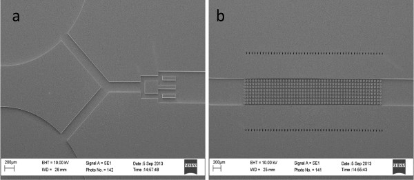
Figure 1. Scanning electron microscope (SEM) images of the microfluidic channel (top-view). a) Inlet section, b) Region containing micro-pillars. Please click here to view a larger version of this figure.
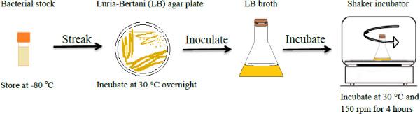
Figure 2. Sequential steps involved in bacterial culture. Please click here to view a larger version of this figure.
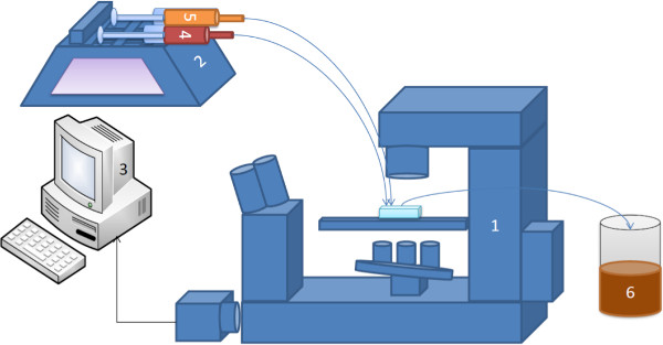
Figure 3. Set up for microfluidic experiments. 1—Optical microscope (inverted), 2—Syringe pump, 3—Image and data acquisition, 4—Syringe containing dye (optional), 5—Syringe containing bacteria, 6—Waste reservoir. Please click here to view a larger version of this figure.
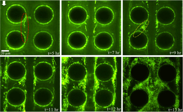
Figure 4. Time-lapse confocal imaging of evolution of streamers. Image plane corresponds to z = 25 μm i.e. middle of device. Dashed ellipses demonstrate biofilm streamers. Please click here to view a larger version of this figure.
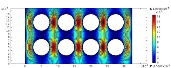
Figure 5. Computational fluid mechanical simulations showing streamlines and velocity contours of flow past micro-pillars. Fluid flow is from top to bottom and velocity scale is in m/sec. Please click here to view a larger version of this figure.
We demonstrated a simple microfluidic device that mimics porous media for studying biofilm development in complex habitats. There are several critical steps that dictate the outcome of the experiments. They include device geometry. While the post geometry can vary, adequate pore-space for streamers to form is necessary. Moreover, Valiei et al.1 have demonstrated that streamer formation occurs only in a certain flow rate range. At flow rates lower than a threshold value, deformation of biofilms into streamers may not be observed. Yet above a certain another threshold flow rate value, biofilm fracture can dominate and not allow formation of streamers. Another issue that can plague these experiments is gas bubbles that can become trapped in the micro-pillar array. Usually these bubbles have to be removed by increasing the flow rate initially and then gradually decreasing it to the desired value.
Microfluidic platforms such as these offer several advantages and few limitations. The platform enables us to work with small culture volumes, and has the flexibility of incorporating user-defined features. For example, different porous structures can be simulated by altering the geometrical parameters of the micro-pillar array. Even structures that mimic the random structure of real porous media can be fabricated on microfluidic platforms18. Moreover, several such channels can be implemented on a single device allowing for collection of significant pertinent data for accurate statistical analysis. However, microfluidic systems typically mimic two-dimensional structure of porous media. Devices that can mimic the three-dimensional nature of porous media are usually quite challenging to fabricate.
Formation and evolution of streamers are not well understood yet, and further research is required in this direction. Understanding of how streamers form and lead to the formation of mature biofilm structures will be relevant to a wide variety of scenarios including clogging of biomedical devices such as heart stents, biofilms in soil, and filtration systems. Our microfluidic platform is a step in that direction.
The authors have nothing to disclose.
The authors would like to thank Professor Howard Ceri from the Biological Sciences Department of the University of Calgary for providing bacterial strains. A. Kumar acknowledges support from NSERC. T. Thundat acknowledges financial support from the Canada Excellence Research Chair (CERC) program. The authors would also like to acknowledge help from Ms. Zahra Nikakhtari for help with videography.
Name of Material/ Equipment | Company | Catalog Number | Comments/Description |
Flourescent Microscope | Nikon | ||
LB agar | Fisher | BP1425-500 | suspend 40 g in 1 L of purified water |
LB broth | Fisher | BP1427-500 | suspend 20 g in 1 L of purified water |
Biosafety hood | Microzone corporation | ||
Petri-dish | Fisher | 875712 | sterile 100mmx15mm polystyrene petri dish |
Incubator shaker | New Brunswick Scientific | Excella E24incubator shaker series | |
50 mL sterilized centrifuge tube | Corning | 430828 | Polypropylene Rnase-/Dnase-free |
Tetracycline free base | MP Biomedicals | 103012 | 50 ug/mL |
SYLGARD 184 silicone | Dow Corning Corporation | 68037-59-2 | Elastomer Base and curing agent |
Positive photoresist (AZ4620) | |||
Plastic tube | Cole- Parmer |