Spinal cord injury is a traumatic condition that causes severe morbidity and high mortality. In this work we describe in detail a contusion model of spinal cord injury in mice followed by a transplantation of neural stem cells.
Spinal cord injury is a devastating clinical condition, characterized by a complex of neurological dysfunctions. Animal models of spinal cord injury can be used both to investigate the biological responses to injury and to test potential therapies. Contusion or compression injury delivered to the surgically exposed spinal cord are the most widely used models of the pathology. In this report the experimental contusion is performed by using the Infinite Horizon (IH) Impactor device, which allows the creation of a reproducible injury animal model through definition of specific injury parameters. Stem cell transplantation is commonly considered a potentially useful strategy for curing this debilitating condition. Numerous studies have evaluated the effects of transplanting a variety of stem cells. Here we demonstrate an adapted method for spinal cord injury followed by tail vein injection of cells in CD1 mice. In short, we provide procedures for: i) cell labeling with a vital tracer, ii) pre-operative care of mice, iii) execution of a contusive spinal cord injury, and iv) intravenous administration of post mortem neural precursors. This contusion model can be utilized to evaluate the efficacy and safety of stem cell transplantation in a regenerative medicine approach.
A spinal cord injury (SCI) is the most common injury caused by high-energy trauma like motor vehicles accidents, falls, sports and violence 1. In severe SCI, the injury force destroys or damages neural tissue, causing sudden loss of neurological function. Traumatic SCI occurs frequently in young adults between 10 and 40 years of age. It greatly affects the patient’s mental and physical condition and causes enormous economic impact to society 2. The treatment approach in the acute phase is often limited to a high-dose of corticosteroid, surgical stabilization and decompression to possibly attenuate further damage 3-4, but the roles of these methods on locomotor recovery after SCI are still controversial. In addition to acute tissue loss, the traumatic injury and the activation of secondary mechanisms of degeneration cause demyelination and death of multiple cell types 5-6. The degree of recovery of function can often be correlated to the extent of spared white matter at the injury site 7.
Animal models of SCI may be used both to investigate the biological responses of the tissue to injury and to test potential therapies. Moreover, a useful animal model of a human pathology not only has to reproduce some aspects of that condition but also must offer advantages over direct clinical observation and experiment. The most widely used models of spinal cord injury involve contusion or compression injury delivered to the surgically exposed spinal cord 8. The development of a controlled weight-drop contusion injury represent an important milestone in the history of SCI research. The Ohio State University spinal cord research center has pursued the technological challenge of a device that can be used to induce a particular compression of the spinal cord with parameters of impact controlled by a computer 9. This was originally designed for use with rats; later it was modified to apply towards mice 10. The advantages of this kind of approach are that the biomechanics of injury can be studied more in depth and the parameters of injury can be defined in a more complete manner in order to obtain a reproducible experimental model, therefore allowing more precise evaluation of the effects of tested treatments on the functional recovery process.
Many studies have evaluated the transplantation effects of a variety of stem cells in SCI models 11. We have recently isolated adult neural stem cells from the Sub-Ventricular Zone (SVZ) several hours after death of the mouse donor 12-13. This procedure provides a population of neural stem cells, called post mortem neural precursors (PM-NPCs), which seem to be advantageous in a regenerative medicine approach for curing SCI. In this paper we will demonstrate: i) the protocol for cell labeling with the vital tracer PKH26, ii) the surgical procedure to perform on traumatic SCI, and iii) the intravenous (i.v.) administration of labeled cells. Moreover, in this work we demonstrate that transplanted cells migrate to spinal cord lesion sites and differentiate mostly into microtubule associated protein (MAP) 2 positive cells. Furthermore, the differentiation is accompanied by the promotion of a stable recovery of hind limb function.
NOTE: All the procedures were approved by the Review Committee of the University of Milan and met the Italian Guidelines for Laboratory Animals in compliance with European Communities Directive dated November 1986 (86/609/EEC).
1. Preparation of Cells for Transplantation
NOTE : Use neural stem cells between the 5th and the 9th passage in culture for these experiments; test the cultures for proliferation and differentiation ability before being labeled for transplantation. Determine the extent of differentiation by immunocytochemistry 12.
2. Preparation for Surgery
3. Preparation of Mice for Surgery and Transplantation
4. Laminectomy
5. IH Impactor Device Protocol (Contusion)
6. Sutures and Post-care
7. Tail Vein Injection of Cells
NOTE: In the following step the procedure for injecting the cells into the tail vein is demonstrated. Cells could be also administered with an intraspinal transplant by using a stereotaxic frame 15-16, or into the cisterna magna 17. It is important to consider that other cell types could be transplanted with this method, such as mesenchymal stromal cells (e.g., bone marrow mesenchymal stem cells, adipose derived stem cells, amniotic fluid cells). Furthermore, other treatment options such as nanoparticles can be injected via the tail vein after the spinal cord injury.
8. Behavioral Tests and Hind Limb Function
9. Perfusion
10. Tissue Collection and Processing, Histology and Iimmunohistochemistry
The total number of transplanted cells is 1 x 10⁶ cells and was divided into three consecutive injections in the tail vein. We administered 3.3 x 10⁵ cells in 50 µl of phosphate buffer solution (PBS). The first injection was performed within 30 min after injury, the second 6 hr later and the last 18 hr after the lesion. The choice of a time limit of 18 hr after SCI for administering PM-NPCs was determined by the optimal permeability of the blood brain barrier at this time 14. To evaluate the effect of stem cells injection it would be useful to have positive control laminectomies animals (n = 14) and PBS injected animals as a negative control (n = 14).
PM-NPCs Improve Recovery of Hind Limb Function, migrate to lesion site and differentiate in MAP-2 positive cells
The T9 contusion caused the transient loss of hind limb function followed by a progressive gradual recovery (Figure 1). Within 2-3 weeks, PBS-treated injured mice improved and hind limb function reached 3 points of BMS (corresponding to plantar placing of the paw with or without weight support or occasional, frequent, or consistent dorsal stepping, but not plantar stepping 18). Instead, within the same observational period, injured mice treated with PM-NPCs showed a higher recovery, reaching 4.5 points of BMS (corresponding to frequent or consistent plantar stepping without coordination, or frequent or consistent plantar stepping with some coordination). The behavioral improvement was particularly evident in the period between day 7 and day 14 after SCI. No signs of allodynia-like forelimb hyper sensibility 19 were recorded at any time in any experimental group throughout the observational period of 30 days.
Most engrafted PM-NPCs, labeled with PKH26 (Figure 2), accumulated at the edges of the lesion forming clusters (Figure 3) from the early days of their administration. Then the transplanted cells migrated along the lesion edges and in a more diffused fashion where they differentiated, gradually assuming the asymmetric cellular conformation of neurons. At 30 days after lesion and transplantation, the cell body of PM-NPCS increased in size and in most cells dendritic-like processes were obvious and fully immunostained by the specific antibodies to MAP-2 (Figure 4). The achievement of morphological complexity and the positivity to MAP2 by transplanted PM-NPCs is likely not due to fusion with surviving host spinal cord neurons, which is evident in their clearly differentiated morphology and the absence of two nuclei in any single labeled cell.
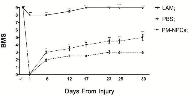
Figure 1. PM-NPCs improve functional recovery in injured animals. The open field locomotion was the test employed for the determination of motor function recovery 18. Animals were tested the day before the contusion and scored 9 points in the BMS scale. On the first day post injury in the lesioned animals, the BMS score decreased to zero. The recovery of hind limb function of lesioned mice showed a remarkable and long lasting improvement when animals were treated with PM-NPCs. The analysis was performed in double blind, and each group was composed of 14 animals. Values represent average ± SEM. We determined the statistical differences by means of ANOVA test followed by Tukey’s post-test. ***P <0.001; **P <0.01 vs PBS.
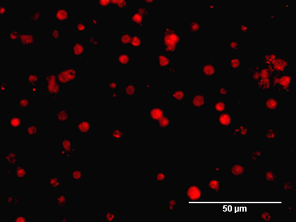
Figure 2. PKH26 labeling of PM-NPCS. After labeling procedure with PKH26, PMNPCS are visualized with the live image microscope EVOS fl and photomicrograph was taken with the same instrument (scale bar = 50 µm). Please click here to view a larger version of this figure.
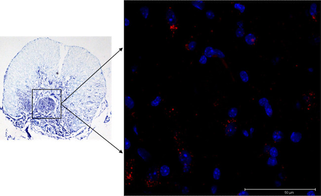
Figure 3. Localization of PM-NPCs in the lesion site. PKH26-labelled PM-NPCs (red) are present throughout the edges of the lesion site at 30 days after their i.v. injection. The image is representative for 1 mouse, but similar images were obtained for at least 5 animals (scale bar = 50 µm). Please click here to view a larger version of this figure.
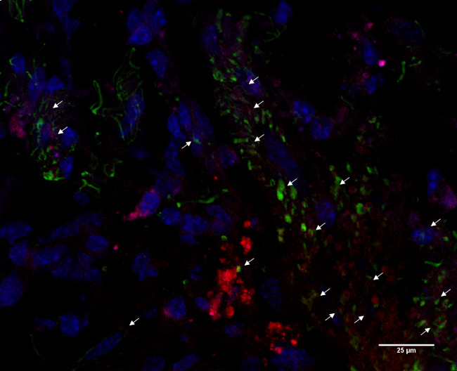
Figure 4. MAP2 expression in transplanted PM-NPCs. Most PKH26-labeled PM-NPCs (red) acquired a neuronal-like shape with dendritic-like processes and had differentiated MAP-2 positive cells (green). Nuclei are stained in blue (DAPI). The image is representative for 1 mouse, but similar images were obtained for at least 5 animals (scale bar = 25 µm). Please click here to view a larger version of this figure.
In this paper we described a method to obtain a reproducible model of traumatic spinal cord injury using an Infinite Horizon Impactor at a force of 70 kdyne (severe). Using a larger force paradigm (80 kdyne), we can cause a more severe injury that unfortunately is associated with higher mice mortality. In order to avoid this problem, we commonly choose a moderate force paradigm (70 kdyne) that is associated to a repeatable lesion with a gradual recovery of function and lower mortality. To produce such a stable injury it is very important to pay particular attention to the correct fixation of the animal on the impactor platform; in particular the spinal cord must be centered at the impactor tip, and the two arms of the impactor must be parallel to each other. Moreover, attention must be taken in blocking the animal with the impactor forceps, when the vertebrae may be crushed and the cord may be damaged by the tips of the forceps. The positioning of the animal after the laminectomy is critical, and the animal handling during this procedure is also very important. Special attention must also be given to the laminectomy procedure, which must always be performed at the same site and for the same extension. When these procedures are performed during laminectomy, another methodological issue to monitor is the reduction of the risk of damaging the cord when using micro-scissors, Rongeur, or forceps to cut bones and free the cord through the removal of the lamina, to eliminate the remaining lateral bone protrusions and fragments, and to remove the periosteum. The use of micro-scissors and Rongeur with tips pointing upwards will reduce the risk of encountering the aforesaid problems.
An important limitation of this method is the high probability that the animals may develop internal and external severe urinary infections in the post injury period. The external infection is due to the inability of the lesioned mice to move with hind limb weight support. In contrast, the internal urinary infection may be caused by the inability of the injured mice to urinate independently. In order to avoid these problems it is absolutely necessary to inject the animals with the indicated antibiotic and to check the size of the bladder twice a day during the animal care procedures of the first week. The hydration status and the weight must be checked carefully during the first three weeks after lesioning.
Applying the described procedures we were able to obtain reproducible deficits of hind limb function that were evaluated through a specific behavioral test developed by Basso and colleagues (BMS) 18. Immediately after the injury the hind limb loss of function is complete, which is followed by a gradual recovery that is most important during the first 2-3 weeks. The behavioral recovery reached a higher level when lesioned mice were treated with adult PM-NPCs (Figure 1). We also serially sectioned spinal cords of cell treated mice at 4 weeks after injury, and detected PKH26 positive PM-NPCs by means of confocal microscopy at the edges of the lesion (Figure 3). We estimated that the total number of vital grafted PM-NPCs is greater than the grafted ESCs and adult NSCs as previously reported by our research group 20-21. At four weeks after lesioning and transplantation, most PM-NPCs have larger bodies and possess extended dendritic-like processes that are fully immunostained by the specific antibodies to MAP-2 (Figure 4).
The major advantage of this model of traumatic spinal cord injury is the standardization of the injury. A reproducible time-related curve of hind limb recovery of function is also achieved. Such reproducibility makes it possible to define proof-of-principle studies for investigated treatments including transplantation of cells for regenerative medicine studies with a reduced number of cases. In addition, several aspects of the pathophysiology of spinal cord injury can be analyzed in greater detail.
The authors have nothing to disclose.
The Authors acknowledge the economic support by FAIP (Federazione Associazioni Italiane Paraplegici), “Neurogel-en-Marche” Foundation (France), Fondazione “La Colonna”.
Name of Material/ Equipment | Company | Catalog Number | Comments/Description |
PKH26GL-1KT | Sigma | 091M0973 | |
Infinite horizon (IH) Impactor device | Precision Systems and Instrumentation, LLC | Model 0400 Serial 0171 | |
Gentamycin 10mg/ml | Euroclone | ECM0011B | 1mg/ml in sterile saline solution |
Isoflurane-Vet 250ml | Merial | B142J12A | |
Blefarolin POM OFT 10g | |||
Slide Warmer | 2Biological Instruments | HB101-sm-402 | |
Scalpel, size 10 | Lance Paragon | 26920 | |
Small Graefe Forceps | 2Biological Instruments | 11023-14 | |
Rongeur | Medicon Instruments | 07 60 07 | |
Micro scissors | 2Biological Instruments | 15000-00 | |
Absorbable sutures (4/0) | Safil Quick | C0046203 | |
Hemostat | 2Biological Instruments | 13014-14 | |
Reflex 7 wound clip applicator | 2Biological Instruments | 12031-07 | |
7mm Reflex wound clips | 2Biological Instruments | 12032-07 | |
NGS | Euroclone | ECS0200D | |
Triton X 100 | Merck Millipore | 1086431000 | |
Anti Microtubule Assocoated Protein (MAP) 2 | Millipore | AB5622 | |
Alexa Fluor 488 | Invitrogen | A11008 | |
FluorSave Reagent | Calbiochem | 345789 | |
Neural stem cells medium | DMEM-F12 medium (Euroclone) containing 2 mm l-glutamine (Euroclone), 0.6% glucose (Sigma-Aldrich), 9.6 gm/ml putrescine (Sigma-Aldrich), 6.3 ng/ml progesterone (Sigma-Aldrich), 5.2 ng/ml sodium selenite (Sigma-Aldrich), 0.025 mg/ml insulin (Sigma-Aldrich), 0.1 mg/ml transferrin (Sigma-Aldrich), and 2 μg/ml heparin (sodium salt, grade II; Sigma-Aldrich), bFGF (human recombinant, 10 ng/mL; Life Technologies) and EGF (human recombinant, 20 ng/mL; Life Technologies) | ||
DMEM-F12 | Euroclone | ASM5002 | |
l-glutamine | Euroclone | ECB3000D | |
glucose | Sigma-Aldrich | G8270-100G | |
putrescine | Sigma-Aldrich | P5780-25G | |
progesterone | Sigma-Aldrich | P6149-1MG | |
Sodium-selenite | Sigma-Aldrich | S9133-1MG | |
transferrin | Sigma-Aldrich | T 5391 | |
Insulin | Sigma-Aldrich | I1882 | |
Heparin sodium-salt | Sigma-Aldrich | H0200000 | |
bFGF | Life Technology | PHG0024 | |
h-EGF | Life Technology | PHG6045 | |
Syringe 0.33cc 29G | Terumo | MYJECTOR | |
buprenorphine | Schering Plough SpA | TEMGESIC | |
eye gel | Bausch & Lomb | LIPOSIC |