Pathology of the aorta can lead to severe morbidity and mortality, therefore research of disease progression and potential therapies is warranted. Here, we present a protocol to isolate and excise the murine aorta to aid researchers in their investigation of cardiovascular disease.
Cardiovascular disease is a broad term describing disease of the heart and/or blood vessels. The main blood vessel supplying the body with oxygenated blood is the aorta. The aorta may become affected in diseases such as atherosclerosis and aneurysm. Researchers investigating these diseases would benefit from direct observation of the aorta to characterize disease progression as well as to evaluate efficacy of potential therapeutics. The goal of this protocol is to describe proper isolation and excision of the aorta to aid investigators researching cardiovascular disease. Isolation and excision of the aorta allows investigators to look at gross morphometric changes as wells as allowing them to preserve and stain the tissue to look at histologic changes if desired. The aorta may be used for molecular studies to evaluate protein and gene expression to discover targets of interest and mechanisms of action. This technique is superior to imaging modalities as they have inherent limitations in technology and cost. Additionally, primary isolated cells from a freshly isolated and excised aorta can allowing researchers to perform further in situ and in vitro assays. The isolation and excision of the aorta has the limitation of having to sacrifice the animal however, in this case the benefits outweigh the harm as it is the most versatile technique in the study of aortic disease.
The aorta plays a pivotal role in cardiovascular function supplying the body with oxygenated blood and similar to others portions of the body, is susceptible to disease. Common aortic diseases include atherosclerosis and aneurysm, which are the results of genetic and environmental factors including diet, smoking and sedentary lifestyle1. Atherosclerosis is the deposition of a calcium- and lipid-based plaque to the aortic wall typically found in patients with chronic hyperlipidemia2. Aneurysms are characterized by degradation of the structural components of the aorta and thinning of the vessel wall, followed by an increased diameter which could ultimately lead to rupture3.
Animal models are important tools used to study the mechanism of disease and efficacy of potential treatments. Common animal models used in cardiovascular research, specifically research investigating vascular and metabolic disorders include genetically modified mice to alter lipid levels such as apolipoprotein E gene knockout and low density lipoprotein receptor gene knockout mice4. The majority of methods to evaluate mechanisms of disease and efficacy of therapy would include the isolation and excision of the aorta.
Once the aorta is localized and isolated, morphometric analysis can be determined, such as presence of aneurysm, typically defined as a greater than 50% increase in diameter5. After necessary in situ analysis is complete, the aorta can be excised for further analysis. An excised aorta can be flash frozen to conduct molecular studies such as protein and/or gene expression assays or fixed using 4% paraformaldehyde and later embedded, sectioned and stained for histological analysis. Histological analysis of the aorta can demonstrate common features of aortic disease such as structural degradation, plaque formation, and infiltration of leukocytes6,7. Additionally, an excised aorta can be used to isolate primary cell lines including endothelial and smooth muscle cells which can subsequently be used for a variety of in vitro studies7.
Currently, there are no other methods to provide this in-depth characterization of aortic disease as well as provide researchers with tools to further study cardiovascular disease. Imaging modalities such as magnetic resonance imaging, computed tomography and ultrasonography are the closest methods to morphologically evaluate the aorta, however this is difficult in small animals and obtaining a device with adequate technology is expensive8. Immortalized cell lines can be purchased to investigate potential mechanisms of disease and efficacy of therapies, however the artificial nature of these cells are limiting in studying the effects on the cell life cycle and apoptosis9.
The overall goal of this manuscript is to demonstrate the sterile isolation and excision of the murine aorta in the investigation of cardiovascular disease.
All animal procedures were performed with the approval of the Institutional Animal Care and Use Committee of the University of Cincinnati and in accordance with the Guide for the Care and Use of Laboratory Animals from the National Institutes of Health (NIH Publication No. 85-23, Revised 1996).
1. Preparing the Mouse
2. Isolation of the Heart and Aorta
3. Perfusion of Heart and Aorta
NOTE: To obtain blood via cardiac puncture, do so just prior to this step.
4. Isolation and Excision of the Aorta
Upon completion of the procedure, there will be an intact aorta originating from the heart, descending into the thoracic and abdominal cavities with the renal arteries still attached (Figure 1A). From here, the aorta can be imaged in situ to quantify morphometric changes which are diagnostic in the study of abdominal aortic aneurysms (Figure 1B and 1C). Subsequently, the aorta can be removed, fixed and stained to look at histological changes. A general and common stain of the aorta is the hematoxylin and eosin stain (Figure 2A and 2B). Additionally, the structural integrity of the aorta can be qualitatively quantified using Verhoff van Geison staining to look at the elastin bands (Figure 2C and 2D). Instead of obtaining a histological analysis, the investigator may flash freeze the aorta for subsequent protein and ribonucleic acid expression. The final option for the investigator is to use the aorta to obtain primary cell isolation. These cells can be grown in culture (Figure 3A) and used for a variety of in vitro studies including viability studies (Figure 3B) and protein localization (Figure 3C).
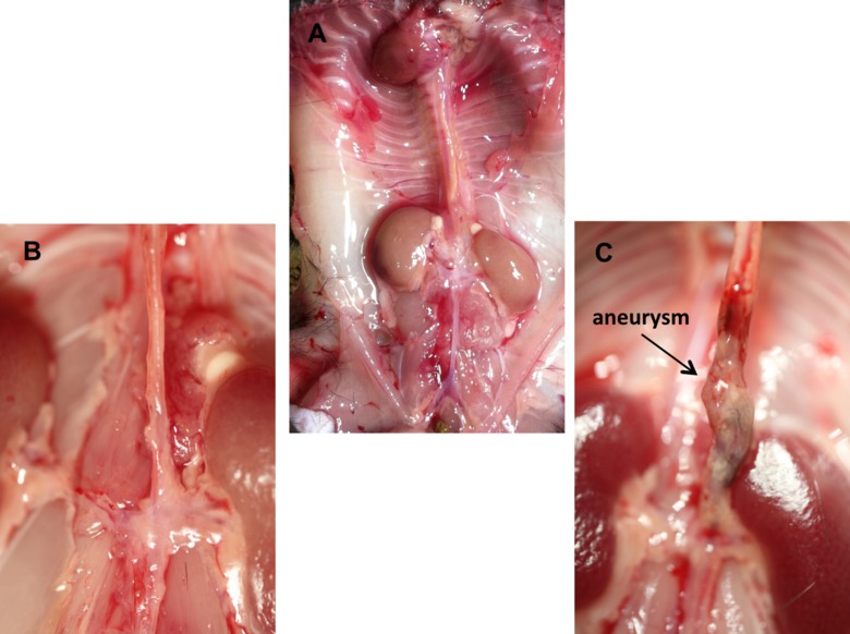
Figure 1. In situ representative images of murine aorta. (A) Depiction of the normal mouse anatomy after the isolation of the aorta including the heart and kidneys. (B) Magnified image of a normal intact murine abdominal aorta. (C) Magnified image of a diseased abdominal aortic with an aneurysm in the suprarenal region. Please click here to view a larger version of this figure.
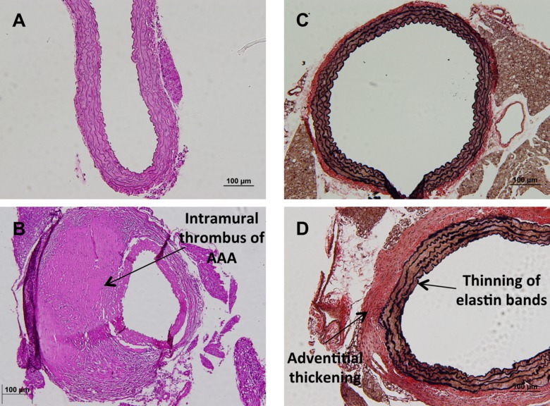
Figure 2. Histological analysis of murine aorta. (A) Representative image of a normal hematoxylin and eosin stain of a healthy suprarenal aorta at 20X magnification. (B) Representative image of an aneurismal suprarenal aorta with intramural thrombus after hematoxylin and eosin stain at 20X magnification. (C) Representative image of a Verhoff van Geison stain, highlighting the elastin bands of a normal suprarenal aorta at 10X magnification. (D) Representative image of a Verhoff van Geison stain highlighting the elastin bands of a suprarenal aorta developing disease noted by the thinning and striating of elastin bands and thickening of the adventitial layer. Please click here to view a larger version of this figure.
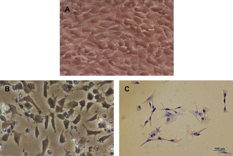
Figure 3. Primary aortic myocytes cultured from aorta. (A) Representative image of primary aortic myocytes in culture at 20X magnification obtained from the abdominal aorta. (B) Representative image of primary aortic myocytes during a Trypan blue exclusion assay after a 500 µM treatment of H2O2 at 20X magnification obtained from the abdominal aorta. (C) Representative image of an immunohistochemistry stain for alpha actin in primary isolate aortic smooth muscle cells from the abdominal aorta. Please click here to view a larger version of this figure.
Aortic disease can lead to significant systemic pathology and possibly death in severe cases. Treatment options are currently limited therefore continued research in the field is necessary10. Murine models are ideal to further study disease progression and potentially therapeutic options4. Therefore, the successful completion of the method described in this manuscript will provide investigators with the tools to measure disease progression and drug efficacy.
This technique can be modified in several ways in order to optimize the results. The use of surgical loupes or a dissecting microscope can aid in the visualization of the murine anatomy and vasculature details and when removing surrounding adipose tissue from the aorta. Additionally, the use of dedicated surgical utensils for specific tasks can increase the longevity of tools; for example, the use of scissors for cutting skin and separate scissors to cut through the ribs would be ideal. The use of a small gauged needle when perfusing the heart is preferred as this will allow the tissue to close around the puncture as opposed to a larger gauge needle where a hole will remain.
After the aorta is successfully removed, it can be utilized for a variety of assays. The aorta can be used for molecular studies such as enzyme activity, protein expression and gene expression profiles. Additionally, the aorta can be fixed for histologic analysis where in situ assays can be utilized as well as characterization of the aortic morphology. Finally, one can isolate smooth muscle and/or endothelial cells from a freshly harvest aorta for in vitro studies with these primary isolated cells.
The major limitation to isolation and excision of the aorta is that the animal must be sacrificed, especially in the cases of longitudinal studies. Currently, there is no single technique comparable to isolation and excision of the murine aorta. Small animal imaging technologies is becoming more advanced and approaching characterization capabilities of direct observation however it continues to have limitations including cost8. Immortalized cells lines can be used for in vitro studies however immortal cells are not as useful as primary isolated cells for in vitro assays especially for viability studies9. Isolation and excision of the murine aorta is currently the most versatile technique that can be used in the study of aortic disease.
The authors have nothing to disclose.
The authors have no acknowledgments.
Name of Reagent/ Equipment | Company | Catalog Number | Comments/Description |
Isoflurane | Med-Vet International | #RXISO-250 | |
70% Ethanol | Fisher | 07-678-001 | |
Phosphate buffered saline | Sigma Aldrich | P5368-10PAK | |
Surgical Tape | 3M | 1527-1 | |
Sterile Gauze | Dukal Corporations | 1312 | |
25 Gauge Needle | BD | 305122 | |
10ml Syringe | BD | 309604 |