Summary

समय-समय पर एसिड शिफ़ धुंधला के साथ परिधीय रक्त कोशिकाओं mononuclear में ग्लाइकोजन का पता लगाने के

Published: December 23, 2014
doi:

Summary

Periodic acid Schiff staining is a technique that visualizes the polysaccharide content of tissues. This article demonstrates periodic acid Schiff staining protocol adapted for use on peripheral blood mononuclear cells purified from human venous blood. Such samples are enriched for lymphocytes and other white blood cells of the immune system.

Abstract

Periodic acid Schiff (PAS) staining is an immunohistochemical technique used on muscle biopsies and as a diagnostic tool for blood samples. Polysaccharides such as glycogen, glycoproteins, and glycolipids stain bright magenta making it easy to enumerate positive and negative cells within the tissue. In muscle cells PAS staining is used to determine the glycogen content in different types of muscle cells, while in blood cell samples PAS staining has been explored as a diagnostic tool for a variety of conditions. Blood contains a proportion of white blood cells that belong to the immune system. The notion that cells of the immune system possess glycogen and use it as an energy source has not been widely explored. Here, we describe an adapted version of the PAS staining protocol that can be applied on peripheral blood mononuclear immune cells from human venous blood. Small cells with PAS-positive granules and larger cells with diffuse PAS staining were observed. Treatment of samples with amylase abrogates these patterns confirming the specificity of the stain. An alternate technique based on enzymatic digestion confirmed the presence and amount of glycogen in the samples. This protocol is useful for hematologists or immunologists studying polysaccharide content in blood-derived lymphocytes.

Introduction

समय-समय पर एसिड शिफ़ (पीए) धुंधला व्यापक रूप से पेशी अनुसंधान और निदान में प्रयोग किया जाता है कि एक immunohistochemical तकनीक है। यह भी रक्त के नमूनों पर एक नैदानिक ​​उपकरण के रूप में उपयोग किया जाता है। तकनीक बेरंग शिफ़ के अभिकर्मक जिससे एक गहरी मैजंटा उत्पाद के उत्पादन के साथ प्रतिक्रिया है कि पोलीसेकेराइड बनाने एल्डिहाइड समूहों के भीतर इकाइयों ऑक्सीकरण नमूना है, जो करने के लिए समय-समय पर एसिड समाधान को लागू करने से काम करता है। इस प्रक्रिया के चरणों का चित्र 1 में दिखाया जाता है। दाग ग्लाइकोजन, ग्लाइकोप्रोटीन, glycolipids, mucins, या पोलीसेकेराइड moieties के साथ अन्य अणुओं सहित polysaccharides Magenta, के साथ कुछ भी बदल जाता है।

पीए धुंधला अक्सर मांसपेशी फाइबर में ग्लाइकोजन के स्तर को मापने के लिए प्रयोग किया जाता है। स्नायु ऊतक वर्गों वे मजबूती से स्लाइड के लिए देते हैं और कई धोने और धुंधला चरणों झेलने के रूप में तकनीक के लिए आदर्श होते हैं। ग्लाइकोजन एक उच्च मांग है जो प्रकार द्वितीय मांसपेशी फाइबर, तेजी से चिकोटी में सबसे मौजूद हैअधिकतम प्रदर्शन 1,2 के लिए ग्लाइकोजन की आवश्यकता तेजी से एटीपी उत्पादन के लिए। ग्लाइकोजन ग्लाइकोजन phosphorylase एन्जाइम की क्रिया के माध्यम से नि: शुल्क ग्लूकोज में तोड़ा जा सकता है कि ग्लूकोज की एक branched बहुलक है। बाकी और पोषक तत्वों की प्रचुरता के समय में, ग्लाइकोजन, glycogenesis की प्रक्रिया के माध्यम से मंगाया जाता है, जबकि पोषण की कमी या उच्च ऊर्जा की मांग के समय में; ग्लाइकोजन glycogenolysis द्वारा ग्लूकोज में टूट गया है। रक्त के नमूनों पर के रूप में जल्दी 1950 के चिकित्सक वैज्ञानिकों का पता लगाया है के रूप में पीए धुंधला से विभिन्न रोगों 3-7 में ग्लाइकोजन सामग्री का विश्लेषण करने के लिए। उदाहरण के लिए, Pompe रोग-एक वास्तविक ग्लाइकोजन भंडारण में रोग सफेद रक्त कोशिकाओं को स्वस्थ नियंत्रण 8 से काफी अलग है कि ग्लाइकोजन की बड़ी मात्रा में जमा है।

इस वीडियो लेख परिधीय रक्त कोशिकाओं mononuclear (PBMC) स्वस्थ मानव विषयों की शिरापरक रक्त से नमूने पर इस्तेमाल के लिए पीए धुंधला के एक अनुकूलित संस्करण को दर्शाता है। पीबीएमसीएस ऐसे प्राकृतिक हत्यारा कोशिकाओं और monocytes के रूप में टी लिम्फोसाइट और बी लिम्फोसाइट परिवारों, साथ ही अन्य प्रतिरक्षा कोशिकाओं के ज्यादातर लिम्फोसाइटों होते हैं। पहले शुद्धि कदम एरिथ्रोसाइट्स, न्यूट्रोफिल, और अन्य granulocytes हटा। इस तकनीक को पूरे रक्त स्मीयरों का उपयोग कर की तुलना में पीए पॉजिटिव कोशिकाओं के और अधिक मजबूत गणन के लिए अनुमति लिम्फोसाइटों का एक केंद्रित अनुपात पर डेटा प्रदान करता है।

चित्रा 1
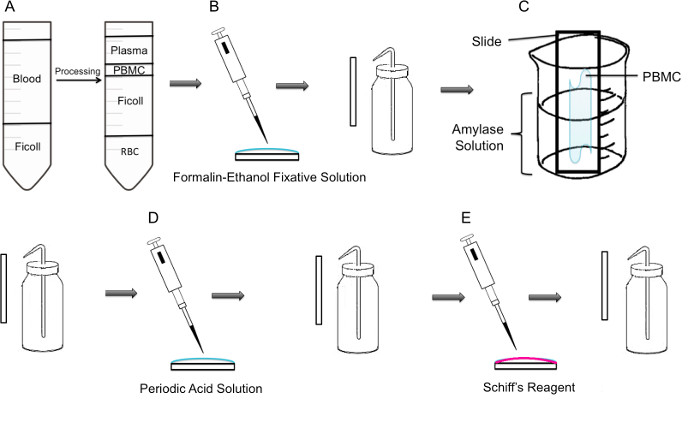
चित्रा 1:। PBMC पर पीए धुंधला के कदम कार्यप्रणाली से कदम (ए) पहले, PBMC के अलगाव Ficoll ढाल के माध्यम से हासिल की है, बाएं पैनल centrifugation के पहले तैयारी से पता चलता है, सही पैनल centrifugation के बाद यह पता चलता है जहां PBMC युक्त buffy कोट ट्यूब के केंद्र में मनाया जाता है। (बी) पृथक PBMCs formalin इथेनॉल लगानेवाला समाधान का उपयोग कर स्लाइड पर तय कर रहे हैंtion। स्लाइड धीरे एक प्लास्टिक धोने की बोतल से आसुत जल के साथ rinsed है। (सी) स्लाइड तो ग्लाइकोजन भंग होगा जो एमिलेज समाधान के साथ भरा एक 100 मिलीलीटर बीकर आधे रास्ते में रखा गया है। स्लाइड धीरे rinsed है। (डी) स्लाइड saccharides के ऑक्सीकरण जगह लेता है, जहां समय-समय पर एसिड समाधान, साथ व्यवहार किया जाता है। स्लाइड्स धीरे rinsed कर रहे हैं; इस अतिरिक्त समय-समय पर एसिड को हटाने और ऑक्सीकरण कदम बंद हो जाएगा। (ई) शिफ़ अभिकर्मक स्लाइड करने के लिए कहा जाता है, यह ऑक्सीकरण चरण के दौरान बनाई गई एल्डीहाइड साथ प्रतिक्रिया होगी। इस बेरंग अभिकर्मक तो एक गहरे लाल मैजंटा उत्पाद में परिणाम होगा। स्लाइड्स धीरे अतिरिक्त शिफ़ अभिकर्मक दूर करने के लिए rinsed हैं।

Protocol

मानव रक्त के नमूनों के साथ रिसर्च Concordia विश्वविद्यालय आचार समीक्षा बोर्ड, प्रमाण पत्र संख्या 10000618. द्वारा Concordia विश्वविद्यालय आचार समीक्षा बोर्ड, प्रमाण पत्र संख्या 2010BERG द्वारा अनुमोदित किया गया था माउस पे…

Representative Results

अभिकर्मकों और बुनियादी तकनीक को मान्य करने के लिए, पीए धुंधला माउस soleus मांसपेशी वर्गों पर निर्माता के निर्देशों के अनुसार किया गया था। धुंधला बलिदान के रूप में एक ही दिन में किया गया था और धुंधला के अंत?…

Discussion

इस वीडियो लेख की महत्वपूर्ण कदम कोशिकाओं की धुलाई और एमिलेज इलाज के दौरान थे। स्लाइड्स धोने, वहीं महत्वपूर्ण कदम एक प्लास्टिक दब्बू बोतल धोने का उपयोग करने और पानी धीरे स्लाइड पर नमूना के माध्यम से चल?…

Divulgazioni

The authors have nothing to disclose.

Acknowledgements

This research was supported by a grant from the NSERC Discovery program grant number RGPIN 418522-2013. We thank R. Kilgour for helpful discussions, and Katelin Gresty and Dr. A. Berghdal for providing the mouse muscle sections.

Materials

Periodic Acid Shiff Kit Sigma-Aldrich 395B Bring to room temperature prior to use. Materials in this kit are toxic and harmful. Use caution 
http://www.sigmaaldrich.com/catalog/product/sigma/395b?lang=en&region=CA
α-Amylase from porcine pancreas Sigma-Aldrich A3176 http://www.sigmaaldrich.com/catalog/product/sigma/a3176?lang=en&region=CA
Binocular Microscope  Carl Zeiss Microscopy Axio Lab A0
Glycogen Assay Kit Sigma-Aldrich MAK016 http://www.sigmaaldrich.com/catalog/product/sigma/mak016?lang=en&region=CA
Ficoll-Paque PLUS VWR, GE Healthcare 17-1440-02 Nonionic synthetic polymer of sucrose
https://us.vwr.com/store/catalog/product.jsp?product_id=4779441
Centrifuge For PBMC isolation, swing buckets were used 

Riferimenti

  1. Rich, P. R. The molecular machinery of keilin’s respiratory chain. Biochem. Soc. Trans. 31 (Pt 6), 1095-1105 (2003).
  2. Peter, J. B., Barnard, R. J., Edgerton, V. R., Gillespie, C. A., Stempel, K. E. Metabolic profiles of three fiber types of skeletal muscle in guinea pigs and rabbits). Biochimica. 11 (14), 2627-2633 (1972).
  3. Jones, R. V., Goffi, G. P., Hutt, M. S. R. Lymphocyte glycogen content in various disease. J. Clin. Pathol. 15 (1), 36-39 (1962).
  4. Scott, R. B. Glycogen in human peripheral blood leukocytes. I. characteristics of the synthesis and turnover of glycogen in vitro. J. Clin. Invest. 47 (2), 344-352 (1968).
  5. Fedele, D., et al. positive index of lymphocytes and metabolic control in insulin-treated and type II diabetes mellitus. Diabete Metab. 9 (3), 188-192 (1983).
  6. Brelińska-Peczalska, R., Mackiewicz, S. Cytochemical studies of peripheral blood granulocytes and lymphocytes in patients with systemic lupus erythematosus. Pol.Med.Sci.Hist.Bull. 15 (2), 231-234 (1976).
  7. Yunis, A. A., Arimura, G. K. Enzymes of glycogen metabolism in white blood cells. I. glycogen phosphorylase in normal and leukemic human leukocytes. Cancer, Res. 24, 489-492 (1964).
  8. Hagemans, M. L., et al. PAS-positive lymphocyte vacuoles can be used as diagnostic screening test for pompe disease. J. Inherit. Metab. Dis. 33 (2), 133-139 (2010).
  9. Totsuka, Y., et al. Physical performance and soleus muscle fiber composition in wild-derived and laboratory inbred mouse strains. 95 (2), 720-727 (2003).
  10. Murat, J. C., Serfaty, A. Simple enzymatic determination of polysaccharide (glycogen) content of animal tissues. Clin. Chem. 20 (12), 1576-1577 (1974).
  11. Arrizabalaga, O., Lacerda, H. M., Zubiaga, A. M., Zugaza, J. L. Rac1 protein regulates glycogen phosphorylase activation and controls interleukin (IL)-2-dependent T cell proliferation. J. Biol. Chem. 287 (15), 11878-11890 (2012).
  12. Pelletier, J., G, J., Mazure, N. M. Biochemical titration of glycogen in vitro. J.Vis.Exp. (81), (2013).
  13. Roach, P. J., Depaoli-Roach, A. A., Hurley, T. D. Tagliabracci V.S. Glycogen and its metabolism: Some new developments and old themes. Biochem.J. 441 (3), 763-787 (2012).
  14. Salmoral, E. M., Tolmasky, D. S., Krisman, C. R. Evidence for the presence of glycogen in rat thymus. Cell Mol.Biol. 36 (2), 163-174 (1990).
  15. Darlington, P. J., et al. Diminished Th17 (not Th1) responses underlie multiple sclerosis disease abrogation after hematopoietic stem cell transplantation. Ann.Neurol. 73 (3), 341-354 (2013).

Play Video

Citazione di questo articolo
Tabatabaei Shafiei, M., Carvajal Gonczi, C. M., Rahman, M. S., East, A., François, J., Darlington, P. J. Detecting Glycogen in Peripheral Blood Mononuclear Cells with Periodic Acid Schiff Staining. J. Vis. Exp. (94), e52199, doi:10.3791/52199 (2014).

View Video