Summary

シングルプラズモニックナノ粒子からの散乱非線形性の測定

Published: January 03, 2016
doi:

Summary

Saturable and reverse saturable scattering were discovered in isolated plasmonic particles and adopted as a novel non-bleaching contrast method in super-resolution microscopy. Here the experimental procedures of detecting and extracting nonlinear scattering are explained in detail, as well as how to enhance resolution with the aid of saturated excitation microscopy.

Abstract

Plasmonics, which are based on the collective oscillation of electrons due to light excitation, involve strongly enhanced local electric fields and thus have potential applications in nonlinear optics, which requires extraordinary optical intensity. One of the most studied nonlinearities in plasmonics is nonlinear absorption, including saturation and reverse saturation behaviors. Although scattering and absorption in nanoparticles are closely correlated by the Mie theory, there has been no report of nonlinearities in plasmonic scattering until very recently.

Last year, not only saturation, but also reverse saturation of scattering in an isolated plasmonic particle was demonstrated for the first time. The results showed that saturable scattering exhibits clear wavelength dependence, which seems to be directly linked to the localized surface plasmon resonance (LSPR). Combined with the intensity-dependent measurements, the results suggest the possibility of a common mechanism underlying the nonlinear behaviors of scattering and absorption. These nonlinearities of scattering from a single gold nanosphere (GNS) are widely applicable, including in super-resolution microscopy and optical switches.

In this paper, it is described in detail how to measure nonlinearity of scattering in a single GNP and how to employ the super-resolution technique to enhance the optical imaging resolution based on saturable scattering. This discovery features the first super-resolution microscopy based on nonlinear scattering, which is a novel non-bleaching contrast method that can achieve a resolution as low as l/8 and will potentially be useful in biomedicine and material studies.

Introduction

プラズモニクスの研究では、様々な分野への応用1-4による大きな関心を集めています。プラズモニクスの中で最も研究さフィールドのうちの1つは、金属と誘電体との界面での外部の電磁波との伝導電子の集団振動カップル表面プラズモニクス、です。表面プラズモニクスは、サブ波長光学、バイオフォトニクス、および顕微鏡5,6におけるその潜在的用途のために検討されています。局在表面プラズモン共鳴(LSPR)による金属ナノ粒子の超小体積の強い場の増強が原因粒子サイズ、粒子形状、及び7周囲媒体の誘電特性に対する卓越した感度だけでなく、広範な関心を集めています-10だけでなく、本質的に弱いため、非線形光学効果11をブーストする能力。 LSPRの例外的な感度バイオセンシングおよび近FIEのための価値がありますLDイメージング技術12,13。一方、プラズモン構造の強化非線形性は、光スイッチング及び全光信号処理14,15のような用途における光集積回路に利用することができます。これはよくプラズモン吸収が低い強度レベルでの励起強度に線形に比例することが知られています。励起が十分に強い場合、吸収が飽和状態に達します。興味深いことに、より高強度で、吸収が再び増加します。これらの非線形効果は、可飽和吸収(SA)15〜17と呼ばれ、それぞれ、可飽和吸収(RSA)18を逆にしています。

これは、LSPRによる、散乱がプラズモニック構造において特に強いことが知られています。基本的な電磁気学に基づいて、入射強度対散乱の応答は線形でなければなりません。しかし、ナノ粒子に、散乱及び吸収が密接Mie理論を介して連結されており、両方が電子であることができます誘電率の実部と虚部の面でxpressed。単一GNSは、光照射下での双極子として振る舞うという仮定の下では、ミー理論による単一プラズモニックナノ粒子からの散乱係数(Qの SCA)と吸収係数(Qの ABS)は 19のように表すことができます。
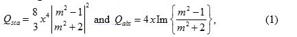
式(1)

xは 2πa/λであり、aが球の半径あり、m 2は 、εM /εdです。ここで、εm εd 、それぞれ、金属のと周囲の誘電体の誘電率に対応しています。散乱係数の形態は、目と同様であるのでEの吸収係数は、従って、単一のプラズモンナノ粒子20で飽和散乱を観察することが期待されます。

最近では、孤立したプラズモニック粒子中の非線形飽和散乱は初めて21実証されました。これは、励起強度が増加したときに深い飽和状態で、実際には散乱強度がわずかに減少していることが顕著です。励起強度は散乱が飽和になった後に増加し続けた場合であってもより顕著に、散乱強度は20散乱 、逆飽和の効果を示す、再び上昇しました。 、波長およびサイズ依存の研究では、21散乱 LSPRと非線形の間に強力な関係を示しています。プラズモン散乱の強度および波長依存性は、これらの非線形行動の根底にある共通のメカニズムを示唆し、吸収のものと非常に似ています。

アプリケーションの観点においては、よく自演さその非線形性WN光学顕微鏡の分解能を改善するのに役立ちます。 2007年には、飽和励起(SAX)顕微鏡は、励起光22の時間的な正弦波変調を経て飽和した信号を抽出することにより、解像度を向上させることができる、提案されました。 SAX顕微鏡は、レーザー焦点のため、強度が周辺部よりも中央で強い概念に基づいています。信号(蛍光または散乱のいずれか)が飽和挙動を示す場合、線形応答は周囲のまま、彩度は、中央から開始する必要があります。唯一の飽和部分を抽出する方法がある場合は、周辺部を排除しつつ、それが効果的に空間分解能を向上させる、中央部のみを残します。原則的に、SAX顕微鏡には低解像度の制限がない限り、深い飽和状態に達しており、強烈な照明のために何のサンプルの損傷はありません。

これはresolutioことが示されています蛍光イメージングのnが著しくSAX技術を利用することによって向上させることができます。しかし、蛍光は光漂白効果に苦しんでいます。非線形散乱の発見およびSAXの概念を組み合わせて、散乱に基づく超解像顕微鏡21を実現することができます。従来の超解像顕微鏡法に比べて、散乱に基づく技術は、新規な非漂白コントラスト方法を提供します。本論文では、ステップバイステップの説明が取得し、プラズモン散乱の非線形性を抽出するために必要な手順の概要を説明するために与えられています。入射強度を変化させることによって導入された散乱非線形性を同定する方法が記載されています。詳細は、これらの非線形性は、単一のナノ粒子の画像をどのように影響するかを解明するために提供され、どのように空間分解能は、SAXの手法により適宜向上させることができます。

Protocol

1. GNSサンプル調製 LSPRピークがシフトする可能性があり、粒子の凝集を防止するために約40キロヘルツ、少なくとも15分間、試料を調製する前に、超音波処理1mLのGNSコロイド溶液。 GNSSを修正するための商用マグネシウムアルミニウムシリケート(MAS)コーティングを施したスライドガラス上にGNSコロイドの100〜200μLをドロップします。 少なくとも1分後、蒸留水で洗浄す…

Representative Results

図6は、80 nmのGNSから測定されたスペクトルを示しています。 Mie理論に基づいて計算された曲線は、優れた一致を示し、同グラフに示されています。 LSPRのピークは580nmで周りです。以下の実験では、レーザー波長は、それがプラズモン効果で光散乱を強化し、飽和21を有効にするために散乱プラズモンバンド内に位置するように選ばれた532 nmでした。 <p class="jove_content"…

Discussion

プロトコルでは、いくつかの重要なステップがあります。サンプルを調製する際にまず、ナノ粒子の密度は、粒子間のプラズモンカップリングを避けるために、高すぎてはなりません。二つ以上の粒子が互いに非常に接近している場合は、LSPR波長の結合の結果、大幅非線形性を減少させる、より長い波長に向かってシフトします。しかし、このイメージング技術は、実際には代わりに粒子自…

Disclosures

The authors have nothing to disclose.

Acknowledgements

This work is supported by Ministry of Science and Technology under NSC-101-2923-M-002-001-MY3 and NSC-102-2112-M-002-018-MY3. This research is also supported by the Japan Society for the Promotion of Science (JSPS) through the “Funding Program for Next Generation World-Leading Researchers (NEXT Program),” initiated by the Council for Science and Technology Policy (CSTP) and JSPS Asian CORE Program.

Materials

microscope body Olympus, Japan BX-51
objective lens Olympus, Japan UPlanSapo, 100X, NA 1.4
80-nm gold colloid BBI Solutions, UK EM.GC80
supercontinuum laser Fianium, United Kingdom SC400-2-PP
broadband dielectric mirrors Thorlabs, USA BB1-E02
field emission SEM JEOL, Japan JSM-6330F optional
spectrometer Andor Technology, UK Shamrock 163
 charge-coupled device Andor Technology, UK iDus DV420A-OE
acousto-optic modulators  IntraAction Corp., USA AOM-402AF1
lock-in amplifier Stanford Research Systems, USA SR-830
MAS-coated slide glass Matsunami Glass, Japan, S9215

References

  1. Fang, N., Lee, H., Sun, C., Zhang, X. Sub-diffraction-limited optical imaging with a silver superlens. Science. 308 (5721), 534-537 (2005).
  2. Lee, B., Kim, S., Kim, H., Lim, Y. The use of plasmonics in light beaming and focusing. 34 (2), 47-87 (2010).
  3. Lal, S., Link, S., Halas, N. J. Nano-optics from sensing to waveguiding. Nature Photon. 1 (11), 641-648 (2007).
  4. Kawata, S., Inouye, Y., Verma, P. Plasmonics for near-field nano-imaging and superlensing. Nature Photon. 3 (7), 388-394 (2009).
  5. Homola, J., Yee, S. S., Gauglitz, G. Surface plasmon resonance sensors: review. Sensor. Actuat. B-Chem. 54 ((1-2)), 3-15 (1999).
  6. Nie, S., Emory, S. R. Probing single molecules and single nanoparticles by surface-enhanced Raman scattering. Science. 275 (5303), 1102-1106 (1997).
  7. Hache, F., Ricard, D., Flytzanis, C. Optical nonlinearities of small metal particles – surface-mediated resonance and quantum size effects. J. Opt. Soc. Am. B. 3 (12), 1647-1655 (1986).
  8. Balamurugan, B., Maruyama, T. Evidence of an enhanced interband absorption in Au nanoparticles: Size-dependent electronic structure and optical properties. Applied Physics Letters. 87 (14), 143105 (2005).
  9. Link, S., El-Sayed, M. A. Size and temperature dependence of the plasmon absorption of colloidal gold nanoparticles. J. Phys. Chem. B. 103 (21), 4212-4217 (1999).
  10. Kelly, K. L., Coronado, E., Zhao, L. L., Schatz, G. C. The optical properties of metal nanoparticles: The influence of size, shape, and dielectric environment. J. Phys. Chem B. 107 (3), 668-677 (2003).
  11. Kauranen, M., Zayats, A. V. Nonlinear plasmonics. Nature Photon. 6 (11), 737-748 (2012).
  12. Homola, J. Present and future of surface plasmon resonance biosensors. Anal Bioanal Chem. 377 (3), 528-539 (2003).
  13. Jain, P. K., El-Sayed, I. H., El-Sayed, M. A. Au nanoparticles target cancer. Nano Today. 2 (1), 18-29 (2007).
  14. Wada, O. Femtosecond all-optical devices for ultrafast communication and signal processing. New J. Phys. 6 (183), .
  15. Elim, H. I., Yang, J., Lee, J. Y., Mi, J., Ji, W. Observation of saturable and reverse-saturable absorption at longitudinal surface plasmon resonance in gold nanorods. Appl. Phys. Lett. 88 (8), 083107 (2006).
  16. Ros, I., Schiavuta, P., Bello, V., Mattei, G., Bozio, R. Femtosecond nonlinear absorption of gold nanoshells at surface plasmon resonance. Phys. Chem. Chem. Phys. 12 (41), 13692-13698 (2010).
  17. De Boni, L., Wood, E. L., Toro, C., Hernandez, F. E. Optical Saturable Absorption in Gold Nanoparticles. Plasmonics. 3 (4), 171-176 (2008).
  18. Gurudas, U., et al. Saturable and reverse saturable absorption in silver nanodots at 532 nm using picosecond laser pulses. J. Appl. Phys. 104 (7), 073107 (2008).
  19. Bohren, C. F., Huffman, D. R. . Absorption and scattering of light by small particles. , (1983).
  20. Chu, S. W., et al. Saturation and reverse saturation of scattering in a single plasmonic nanoparticle. ACS Photon. 1 (1), 32-37 (2014).
  21. Chu, S. W., et al. Measurement of a saturated emission of optical radiation from gold nanoparticles: application to an ultrahigh resolution microscope. Phys. Rev. Lett. 112 (1), 017402 (2014).
  22. Fujita, K., Kobayashi, M., Kawano, S., Yamanaka, M., Kawata, S. High-resolution confocal microscopy by saturated excitation of fluorescence. Phys. Rev. Lett. 99 (22), 228105 (2007).
  23. Smith, K. C. A., Oatley, C. W. The scanning electron microscope and its fields of application. Brit. J.Appl. Phys. 6 (11), (1955).
  24. Yu, J. Y., et al. A diffraction-limited scanning system providing broad spectral range for laser scanning microscopy. Rev. Sci. Instru. 80 (11), 113704 (2009).
  25. Lee, H., et al. Point spread function analysis with saturable and reverse saturable scattering. Opt. Express. 22 (21), 26016-26022 (2014).
  26. Gustafsson, M. G. L. Nonlinear structured-illumination microscopy: Wide-field fluorescence imaging with theoretically unlimited resolution. Proc. Natl. Acad. Sci. U. S. A. 102 (37), 13081-13086 (2005).
  27. Yonemaru, Y., Yamanaka, M., Smith, N. I., Kawata, S., Fujita, K. Saturated Excitation Microscopy with Optimized Excitation Modulation. ChemPhysChem. 15 (4), 743-749 (2014).
  28. Betzig, E., et al. Imaging intracellular fluorescent proteins at nanometer resolution. Science. 313 (5793), 1642-1645 (2006).
  29. Hell, S. W. Far-field optical nanoscopy. Science. 316 (5828), 1153-1158 (2007).
  30. Huang, B., Wang, W. Q., Bates, M., Zhuang, X. W. Three-dimensional super-resolution imaging by stochastic optical reconstruction microscopy. Science. 319 (5864), 810-813 (2008).
  31. Heintzmann, R., Jovin, T. M., Cremer, C. Saturated patterned excitation microscopy – a concept for optical resolution improvement. J. Opt. Soc. Am. A. 19 (8), 1599-1609 (2002).
  32. Tzang, O., Pevzner, A., Marvel, R. E., Haglund, R. F., Cheshnovsky, O. Super-Resolution in Label-Free Photomodulated Reflectivity. Nano Lett. 15 (2), 1362-1367 (2015).
check_url/53338?article_type=t

Play Video

Cite This Article
Lee, H., Li, K., Huang, Y., Shen, P., Deka, G., Oketani, R., Yonemaru, Y., Yamanaka, M., Fujita, K., Chu, S. Measurement of Scattering Nonlinearities from a Single Plasmonic Nanoparticle. J. Vis. Exp. (107), e53338, doi:10.3791/53338 (2016).

View Video