Summary

Meting van Verstrooiing lineariteiten van een Single Plasmonische Nanodeeltje

Published: January 03, 2016
doi:

Summary

Saturable and reverse saturable scattering were discovered in isolated plasmonic particles and adopted as a novel non-bleaching contrast method in super-resolution microscopy. Here the experimental procedures of detecting and extracting nonlinear scattering are explained in detail, as well as how to enhance resolution with the aid of saturated excitation microscopy.

Abstract

Plasmonics, which are based on the collective oscillation of electrons due to light excitation, involve strongly enhanced local electric fields and thus have potential applications in nonlinear optics, which requires extraordinary optical intensity. One of the most studied nonlinearities in plasmonics is nonlinear absorption, including saturation and reverse saturation behaviors. Although scattering and absorption in nanoparticles are closely correlated by the Mie theory, there has been no report of nonlinearities in plasmonic scattering until very recently.

Last year, not only saturation, but also reverse saturation of scattering in an isolated plasmonic particle was demonstrated for the first time. The results showed that saturable scattering exhibits clear wavelength dependence, which seems to be directly linked to the localized surface plasmon resonance (LSPR). Combined with the intensity-dependent measurements, the results suggest the possibility of a common mechanism underlying the nonlinear behaviors of scattering and absorption. These nonlinearities of scattering from a single gold nanosphere (GNS) are widely applicable, including in super-resolution microscopy and optical switches.

In this paper, it is described in detail how to measure nonlinearity of scattering in a single GNP and how to employ the super-resolution technique to enhance the optical imaging resolution based on saturable scattering. This discovery features the first super-resolution microscopy based on nonlinear scattering, which is a novel non-bleaching contrast method that can achieve a resolution as low as l/8 and will potentially be useful in biomedicine and material studies.

Introduction

De studie van plasmonics trok veel belangstelling vanwege de toepassingen in vele verschillende gebieden 1-4. Een van de meest onderzochte velden plasmonics een oppervlaktebehandeling plasmonics, waarbij de collectieve oscillatie van geleidingselektronen paren met een externe electromagnetische golf bij een grensvlak tussen een metaal en diëlektricum. Oppervlak Plasmonics is onderzocht voor zijn potentiële toepassingen in subwavelength optica, biofotonica en microscopie 5,6. De sterke veldversterking in de ultra-kleine hoeveelheid metallische nanodeeltjes als gevolg van lokale surface plasmon resonance (LSPR) trok veel aandacht, niet alleen vanwege de uitzonderlijke gevoeligheid voor deeltjesgrootten, deeltjesvormen en de diëlektrische eigenschappen van het omringende medium 7 -10, maar ook vanwege zijn vermogen om inherent zwak niet-lineaire optische effecten 11 te verhogen. De uitzonderlijke gevoeligheid van LSPR is waardevol voor bio-sensing en bijna-field beeldvormende technieken 12,13. Aan de andere kant kan de verbeterde lineariteit van plasmon structuren worden gebruikt in fotonische geïntegreerde schakelingen in toepassingen zoals optische switching en volledig optische signaalverwerking 14,15. Het is bekend dat de plasmon absorptie lineair evenredig aan de excitatie-intensiteit bij lage intensiteit. Wanneer de excitatie sterk genoeg is, de absorptie bereikt verzadiging. Intrigerend, bij hogere intensiteit, de absorptie verhoogt opnieuw. Deze niet-lineaire effecten zijn verzadigbaar absorptie (SA) 15-17 genoemd en achteruit verzadigbaar absorptie (RSA) 18, respectievelijk.

Het is bekend dat als gevolg van de LSPR, verstrooiing is bijzonder sterk in plasmonische structuren. Op basis van fundamentele elektromagnetisme, moet de respons van verstrooiing versus incident intensiteit lineair zijn. Echter, in nanodeeltjes, verstrooiing en absorptie zijn nauw met elkaar verbonden via de Mie theorie, en beide kunnen worden expressed in termen van reële en imaginaire delen van de diëlektrische constante. In de veronderstelling dat een enkele GNS zich gedraagt ​​als een dipool onder licht verlichting, kan de verstrooiing coëfficiënt (Q SCA) en absorptie coëfficiënt (Q abs) uit één plasmonische nanodeeltje volgens de Mie theorie worden uitgedrukt als 19

Vergelijking 1

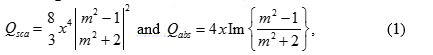
waarin x 2 πa / λ, a de straal van de bol en m 2 is ε m / ε d. Hier, ε m en ε d overeen met de diëlektrische constanten van het metaal en de omringende diëlektrische resp. Aangezien de vorm van de verstrooiingscoëfficiënt is vergelijkbaar met die van the absorptiecoëfficiënt, wordt daarom verwacht dat verzadigbaar verstrooiing observeren in één plasmonische nanodeeltje 20.

Onlangs lineaire verzadigbare verstrooiing in een geïsoleerde plasmonische deeltjes werd aangetoond voor het eerst 21. Het is opmerkelijk dat in diepe verzadiging, de verstrooiingsintensiteit in feite daalde licht wanneer de excitatie-intensiteit verhoogd. Nog meer opvallend wanneer de excitatie-intensiteit een intensievere na verstrooiing verzadigd raakte, de verstrooiingsintensiteit opgestaan, die het effect van omgekeerde verzadigbaar 20 verstrooiing. Wavelength- en grootte-afhankelijke studies hebben aangetoond een sterke relatie tussen LSPR en niet-lineaire verstrooiing 21. De intensiteit en de golflengte afhankelijkheid van plasmon verstrooiing is vergelijkbaar met de absorptie, suggereert een gemeenschappelijk mechanisme achter deze niet-lineair gedrag.

In termen van toepassingen, is het goed known lineariteit die helpt optische microscopie resolutie te verbeteren. In 2007, verzadigd excitatie (SAX) microscopie werd voorgesteld, die resolutie kan verbeteren door het extraheren van de verzadigde signaal via een tijdelijke sinusvormige modulatie van de excitatiebundel 22. SAX microscopie is gebaseerd op het concept dat een laser brandpunt, de intensiteit sterker in het midden dan aan de rand. Als het signaal (ofwel fluorescentie of verstrooiing) vertoont saturatiegedrag, moet de verzadiging starten van het centrum, terwijl de lineaire respons blijft aan de periferie. Daarom, als er een methode om alleen het gedeelte verzadigde extract, het zal alleen laat het middengedeelte en de verwerping het randdeel, dus effectief verbeteren van de ruimtelijke resolutie. In principe is er geen lagere resolutie limiet in SAX microscopie, mits diepe verzadiging bereikt is en er geen monster schade als gevolg van de intense verlichting.

Aangetoond is dat de resolution fluorescentiebeeldvorming kan aanzienlijk worden verbeterd door gebruikmaking van de SAX techniek. Echter, lijdt de fluorescentie fotobleken effect. Het combineren van de ontdekking van verstrooiing lineariteit en het concept van SAX, kan superresolutietechnieken basis van verstrooiing worden gerealiseerd 21. Vergeleken met conventionele superresolutie microscopie, de verstrooiing gebaseerde techniek verschaft een nieuwe niet-blekende contrast methode. In dit artikel wordt een stap-voor-stap beschrijving gegeven om de vereiste te verkrijgen en halen de niet-lineariteit van plasmonische verstrooiing procedures te schetsen. Werkwijzen voor het identificeren verstrooiing lineariteiten geïntroduceerd door het veranderen van de invallende intensiteit beschreven. Meer informatie zal worden verstrekt om te ontrafelen hoe deze lineariteiten invloed beelden van enkele nanodeeltjes en hoe ruimtelijke resolutie kan dienovereenkomstig door de SAX techniek worden verbeterd.

Protocol

1. GNS Monstervoorbereiding Voor de monsterbereiding, ultrasone trillingen 1 ml GNS colloïdale oplossing ten minste 15 minuten bij ongeveer 40 kHz tot deeltjesaggregatie, wat kan leiden tot de LSPR piek verschuiven voorkomen. Drop 100-200 ul van GNS colloïden op een dia glas met commerciële magnesiumaluminiumsilicaat (MAS) coating aan het GNSS te lossen. Na ten minste 1 min, verwijder de extra colloïde door te spoelen met gedestilleerd water. De wachttijd is afhankelijk van de gewenste…

Representative Results

Figuur 6 toont het gemeten spectrum van een 80 nm GNS. Een berekende curve op basis van de Mie theorie wordt gegeven in dezelfde plot, toont uitstekende overeenkomst. De LSPR piek ligt rond de 580 nm. In het volgende experiment, de laser golflengte was 532 nm, die werd gekozen omdat het zich in het plasmonische band optische verstrooiing met plasmon effect te versterken en mogelijk verstrooiing verzadiging 21. Figuur 7 presenteert verstrooiing bee…

Discussion

In het protocol zijn er verschillende kritische stappen. Ten eerste, de voorbereiding van de monsters, de dichtheid van nanodeeltjes moet niet te hoog zijn, plasmon koppeling tussen deeltjes te voorkomen. Als twee of meer deeltjes zeer dicht bij elkaar, de koppeling resulteert in de LSPR golflengteverschuivende richting van langere golflengten, waardoor de lineariteit aanzienlijk verminderen. Echter, deze beeldvormende techniek daadwerkelijk brengt de verdeling van plasmonische wijzen, in plaats van de deeltjes zelf. Da…

Divulgazioni

The authors have nothing to disclose.

Acknowledgements

This work is supported by Ministry of Science and Technology under NSC-101-2923-M-002-001-MY3 and NSC-102-2112-M-002-018-MY3. This research is also supported by the Japan Society for the Promotion of Science (JSPS) through the “Funding Program for Next Generation World-Leading Researchers (NEXT Program),” initiated by the Council for Science and Technology Policy (CSTP) and JSPS Asian CORE Program.

Materials

microscope body Olympus, Japan BX-51
objective lens Olympus, Japan UPlanSapo, 100X, NA 1.4
80-nm gold colloid BBI Solutions, UK EM.GC80
supercontinuum laser Fianium, United Kingdom SC400-2-PP
broadband dielectric mirrors Thorlabs, USA BB1-E02
field emission SEM JEOL, Japan JSM-6330F optional
spectrometer Andor Technology, UK Shamrock 163
 charge-coupled device Andor Technology, UK iDus DV420A-OE
acousto-optic modulators  IntraAction Corp., USA AOM-402AF1
lock-in amplifier Stanford Research Systems, USA SR-830
MAS-coated slide glass Matsunami Glass, Japan, S9215

Riferimenti

  1. Fang, N., Lee, H., Sun, C., Zhang, X. Sub-diffraction-limited optical imaging with a silver superlens. Science. 308 (5721), 534-537 (2005).
  2. Lee, B., Kim, S., Kim, H., Lim, Y. The use of plasmonics in light beaming and focusing. 34 (2), 47-87 (2010).
  3. Lal, S., Link, S., Halas, N. J. Nano-optics from sensing to waveguiding. Nature Photon. 1 (11), 641-648 (2007).
  4. Kawata, S., Inouye, Y., Verma, P. Plasmonics for near-field nano-imaging and superlensing. Nature Photon. 3 (7), 388-394 (2009).
  5. Homola, J., Yee, S. S., Gauglitz, G. Surface plasmon resonance sensors: review. Sensor. Actuat. B-Chem. 54 ((1-2)), 3-15 (1999).
  6. Nie, S., Emory, S. R. Probing single molecules and single nanoparticles by surface-enhanced Raman scattering. Science. 275 (5303), 1102-1106 (1997).
  7. Hache, F., Ricard, D., Flytzanis, C. Optical nonlinearities of small metal particles – surface-mediated resonance and quantum size effects. J. Opt. Soc. Am. B. 3 (12), 1647-1655 (1986).
  8. Balamurugan, B., Maruyama, T. Evidence of an enhanced interband absorption in Au nanoparticles: Size-dependent electronic structure and optical properties. Applied Physics Letters. 87 (14), 143105 (2005).
  9. Link, S., El-Sayed, M. A. Size and temperature dependence of the plasmon absorption of colloidal gold nanoparticles. J. Phys. Chem. B. 103 (21), 4212-4217 (1999).
  10. Kelly, K. L., Coronado, E., Zhao, L. L., Schatz, G. C. The optical properties of metal nanoparticles: The influence of size, shape, and dielectric environment. J. Phys. Chem B. 107 (3), 668-677 (2003).
  11. Kauranen, M., Zayats, A. V. Nonlinear plasmonics. Nature Photon. 6 (11), 737-748 (2012).
  12. Homola, J. Present and future of surface plasmon resonance biosensors. Anal Bioanal Chem. 377 (3), 528-539 (2003).
  13. Jain, P. K., El-Sayed, I. H., El-Sayed, M. A. Au nanoparticles target cancer. Nano Today. 2 (1), 18-29 (2007).
  14. Wada, O. Femtosecond all-optical devices for ultrafast communication and signal processing. New J. Phys. 6 (183), .
  15. Elim, H. I., Yang, J., Lee, J. Y., Mi, J., Ji, W. Observation of saturable and reverse-saturable absorption at longitudinal surface plasmon resonance in gold nanorods. Appl. Phys. Lett. 88 (8), 083107 (2006).
  16. Ros, I., Schiavuta, P., Bello, V., Mattei, G., Bozio, R. Femtosecond nonlinear absorption of gold nanoshells at surface plasmon resonance. Phys. Chem. Chem. Phys. 12 (41), 13692-13698 (2010).
  17. De Boni, L., Wood, E. L., Toro, C., Hernandez, F. E. Optical Saturable Absorption in Gold Nanoparticles. Plasmonics. 3 (4), 171-176 (2008).
  18. Gurudas, U., et al. Saturable and reverse saturable absorption in silver nanodots at 532 nm using picosecond laser pulses. J. Appl. Phys. 104 (7), 073107 (2008).
  19. Bohren, C. F., Huffman, D. R. . Absorption and scattering of light by small particles. , (1983).
  20. Chu, S. W., et al. Saturation and reverse saturation of scattering in a single plasmonic nanoparticle. ACS Photon. 1 (1), 32-37 (2014).
  21. Chu, S. W., et al. Measurement of a saturated emission of optical radiation from gold nanoparticles: application to an ultrahigh resolution microscope. Phys. Rev. Lett. 112 (1), 017402 (2014).
  22. Fujita, K., Kobayashi, M., Kawano, S., Yamanaka, M., Kawata, S. High-resolution confocal microscopy by saturated excitation of fluorescence. Phys. Rev. Lett. 99 (22), 228105 (2007).
  23. Smith, K. C. A., Oatley, C. W. The scanning electron microscope and its fields of application. Brit. J.Appl. Phys. 6 (11), (1955).
  24. Yu, J. Y., et al. A diffraction-limited scanning system providing broad spectral range for laser scanning microscopy. Rev. Sci. Instru. 80 (11), 113704 (2009).
  25. Lee, H., et al. Point spread function analysis with saturable and reverse saturable scattering. Opt. Express. 22 (21), 26016-26022 (2014).
  26. Gustafsson, M. G. L. Nonlinear structured-illumination microscopy: Wide-field fluorescence imaging with theoretically unlimited resolution. Proc. Natl. Acad. Sci. U. S. A. 102 (37), 13081-13086 (2005).
  27. Yonemaru, Y., Yamanaka, M., Smith, N. I., Kawata, S., Fujita, K. Saturated Excitation Microscopy with Optimized Excitation Modulation. ChemPhysChem. 15 (4), 743-749 (2014).
  28. Betzig, E., et al. Imaging intracellular fluorescent proteins at nanometer resolution. Science. 313 (5793), 1642-1645 (2006).
  29. Hell, S. W. Far-field optical nanoscopy. Science. 316 (5828), 1153-1158 (2007).
  30. Huang, B., Wang, W. Q., Bates, M., Zhuang, X. W. Three-dimensional super-resolution imaging by stochastic optical reconstruction microscopy. Science. 319 (5864), 810-813 (2008).
  31. Heintzmann, R., Jovin, T. M., Cremer, C. Saturated patterned excitation microscopy – a concept for optical resolution improvement. J. Opt. Soc. Am. A. 19 (8), 1599-1609 (2002).
  32. Tzang, O., Pevzner, A., Marvel, R. E., Haglund, R. F., Cheshnovsky, O. Super-Resolution in Label-Free Photomodulated Reflectivity. Nano Lett. 15 (2), 1362-1367 (2015).
check_url/it/53338?article_type=t

Play Video

Citazione di questo articolo
Lee, H., Li, K., Huang, Y., Shen, P., Deka, G., Oketani, R., Yonemaru, Y., Yamanaka, M., Fujita, K., Chu, S. Measurement of Scattering Nonlinearities from a Single Plasmonic Nanoparticle. J. Vis. Exp. (107), e53338, doi:10.3791/53338 (2016).

View Video