Summary

מדידת פיזור אי-לינאריות מNanoparticle Plasmonic יחיד

Published: January 03, 2016
doi:

Summary

Saturable and reverse saturable scattering were discovered in isolated plasmonic particles and adopted as a novel non-bleaching contrast method in super-resolution microscopy. Here the experimental procedures of detecting and extracting nonlinear scattering are explained in detail, as well as how to enhance resolution with the aid of saturated excitation microscopy.

Abstract

Plasmonics, which are based on the collective oscillation of electrons due to light excitation, involve strongly enhanced local electric fields and thus have potential applications in nonlinear optics, which requires extraordinary optical intensity. One of the most studied nonlinearities in plasmonics is nonlinear absorption, including saturation and reverse saturation behaviors. Although scattering and absorption in nanoparticles are closely correlated by the Mie theory, there has been no report of nonlinearities in plasmonic scattering until very recently.

Last year, not only saturation, but also reverse saturation of scattering in an isolated plasmonic particle was demonstrated for the first time. The results showed that saturable scattering exhibits clear wavelength dependence, which seems to be directly linked to the localized surface plasmon resonance (LSPR). Combined with the intensity-dependent measurements, the results suggest the possibility of a common mechanism underlying the nonlinear behaviors of scattering and absorption. These nonlinearities of scattering from a single gold nanosphere (GNS) are widely applicable, including in super-resolution microscopy and optical switches.

In this paper, it is described in detail how to measure nonlinearity of scattering in a single GNP and how to employ the super-resolution technique to enhance the optical imaging resolution based on saturable scattering. This discovery features the first super-resolution microscopy based on nonlinear scattering, which is a novel non-bleaching contrast method that can achieve a resolution as low as l/8 and will potentially be useful in biomedicine and material studies.

Introduction

המחקר של plasmonics משך עניין רב בשל היישומים שלה בתחומים רבים ושונים 1-4. אחד התחומים הנחקרים ביותר בplasmonics הוא plasmonics פני השטח, שבו התנודה הקולקטיבית של זוגות אלקטרונים הולכה עם גל אלקטרומגנטים חיצוני בממשק שבין מתכת ודיאלקטרי. plasmonics המשטח נחקר עבור היישומים הפוטנציאליים שלה באופטיקה subwavelength, biophotonics, ומיקרוסקופיה 5,6. שיפור השדה החזק בנפח קטן במיוחד של חלקיקים מתכתיים בשל תהודת plasmon המשטח מקומי (LSPR) משך תשומת לב רבה, לא רק בגלל הרגישות יוצאת דופן שלה לגדלי חלקיקים, צורות חלקיקים, ומאפיינים דיאלקטרי של המדיום סביב 7 -10, אלא גם בגלל היכולת שלה כדי להגביר את האפקטים אופטיים קוי חלשים מטבעם 11. הרגישות יוצאת דופן של LSPR היא בעל ערך ליו-חישה וליד-אוףטכניקות הדמיה LD 12,13. מצד השני, הליניאריות המשופרת של מבני plasmonic יכולה להיות מנוצלת במעגלים משולבים פוטוניים ביישומים כגון מיתוג אופטי ועיבוד אותות כל-אופטי 14,15. זה ידוע היטב כי קליטת plasmonic היא ליניארי פרופורציונאלי לעוצמת העירור ברמות עצימות נמוכות. כאשר העירור הוא מספיק חזק, הקליטה מגיעה לרוויה. מסקרן, בעוצמות גבוהות יותר, הקליטה מגדילה שוב. השפעות קוי אלה נקראות קליטת saturable (SA) 15-17 ולהפוך קליטת saturable (RSA) 18, בהתאמה.

זה ידוע כי בשל LSPR, פיזור הוא חזק במיוחד במבני plasmonic. בהתבסס על אלקטרומגנטיות בסיסית, התגובה של פיזור לעומת עוצמת אירוע צריכה להיות ליניארי. עם זאת, בחלקיקים, פיזור וקליטה קשור קשר הדוק באמצעות תיאורית מ.י., ושניהם יכולים להיות דוארxpressed במונחים של חלקים אמיתיים ומדומים של קבוע דיאלקטרי. בהנחה שGNS אחת מתנהג כמו דיפול תחת תאורת אור, מקדם פיזור (SCA Q) ומקדם קליטה (שרירי בטן) Q מננו-חלקיקי plasmonic אחת על פי תיאורית מ.י. יכולים לבוא לידי ביטוי כ19

משוואת 1

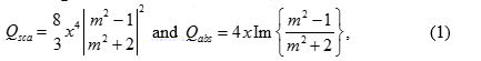
כאשר x הוא 2 πa / λ, הוא הרדיוס של הכדור, והמטר 2 הוא ד מ '/ ε ε. הנה, מ וד ε מתאים לקבועי דיאלקטרי של המתכת ושל החומרים דיאלקטריים שמסביב, בהתאמה. מאז בצורה של מקדם הפיזור דומה לזה של המקדם קליטת דואר, זה צפוי ולכן להתבונן פיזור saturable בננו-חלקיקי plasmonic אחת 20.

לאחרונה, פיזור saturable קוי בחלקיקי plasmonic מבודדים הודגם לראשונה 21. זה מדהים כי ברוויה עמוקה, עוצמת הפיזור למעשה ירדה מעט כאשר עוצמת העירור מוגברת. עוד יותר להפליא, כאשר עוצמת העירור המשיכה להגדיל לאחר הפיזור הפך רווי, עוצמת הפיזור עלתה שוב, מראה את ההשפעה של הפוך saturable פיזור 20. Wavelength- ומחקרי גודל תלוי הראו קשר חזק בין LSPR וקוי פיזור 21. Dependences העצמה ואורך הגל של פיזור plasmonic מאוד דומה לאלה של ספיגה, המצביעים על מנגנון משותף שבסיס התנהגות לא לינארית אלה.

במונחים של יישומים, זה kno גםwn הליניאריות שמסייעת לשפר את הרזולוציה מיקרוסקופיה אופטית. בשינה 2007, עירור רווי מיקרוסקופיה (SAX) הוצע, אשר יכולה לשפר את הרזולוציה על ידי חילוץ האות הרווי באמצעות אפנון סינוסי זמני של קרן עירור 22. מיקרוסקופיה SAX מבוססת על הרעיון כי, לנקודת מוקד לייזר, העצמה היא חזקה במרכז מאשר בפריפריה. אם האות (או הקרינה או פיזור) מציגה הרוויה התנהגות, הרוויה חייבת להתחיל מהמרכז, ואילו התגובה ליניארי נשארה בפריפריה. לכן, אם יש שיטה כדי לחלץ רק חלק הרווי, זה ישאיר רק את החלק המרכזי תוך דחיית החלק ההיקפי, ובכך למעשה שיפור הרזולוציה מרחבית. בעיקרון, יש גבול רזולוציה לא נמוך במיקרוסקופ SAX, כל עוד הוא הגיעה לרוויה עמוקה ואין נזק מדגם בשל התאורה החזקה.

הוכח שresolution של הדמיה הקרינה ניתן לשפר באופן משמעותי על ידי שימוש בטכניקת SAX. עם זאת, הקרינה סובלת מאפקט photobleaching. שילוב של גילוי הליניאריות פיזור ואת הרעיון של SAX, יכול להתממש במיקרוסקופ ברזולוציה הסופר מבוסס על פיזור 21. בהשוואה לmicroscopies ברזולוציה סופר הקונבנציונלי, הטכניקה מבוססת הפיזור מספקת שיטת ניגוד הלא הלבנת רומן. במאמר זה, תיאור צעד-אחר-צעד ניתן להתוות את ההליכים הנדרשים על מנת להשיג ולחלץ את הליניאריות של פיזור plasmonic. שיטות לזיהוי אי-לינאריות פיזור הציגה על ידי שינוי עוצמת האירוע מתוארות. פרטים נוספים יינתנו לפענח איך אי-לינאריות אלה משפיעות תמונות של חלקיקים בודדים וכיצד מרחבי רזולוציה ניתן לשפר בהתאם על ידי טכניקת SAX.

Protocol

1. GNS הכנת דוגמאות לפני הכנת המדגם, פתרון קולואיד 1 מיליליטר GNS sonicate לפחות 15 דקות בכ -40 קילוהרץ כדי למנוע אגרגציה, אשר עלול לגרום לשיא LSPR להעביר. זרוק 100-200 μl של קולואיד GNS על זכוכית שקופית עם ציפו…

Representative Results

איור 6 מציג את הספקטרום הנמדד מGNS 80 ננומטר. עקומה המחושבת על בסיס תיאורית מ.י. ניתנת באותה החלקה, מראה הסכם מצוין. שיא LSPR הוא סביב 580 ננומטר. בניסוי הבא, באורך גל הלייזר היה 532 ננומטר, אשר נבחר כהוא ממוקם בתוך להקת plasmonic כדי לשפר את הפיזור אופטי עם אפקט plasmonic ולאפשר…

Discussion

בפרוטוקול, יש כמה שלבים קריטיים. ראשית, בעת הכנת הדגימות, הצפיפות של חלקיקים לא צריכה להיות גבוהה מדי, כדי להימנע מצימוד plasmonic בין חלקיקים. אם שניים או יותר חלקיקים קרובים מאוד זה לזה, תוצאות הצימוד באורך גל LSPR הסטה לכיוון אורכי גל ארוכים יותר, ובכך להקטין באופן משמעותי…

Divulgazioni

The authors have nothing to disclose.

Acknowledgements

This work is supported by Ministry of Science and Technology under NSC-101-2923-M-002-001-MY3 and NSC-102-2112-M-002-018-MY3. This research is also supported by the Japan Society for the Promotion of Science (JSPS) through the “Funding Program for Next Generation World-Leading Researchers (NEXT Program),” initiated by the Council for Science and Technology Policy (CSTP) and JSPS Asian CORE Program.

Materials

microscope body Olympus, Japan BX-51
objective lens Olympus, Japan UPlanSapo, 100X, NA 1.4
80-nm gold colloid BBI Solutions, UK EM.GC80
supercontinuum laser Fianium, United Kingdom SC400-2-PP
broadband dielectric mirrors Thorlabs, USA BB1-E02
field emission SEM JEOL, Japan JSM-6330F optional
spectrometer Andor Technology, UK Shamrock 163
 charge-coupled device Andor Technology, UK iDus DV420A-OE
acousto-optic modulators  IntraAction Corp., USA AOM-402AF1
lock-in amplifier Stanford Research Systems, USA SR-830
MAS-coated slide glass Matsunami Glass, Japan, S9215

Riferimenti

  1. Fang, N., Lee, H., Sun, C., Zhang, X. Sub-diffraction-limited optical imaging with a silver superlens. Science. 308 (5721), 534-537 (2005).
  2. Lee, B., Kim, S., Kim, H., Lim, Y. The use of plasmonics in light beaming and focusing. 34 (2), 47-87 (2010).
  3. Lal, S., Link, S., Halas, N. J. Nano-optics from sensing to waveguiding. Nature Photon. 1 (11), 641-648 (2007).
  4. Kawata, S., Inouye, Y., Verma, P. Plasmonics for near-field nano-imaging and superlensing. Nature Photon. 3 (7), 388-394 (2009).
  5. Homola, J., Yee, S. S., Gauglitz, G. Surface plasmon resonance sensors: review. Sensor. Actuat. B-Chem. 54 ((1-2)), 3-15 (1999).
  6. Nie, S., Emory, S. R. Probing single molecules and single nanoparticles by surface-enhanced Raman scattering. Science. 275 (5303), 1102-1106 (1997).
  7. Hache, F., Ricard, D., Flytzanis, C. Optical nonlinearities of small metal particles – surface-mediated resonance and quantum size effects. J. Opt. Soc. Am. B. 3 (12), 1647-1655 (1986).
  8. Balamurugan, B., Maruyama, T. Evidence of an enhanced interband absorption in Au nanoparticles: Size-dependent electronic structure and optical properties. Applied Physics Letters. 87 (14), 143105 (2005).
  9. Link, S., El-Sayed, M. A. Size and temperature dependence of the plasmon absorption of colloidal gold nanoparticles. J. Phys. Chem. B. 103 (21), 4212-4217 (1999).
  10. Kelly, K. L., Coronado, E., Zhao, L. L., Schatz, G. C. The optical properties of metal nanoparticles: The influence of size, shape, and dielectric environment. J. Phys. Chem B. 107 (3), 668-677 (2003).
  11. Kauranen, M., Zayats, A. V. Nonlinear plasmonics. Nature Photon. 6 (11), 737-748 (2012).
  12. Homola, J. Present and future of surface plasmon resonance biosensors. Anal Bioanal Chem. 377 (3), 528-539 (2003).
  13. Jain, P. K., El-Sayed, I. H., El-Sayed, M. A. Au nanoparticles target cancer. Nano Today. 2 (1), 18-29 (2007).
  14. Wada, O. Femtosecond all-optical devices for ultrafast communication and signal processing. New J. Phys. 6 (183), .
  15. Elim, H. I., Yang, J., Lee, J. Y., Mi, J., Ji, W. Observation of saturable and reverse-saturable absorption at longitudinal surface plasmon resonance in gold nanorods. Appl. Phys. Lett. 88 (8), 083107 (2006).
  16. Ros, I., Schiavuta, P., Bello, V., Mattei, G., Bozio, R. Femtosecond nonlinear absorption of gold nanoshells at surface plasmon resonance. Phys. Chem. Chem. Phys. 12 (41), 13692-13698 (2010).
  17. De Boni, L., Wood, E. L., Toro, C., Hernandez, F. E. Optical Saturable Absorption in Gold Nanoparticles. Plasmonics. 3 (4), 171-176 (2008).
  18. Gurudas, U., et al. Saturable and reverse saturable absorption in silver nanodots at 532 nm using picosecond laser pulses. J. Appl. Phys. 104 (7), 073107 (2008).
  19. Bohren, C. F., Huffman, D. R. . Absorption and scattering of light by small particles. , (1983).
  20. Chu, S. W., et al. Saturation and reverse saturation of scattering in a single plasmonic nanoparticle. ACS Photon. 1 (1), 32-37 (2014).
  21. Chu, S. W., et al. Measurement of a saturated emission of optical radiation from gold nanoparticles: application to an ultrahigh resolution microscope. Phys. Rev. Lett. 112 (1), 017402 (2014).
  22. Fujita, K., Kobayashi, M., Kawano, S., Yamanaka, M., Kawata, S. High-resolution confocal microscopy by saturated excitation of fluorescence. Phys. Rev. Lett. 99 (22), 228105 (2007).
  23. Smith, K. C. A., Oatley, C. W. The scanning electron microscope and its fields of application. Brit. J.Appl. Phys. 6 (11), (1955).
  24. Yu, J. Y., et al. A diffraction-limited scanning system providing broad spectral range for laser scanning microscopy. Rev. Sci. Instru. 80 (11), 113704 (2009).
  25. Lee, H., et al. Point spread function analysis with saturable and reverse saturable scattering. Opt. Express. 22 (21), 26016-26022 (2014).
  26. Gustafsson, M. G. L. Nonlinear structured-illumination microscopy: Wide-field fluorescence imaging with theoretically unlimited resolution. Proc. Natl. Acad. Sci. U. S. A. 102 (37), 13081-13086 (2005).
  27. Yonemaru, Y., Yamanaka, M., Smith, N. I., Kawata, S., Fujita, K. Saturated Excitation Microscopy with Optimized Excitation Modulation. ChemPhysChem. 15 (4), 743-749 (2014).
  28. Betzig, E., et al. Imaging intracellular fluorescent proteins at nanometer resolution. Science. 313 (5793), 1642-1645 (2006).
  29. Hell, S. W. Far-field optical nanoscopy. Science. 316 (5828), 1153-1158 (2007).
  30. Huang, B., Wang, W. Q., Bates, M., Zhuang, X. W. Three-dimensional super-resolution imaging by stochastic optical reconstruction microscopy. Science. 319 (5864), 810-813 (2008).
  31. Heintzmann, R., Jovin, T. M., Cremer, C. Saturated patterned excitation microscopy – a concept for optical resolution improvement. J. Opt. Soc. Am. A. 19 (8), 1599-1609 (2002).
  32. Tzang, O., Pevzner, A., Marvel, R. E., Haglund, R. F., Cheshnovsky, O. Super-Resolution in Label-Free Photomodulated Reflectivity. Nano Lett. 15 (2), 1362-1367 (2015).
check_url/it/53338?article_type=t

Play Video

Citazione di questo articolo
Lee, H., Li, K., Huang, Y., Shen, P., Deka, G., Oketani, R., Yonemaru, Y., Yamanaka, M., Fujita, K., Chu, S. Measurement of Scattering Nonlinearities from a Single Plasmonic Nanoparticle. J. Vis. Exp. (107), e53338, doi:10.3791/53338 (2016).

View Video