Summary

단일 플라즈몬 나노 입자에서 비선형 산란 측정

Published: January 03, 2016
doi:

Summary

Saturable and reverse saturable scattering were discovered in isolated plasmonic particles and adopted as a novel non-bleaching contrast method in super-resolution microscopy. Here the experimental procedures of detecting and extracting nonlinear scattering are explained in detail, as well as how to enhance resolution with the aid of saturated excitation microscopy.

Abstract

Plasmonics, which are based on the collective oscillation of electrons due to light excitation, involve strongly enhanced local electric fields and thus have potential applications in nonlinear optics, which requires extraordinary optical intensity. One of the most studied nonlinearities in plasmonics is nonlinear absorption, including saturation and reverse saturation behaviors. Although scattering and absorption in nanoparticles are closely correlated by the Mie theory, there has been no report of nonlinearities in plasmonic scattering until very recently.

Last year, not only saturation, but also reverse saturation of scattering in an isolated plasmonic particle was demonstrated for the first time. The results showed that saturable scattering exhibits clear wavelength dependence, which seems to be directly linked to the localized surface plasmon resonance (LSPR). Combined with the intensity-dependent measurements, the results suggest the possibility of a common mechanism underlying the nonlinear behaviors of scattering and absorption. These nonlinearities of scattering from a single gold nanosphere (GNS) are widely applicable, including in super-resolution microscopy and optical switches.

In this paper, it is described in detail how to measure nonlinearity of scattering in a single GNP and how to employ the super-resolution technique to enhance the optical imaging resolution based on saturable scattering. This discovery features the first super-resolution microscopy based on nonlinear scattering, which is a novel non-bleaching contrast method that can achieve a resolution as low as l/8 and will potentially be useful in biomedicine and material studies.

Introduction

플라즈몬의 연구로 인해 다양한 분야 1-4의 응용 프로그램에 큰 관심을 끌고있다. 플라즈몬 가장 연구 분야 중 하나는, 표면 플라즈몬,되는 금속과 유전체 사이의 계면에서 외부와 전자파 전도 전자 커플 집단적 진동. 표면 플라즈몬은 서브 파장 광학, 바이오 포토닉스 및 현미경 5,6에서의 응용 가능성에 대해 탐구하고있다. 국소 표면 플라즈몬 공명 (LSPR)에 의한 금속 나노 입자의 매우 작은 체적 강전 향상뿐만 아니라 때문에 입자 크기, 입자 형상, 및 주변 매질 (7)의 유전 특성에 탁월한 감도, 광범위한 주목을 받고 -10뿐만 아니라, 때문에 본질적으로 약한 비선형 광학 효과 (11)을 높일 수있는 능력. 저 채도의 뛰어난 감도는 바이오 센싱과 가까운 헛소리에 대한 가치가있다LD 이미징 기술 (12, 13). 한편, 구조물의 플라즈몬 강화 된 비선형 광학 스위칭 및 전광 신호 처리 애플리케이션 (14, 15) 등의 광 집적 회로에 사용될 수있다. 이것은 잘 플라즈몬 흡수가 낮은 세기 레벨의 여진 강도에 정비례하는 것으로 알려져있다. 여진이 충분히 강한 경우, 흡수가 포화에 도달한다. 흥미롭게도, 높은 강도에서 흡수가 다시 증가한다. 이러한 비선형 효과는 포화 흡수 (SA) 15 ~ 17이라고 각각 포화 흡수 (RSA) (18)을 반대로한다.

그것은 LSPR 때문에, 산란 플라즈몬 구조에서 특히 강한 것으로 알려져있다. 기본적인 전자기학에 기초하여, 입사광의 산란 강도에 비해 응답은 선형이어야한다. 그러나, 나노 입자, 산란 및 흡수 밀접 Mie 이론을 통해 연결되어, 두 전자 일 수있다비유 전율의 실수 부 및 허수 부분의 관점에서 xpressed. 단일 GNS 광 조명하 다이폴로서 동작하는 것으로 가정하여, Mie 이론에 따른 단일 플라즈몬 나노 입자로부터의 산란 계수 (Q의 SCA) 및 흡수 계수 (Q의 절대치)를 19로 표현 될 수있다

식 (1)

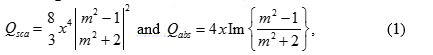
x가 2이고 πa / λ,이 구 반경이며, m은 2 ε m / ε의 D이다. 여기서, ε를 ε mD는 각각 둘러싸는 금속 절연체의 유전 상수와의 대응. 산란 계수의 형태 번째 이후의 것과 유사전자 흡수 계수, 그것은 따라서 단일 플라즈몬 나노 입자 (20) 가포 산란을 관찰 할 것으로 예상된다.

최근, 고립 된 플라즈몬 입자의 비선형 포화 산란는 처음으로 21 증명되었다. 이 여진 강도가 증가 할 때 포화에서 깊은 사실 산란 강도는 약간 감소하는 것이 현저하다. 여진 강도가 산란을 포화 낳은 후에 계속 증가 할 때 더욱 현저하게, 산란 강도는 가포 리버스 20 산란의 효과를 나타내는, 부활. 파장 – 크기에 의존하는 연구는 21 산란 저 채도 및 비선형 사이에 강한 관계를 보여 주었다. 플라즈몬 산란 강도와 파장 의존성이 비선​​형 행동을 기본 공통 메커니즘을 제안 흡수의 것과 매우 유사하다.

애플리케이션의 관점에서는 물론이다 KNOWN 그 비선형 광학 현미경의 해상도를 개선하는 데 도움이됩니다. 2007에서, 포화가 여진 (SAX) 현미경은 여기 빔 (22)의 시간적 정현파 변조를 통해 포화 신호를 추출함으로써 해상도를 향상시킬 수있는 제안되었다. SAX 현미경은 레이저 초점 스폿, 강도가 주변부보다 중심에서 더 강한 개념에 기초한다. 신호 (형광 또는 산란 중)가 포화 동작을 나타내는 경우 선형 응답 주위에 남아있는 동안, 채도, 중심에서 시작해야한다. 단지 포화 된 부분을 추출하는 방법이 있으면 주변 부분을 차단하면서도 따라서, 그것은 따라서 효과적으로 공간 분해능을 향상에만 중앙부를 떠날 것이다. 원칙적으로, 깊은만큼 포화에 도달 SAX 현미경없이 낮은 해상도 한계가 있고 강한 조명에 의한 시료에는 손상이 없다.

그것은 resolutio 것으로 나타났다형광 이미징의 N 크게 SAX 기술을 이용함으로써 향상 될 수있다. 그러나, 형광 광표백 효과에서 겪고있다. 산란 비선형의 발견과 SAX의 개념을 조합 산란에 기초하여 수퍼 – 해상도 현미경 (21)을 실현할 수있다. 종래의 수퍼 – 해상도 microscopies에 비해, 산란 기반 기술은 신규 한 비 표백 조영 방법을 제공한다. 본 논문에서는 단계별 설명은 획득 및 플라즈몬 산란 비선형를 추출하기 위해 필요한 절차를 간략하게 설명하기 위해 제공된다. 입사 광량 변경에 의해 도입 비선형 산란을 식별하는 방법은 기술된다. 더 자세한 내용은 이러한 비선형 단일 나노 입자의 이미지에 영향을 미치는 방법 및 공간 해상도 것은 SAX 기술에 의해 적절히 강화 될 수있는 방법을 해명하기 위해 제공 될 것이다.

Protocol

1. GNS 샘플 준비 LSPR 피크 시프트가 발생할 수의 입자 응집을 방지하기 위해 약 40 kHz에서 적어도 15 분 동안 샘플을 초음파 처리 GNS 1ml의 콜로이드 용액을 준비하기 전에. GNSS를 해결하기 위해 상업 마그네슘 알루미늄 실리케이트 (MAS) 코팅 된 슬라이드 글라스에 GNS 콜로이드의 100 ~ 200 μl를 삭제합니다. 적어도 1 분 후, 증류수로 세척하여 여분의 콜로이드를 제거합니다. 대기 …

Representative Results

그림 6은 80 nm의 GNS에서 측정 된 스펙트럼을 보여줍니다. Mie 이론에 기초하여 계산 된 곡선이 잘 일치를 보여주는, 같은 플롯에 제시되어있다. LSPR 피크는 580 nm의 주위이다. 다음 실험에서, 레이저는 파장이 플라즈몬 광 산란 효과를 향상시키고 포화 21 산란 활성화 플라즈몬 밴드 안에 위치로 선정 된 532 나노 미터였다. 그림 7 다른 여기 강도?…

Discussion

프로토콜에서, 몇 가지 중요한 단계가 있습니다. 샘플을 준비 할 때 먼저, 나노 입자 밀도는 입자 사이 플라즈몬 결합을 피하기 위해 너무 높은이어야한다. 두 개 이상의 입자가 서로 매우 근접 할 경우, LSPR 파장 결합 결과는 따라서 크게 비선형 성을 감소시키는, 장파장 측으로 시프트. 그러나,이 영상 법은 실제로 입자 자체 대신에, 플라즈몬 모드의 분포를 매핑한다. 따라서, 적합한 여기 파장과…

Divulgazioni

The authors have nothing to disclose.

Acknowledgements

This work is supported by Ministry of Science and Technology under NSC-101-2923-M-002-001-MY3 and NSC-102-2112-M-002-018-MY3. This research is also supported by the Japan Society for the Promotion of Science (JSPS) through the “Funding Program for Next Generation World-Leading Researchers (NEXT Program),” initiated by the Council for Science and Technology Policy (CSTP) and JSPS Asian CORE Program.

Materials

microscope body Olympus, Japan BX-51
objective lens Olympus, Japan UPlanSapo, 100X, NA 1.4
80-nm gold colloid BBI Solutions, UK EM.GC80
supercontinuum laser Fianium, United Kingdom SC400-2-PP
broadband dielectric mirrors Thorlabs, USA BB1-E02
field emission SEM JEOL, Japan JSM-6330F optional
spectrometer Andor Technology, UK Shamrock 163
 charge-coupled device Andor Technology, UK iDus DV420A-OE
acousto-optic modulators  IntraAction Corp., USA AOM-402AF1
lock-in amplifier Stanford Research Systems, USA SR-830
MAS-coated slide glass Matsunami Glass, Japan, S9215

Riferimenti

  1. Fang, N., Lee, H., Sun, C., Zhang, X. Sub-diffraction-limited optical imaging with a silver superlens. Science. 308 (5721), 534-537 (2005).
  2. Lee, B., Kim, S., Kim, H., Lim, Y. The use of plasmonics in light beaming and focusing. 34 (2), 47-87 (2010).
  3. Lal, S., Link, S., Halas, N. J. Nano-optics from sensing to waveguiding. Nature Photon. 1 (11), 641-648 (2007).
  4. Kawata, S., Inouye, Y., Verma, P. Plasmonics for near-field nano-imaging and superlensing. Nature Photon. 3 (7), 388-394 (2009).
  5. Homola, J., Yee, S. S., Gauglitz, G. Surface plasmon resonance sensors: review. Sensor. Actuat. B-Chem. 54 ((1-2)), 3-15 (1999).
  6. Nie, S., Emory, S. R. Probing single molecules and single nanoparticles by surface-enhanced Raman scattering. Science. 275 (5303), 1102-1106 (1997).
  7. Hache, F., Ricard, D., Flytzanis, C. Optical nonlinearities of small metal particles – surface-mediated resonance and quantum size effects. J. Opt. Soc. Am. B. 3 (12), 1647-1655 (1986).
  8. Balamurugan, B., Maruyama, T. Evidence of an enhanced interband absorption in Au nanoparticles: Size-dependent electronic structure and optical properties. Applied Physics Letters. 87 (14), 143105 (2005).
  9. Link, S., El-Sayed, M. A. Size and temperature dependence of the plasmon absorption of colloidal gold nanoparticles. J. Phys. Chem. B. 103 (21), 4212-4217 (1999).
  10. Kelly, K. L., Coronado, E., Zhao, L. L., Schatz, G. C. The optical properties of metal nanoparticles: The influence of size, shape, and dielectric environment. J. Phys. Chem B. 107 (3), 668-677 (2003).
  11. Kauranen, M., Zayats, A. V. Nonlinear plasmonics. Nature Photon. 6 (11), 737-748 (2012).
  12. Homola, J. Present and future of surface plasmon resonance biosensors. Anal Bioanal Chem. 377 (3), 528-539 (2003).
  13. Jain, P. K., El-Sayed, I. H., El-Sayed, M. A. Au nanoparticles target cancer. Nano Today. 2 (1), 18-29 (2007).
  14. Wada, O. Femtosecond all-optical devices for ultrafast communication and signal processing. New J. Phys. 6 (183), .
  15. Elim, H. I., Yang, J., Lee, J. Y., Mi, J., Ji, W. Observation of saturable and reverse-saturable absorption at longitudinal surface plasmon resonance in gold nanorods. Appl. Phys. Lett. 88 (8), 083107 (2006).
  16. Ros, I., Schiavuta, P., Bello, V., Mattei, G., Bozio, R. Femtosecond nonlinear absorption of gold nanoshells at surface plasmon resonance. Phys. Chem. Chem. Phys. 12 (41), 13692-13698 (2010).
  17. De Boni, L., Wood, E. L., Toro, C., Hernandez, F. E. Optical Saturable Absorption in Gold Nanoparticles. Plasmonics. 3 (4), 171-176 (2008).
  18. Gurudas, U., et al. Saturable and reverse saturable absorption in silver nanodots at 532 nm using picosecond laser pulses. J. Appl. Phys. 104 (7), 073107 (2008).
  19. Bohren, C. F., Huffman, D. R. . Absorption and scattering of light by small particles. , (1983).
  20. Chu, S. W., et al. Saturation and reverse saturation of scattering in a single plasmonic nanoparticle. ACS Photon. 1 (1), 32-37 (2014).
  21. Chu, S. W., et al. Measurement of a saturated emission of optical radiation from gold nanoparticles: application to an ultrahigh resolution microscope. Phys. Rev. Lett. 112 (1), 017402 (2014).
  22. Fujita, K., Kobayashi, M., Kawano, S., Yamanaka, M., Kawata, S. High-resolution confocal microscopy by saturated excitation of fluorescence. Phys. Rev. Lett. 99 (22), 228105 (2007).
  23. Smith, K. C. A., Oatley, C. W. The scanning electron microscope and its fields of application. Brit. J.Appl. Phys. 6 (11), (1955).
  24. Yu, J. Y., et al. A diffraction-limited scanning system providing broad spectral range for laser scanning microscopy. Rev. Sci. Instru. 80 (11), 113704 (2009).
  25. Lee, H., et al. Point spread function analysis with saturable and reverse saturable scattering. Opt. Express. 22 (21), 26016-26022 (2014).
  26. Gustafsson, M. G. L. Nonlinear structured-illumination microscopy: Wide-field fluorescence imaging with theoretically unlimited resolution. Proc. Natl. Acad. Sci. U. S. A. 102 (37), 13081-13086 (2005).
  27. Yonemaru, Y., Yamanaka, M., Smith, N. I., Kawata, S., Fujita, K. Saturated Excitation Microscopy with Optimized Excitation Modulation. ChemPhysChem. 15 (4), 743-749 (2014).
  28. Betzig, E., et al. Imaging intracellular fluorescent proteins at nanometer resolution. Science. 313 (5793), 1642-1645 (2006).
  29. Hell, S. W. Far-field optical nanoscopy. Science. 316 (5828), 1153-1158 (2007).
  30. Huang, B., Wang, W. Q., Bates, M., Zhuang, X. W. Three-dimensional super-resolution imaging by stochastic optical reconstruction microscopy. Science. 319 (5864), 810-813 (2008).
  31. Heintzmann, R., Jovin, T. M., Cremer, C. Saturated patterned excitation microscopy – a concept for optical resolution improvement. J. Opt. Soc. Am. A. 19 (8), 1599-1609 (2002).
  32. Tzang, O., Pevzner, A., Marvel, R. E., Haglund, R. F., Cheshnovsky, O. Super-Resolution in Label-Free Photomodulated Reflectivity. Nano Lett. 15 (2), 1362-1367 (2015).
check_url/it/53338?article_type=t

Play Video

Citazione di questo articolo
Lee, H., Li, K., Huang, Y., Shen, P., Deka, G., Oketani, R., Yonemaru, Y., Yamanaka, M., Fujita, K., Chu, S. Measurement of Scattering Nonlinearities from a Single Plasmonic Nanoparticle. J. Vis. Exp. (107), e53338, doi:10.3791/53338 (2016).

View Video