1. ICC Scaffold Fabrication (Figure 1)
- Prepare the polystyrene (PS) lattices (diameter = 6 mm; 8-13 layers of beads).
- To prepare the mold, cut the tips off from 0.2 ml boil-proof microcentrifuge tubes at the 40 µl level. Adhere the top of the cut-tubes to 24 x 60 mm2 microscope cover glass slips with water-proof glue.
- Put the PS spheres (diameter = 140 µm) contained within a water suspension into a 20 ml vial, carefully pipette out the water suspension, and add 18 ml of 70% ethanol solution into the vial. Put the sphere solution into an ultrasonic bath to loosen aggregated spheres. Repeat this washing step several times in order to remove water and water-soluble components completely.
- Pipette 100 µl of ethanol into the molds.
- Cut the top of a 200 µl micropipette tip by 4 mm. Pipette 25 µl of the spheres into the mold twice using the 200 µl micropipette to achieve a total volume of 50 µl in each mold.
- Place the molds on a rocking shaker at 120 rpm overnight.
- Check the arrangement of the spheres in each mold under an optical microscope. If the spheres are not ordered hexagonally, add 50 µl of 70% ethanol and shake manually in the longitudinal and lateral axis direction to correct the arrangement.
- Let the ethanol evaporate at room temperature (RT) for two nights. Place the mold and bead complex in a 130 °C furnace for 6 hr to anneal the PS beads.
- Preparing bare and ECM-coated PEGDA scaffolds.
- Synthesize PEGDA macromers using established protocols45,46 for acrylating linear PEG macromers (Mw = 4.6 kDa).
- Prepare 50% (w/v) PEGDA solution in de-ionized (DI) water and allow the macromer to properly dissolve by centrifuging at 4,713 x g until it is completely dissolved.
- For ECM conjugated ICC scaffolds, dissolve an additional 10% (w/v) Acryloyl-PEG-NHS (Mw=3.4 kDa) in the 50% PEGDA solution.
- Prepare a 20% (w/v) stock solution of 2-hydroxy-4'-(2-hydroxyethoxy)-2-methylpropiophenone (PI) in 70% ethanol.
- Add 50 µl of 20% (w/v) PI stock solution per 1 ml of 50 % (w/v) of PEGDA. Adjust the needed amount of PI stock solution based on the molecular weight of PEGDA.
- Vortex the mixture in centrifuge tube for 1 min to reach a homogenous solution.
- Peel the molds from the glass slide (from step 1.1.7), remove the glue from the molds, push the lattices out carefully using a spatula and place each of them into a 1.5 ml tube. Pipette 300 µl of the PEGDA solution and centrifuge at 845 x g for 5 min to allow proper PEGDA solution infiltration into the lattice.
- Remove the lattice from the tube using tweezers and carefully blot dry excess PEGDA solution on gloves. Place the lattice on a paraffin film-covered glass with the flat circular surface facing up.
- Expose the PEGDA solution infiltrated scaffold to 365 nm ultraviolet (UV) light (10.84 mW/cm2) for 5 min using a UV spot lamp.
- Place PEGDA-polymerized crystal lattices in new vials (around 10 lattices per vial) and add 20 ml of tetrahydrofuran (THF). Shake the vials on an orbital shaker at 300 rpm. Change THF at least 3 times with an interval of 1-2 hr.
Note: Do not remove THF completely when changing the THF in order to prevent bubbles from entering the scaffolds, which in turn can cause incomplete removal of PS. Leave enough solution to cover the lattices and add new THF.
Caution: THF is toxic. Wear gloves, a lab coat and goggles. Avoid inhalation by operating under the fume hood. - Check if PS spheres are dissolved by putting water into the used THF solution and observing the solution color. Repeat step 1.2.9 if the PS spheres are not properly dissolved.
Note: The solution color will change to white if there are any remaining PS spheres.
- Clean the scaffolds in the biosafety cabinet (BSC).
- To sterilize the scaffolds, prepare a 50 ml centrifuge tube with 2 ml of 70% ethanol per scaffold and place the scaffolds in the tube using a spatula. Allow the scaffolds to soak in ethanol for 1 hr. From this step forward, conduct all the procedures in the BSC.
- Carefully pour the ethanol out and replace with phosphate buffer saline (PBS) (2 ml per scaffold) and centrifuge at 524 x g for 3 min to remove bubbles. Keep it in the refrigerator and change the PBS a few times with an interval of 1-2 hr.
- For Collagen type I-coated scaffolds, prepare another 50 ml centrifuge tube containing Collagen type 1 stock solution (1 ml per scaffold), transfer the sterilized scaffolds to this tube using a spatula, and centrifuge at 524 x g for 3 min. Shake the scaffolds at 400 rpm on an orbital shaker for 30 min and keep the tube in the refrigerator overnight.
- Wash the scaffolds with PBS twice before use by submerging the scaffolds in fresh PBS and then aspirating the PBS.
Note: Other ECM proteins can also be used instead of Collagen type I because the NHS chemistry requires an amine group to form the bond (Figure 2).
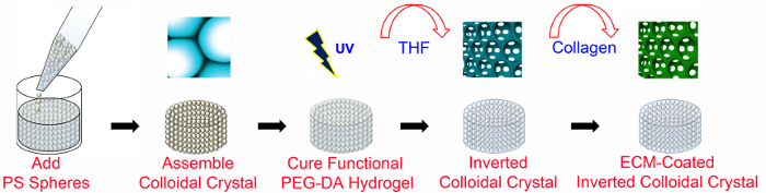
Figure 1. Overview of ICC fabrication. PEG-based ICC scaffolds are fabricated using microfabrication techniques with and without ECM-functionalization. ECM-coated ICC scaffolds require PEG-NHS as well as PEGDA (as detailed in Figure 2). The PS lattice has a diameter of 6 mm and a height of 8-13 bead layers. PS, polystyrene; PEGDA, poly (ethylene glycol) diacrylate; UV, ultraviolet; THF, tetrahydrofuran; ECM, extracellular matrix. This figure has been modified and used with permission from Wiley47. Please click here to view a larger version of this figure.
2. ICC Structure Characterization
- To analyze ICC structure with or without conjugated proteins, use scanning electron microscopy (SEM)47.
- Fix the scaffolds with 4% paraformaldehyde (PFA), serially dehydrate them in 25, 50, 75, 95 and 100% ethanol solutions, and store them at -80 °C until the ethanol evaporates completely.
- Dry samples in a freeze drier for 48 hr.
- Affix the sample onto sample holder using carbon tape and place in a sputter coater.
- After automatic vacuuming, coat it with a Pt film of 10 nm thickness by sputtering for 60 sec at 20 mA.
- Image ICC scaffolds using SEM at a voltage of 5 kV (Figure 3A, Figure 4A).
- To measure the pore and interconnection diameter of cavities, analyze SEM micrographs using image analysis software48 (e.g., ImageJ; Figure 3B,C).
- To visualize the conjugated collagen to the scaffold without cells, fluorescently tag the collagen using antibodies (1:100) against the collagen type I and image with confocal laser scanning microscopy47 (CLSM; Figure 4B).
3. Huh-7.5 Cell Culture and Seeding
- Culture Huh-7.5 cells at a seeding density of 2-2.5 x 10⁶ cells/ml in 100 mm cell culture dishes with 10 ml Dulbecco’s Modified Eagle’s Medium (DMEM) supplemented with 10% fetal bovine serum (FBS) and 100 U/ml penicillin-streptomycin (growth media) at 37 °C and 5% CO2. Change the media every three days in the BSC until they have reached 75-80% confluency.
- Prepare the scaffolds for cell seeding in the BSC.
- Carefully place the scaffolds in a 24-well plate with the flat surface facing up.
- To wash the scaffold, pipette 2 ml PBS to each well containing a scaffold. Aspirate the PBS and pipette 2 ml fresh PBS into each well.
- Aspirate the PBS and pipette 2 ml of growth media (see step 3.1) and leave for 30 min. Aspirate the media and allow the scaffold to dry for 1 hr.
- Detach confluent Huh-7.5 cells (from step 3.1) from the culture plate in the BSC using the trypsin digestion method.
- Aspirate media from plate, add 4 ml PBS to wash adherent cells and then aspirate PBS.
- Pipette 0.75-1 ml 0.25% trypsin and place in an incubator at 37 °C, 5% CO2 for 3 min.
- Remove plate from incubator and pipette 5 ml media to stop the trypsin reaction. Pipette media, detached cells, and trypsin mixture into a 15 ml tube.
- Centrifuge at 524 x g for 3 min, remove the supernatant and resuspend the pellet in 5 ml media.
- Count cells using a hemocytometer and calculate the volume of the cell suspension that contains cells at target number, N0, per 25 µl (for standard experiment, N0 is 1 x 10⁶ cells).
Volume of cell suspension=(target number of cells)/(concentration of cell suspension) - Slowly pipette 25 µl of cell suspension (containing N0 cells) directly on top of each scaffold (from step 3.1.4). Place the 24-well plate in the incubator.
- After 12 hr, transfer the scaffolds carefully with the use of a spatula into a new 24-well plate and pipette 2 ml of media into each well. Place the 24-well plate in the incubator.
- Change the media every 3 days or depending on when media is collected for protein secretion analysis (see Step 5.1).
4. Cell Viability
- To qualitatively analyze cell viability, use fluorescent live/dead staining kits to stain the cells and image using CLSM.
- Following kit instruction, prepare a solution with 4 µM calcein AM and 8 µM ethidium homo-dimer-1 in media (see step 3.1.3).
Note: Optimize depending on the cell number. Use double the amount of reagent if cells proliferate and double in number (around 2 million). - In the BSC, aspirate media in each well with a scaffold (step 3.7) and pipette 500 µl of the prepared solution. Incubate samples in a 37 °C incubator for 1 hr.
- Cover the plates in foil to protect the samples from light when removing the plate out of the incubator. Image samples using CLSM49.
- Following kit instruction, prepare a solution with 4 µM calcein AM and 8 µM ethidium homo-dimer-1 in media (see step 3.1.3).
- To quantitatively assess cell viability, measure enzymatic activity (in live cells) using colorimetric assays50 (i.e., 2-(2-methoxy-4-nitrophenyl)-3-(4-nitrophenyl)-5-(2,4-disulfophenyl)-2H-tetrazolium (monosodium-salt reagent)).
- Create a standard curve for the ICC platform (i.e., a graph of absorbance (OD) versus given cell number).
Note: Cell seeding is not 100% efficient in comparison to other platforms, since cells can pass through the cavities of scaffolds.- Determine the cell seeding numbers, N0 that will be used to make the standard curve.
Note: Choose a range that includes cell numbers that are estimated in the experiment. For example, if the initial cell number is 5 x 10⁵ cells and there is an estimated ~3 fold increase by the last day of the experiment, choose 2.5 x 10⁵, 5 x 10⁵, 1 x 10⁶, and 2 x 10⁶ cells as N0. - Perform cell seeding in the ICC scaffolds (one N0 per scaffold with seeding volume of 25 µl) as described in steps 3.1-3.6.
- After 6 hr, transfer the scaffolds to another 24-well plate well. Select a time that allows cell adherence but not cell proliferation.
- Perform cell counting on the transferred ICC scaffold.
- Dilute 10x monosodium-salt reagent (MSR) solution to 1x with media in the BSC and pipette 500 µl 1x MSR solution into each well with a scaffold. Incubate the 24-well plate at 37 °C for 1 hr.
- From each well, transfer 100 µl into a 96-well plate well. As a blank, pipette 100 µl of fresh 1x monosodium-salt reagent solution into different wells on the 96-well plate. Manually remove any bubbles present using a dry pipette tip and cover the 96-well plate in foil to protect it from light.
- Measure OD at λ = 450 nm reading using a spectrophotometer. Subtract the blank OD from other values to find the accurate OD51.
- Count the number of cells (NL) remaining in the well after transferring the scaffold (step 4.2.1.3) using a hemocytometer.
Note: Use 300 µl of trypsin to trypsinize the cells. - Calculate the actual cell number, NA.
actual = initial – left in well
NA=N0-NL - Make standard curve by plotting OD obtained in Step 4.2.1.4.3 vs. actual cell number (NA) and use this to estimate the cell number in the experiments.
Note: Make a new standard curve if any ICC parameters (i.e., porogen size, dimensions of the scaffold, ECM protein, etc.) are changed.
- Determine the cell seeding numbers, N0 that will be used to make the standard curve.
- Create a standard curve for the ICC platform (i.e., a graph of absorbance (OD) versus given cell number).
5. Cell Function
- Analyze protein secretion by the Huh-7.5 cells (i.e., albumin, urea) from the collected media (from step 3.7) by enzyme linked immunosorbent assay (ELISA)52.
Note: Dilute the media, depending on the number of cells seeded and the amount of media collected. For 5 x 10⁵ cells seeded in ICC, use a ~1:25 ratio, before introducing it to the antibody-precoated wells. - To qualitatively analyze cell function, immunostain specific intracellular proteins (i.e., albumin), enzymes (i.e., CYP450), stain structural components (i.e., cellular actin) as well as the nuclei and image using CLSM49.
- Aspirate media (from step 3.7) and pipette 2 ml PBS to wash the cell-laden ICC scaffolds.
- Pipette 1 ml of 4% PFA and incubate for 5 min at room temperature for fixation.
- Wash 3x with 2 ml PBS.
- Permeabilize membranes by incubating the scaffolds in 1 ml of 0.1% 4-(1,1,3,3-tetramethylbutyl)phenyl-polyethylene glycol (surfactant) for 30 min.
- Wash 3x with 2 ml PBS to remove any leaking proteins.
- Pipette 500 µl 1% bovine serum albumin (BSA) and incubate at RT for 1 hr to block non-specific binding.
- Prepare diluted primary antibody (i.e., albumin, CYP450) solution.
- Pipette 500 µl of 1% BSA solution into a 15 ml tube and add 4.5 ml of 0.1% surfactant solution to prepare a total 5 ml of 0.1% BSA solution.
- Pipette 98 µl of the 0.1% BSA solution into a 200 µl microcentrifuge tube and 2 µl of the primary antibody to produce a 1:50 (primary antibody: 0.1% BSA) primary antibody solution.
- Pipette 40 µl of the primary antibody solution on the scaffold and cover the substrate with paraffin film. Wrap the 24 well plate with aluminum foil and store the dish at 4 °C overnight.
- Wash 3x with 2 ml PBS and shake the plate gently in between washing.
- Prepare diluted biotinylated secondary antibody (i.e., Anti-mouse antibody) stock solution.
- Pipette 198 µl 0.1% BSA solution and 2 µl second antibody to produce a 1:100 (secondary antibody: 0.1% BSA) secondary antibody solution.
- Prepare a 0.1% stock solution of rhodamine or fluorescein labeled phalloidin (to stain the cellular actin filaments) in the 0.1% BSA solution.
- Pipette 25 µl of each solution in a tube and mix well.
- Pipette 50 µl of the secondary antibody stock solution on the scaffold. Cover the scaffold with paraffin film, wrap the dish with aluminum foil and store at RT for 2 hr.
- Wash 3x with 2 ml PBS.
- Pipette 200 µl of 0.2% 4'-6-diamidino-2-phenylindole (DAPI; a nucleus stain) solution on the scaffold and keep at RT for 2-3 min. Cover the plate with aluminum foil.
- Wash 2x with 2 ml PBS.
- Using a dropper, place a drop of mounting media on the substrate.
- Carefully put the scaffold on a glass slide and image using CLSM47.
- Assess the gene expression by real-time polymerase chain reaction (qPCR). Use standard kits for reverse transcriptase PCR (RT-PCR)53 and qPCR54 as per manufacturer's instructions. Extract the RNA from the cells as described below.
- Place the scaffold (from step 3.7) in a 1.5 ml microcentrifuge tube.
- Pipette 1 ml of an RNA extraction solution into the tube and keep in a sonicator for 5 min at RT.
- Pipette 200 µl of chloroform into each microcentrifuge tube and shake the tube vigorously in hand for 15-20 sec. Keep the tubes at RT for ~3 min until the phases separate.
- Centrifuge the sample at 13,000 x g, 4 °C for 15 min and remove the tubes carefully so that phases do not mix.
- Carefully pipette 500-600 µl of the upper aqueous phase from the first tube into a second microcentrifuge tube.
- Add an equivalent volume (500-600 µl) of isopropanol to this second tube.
- Invert the tube 3-5 times and leave the tube standing at RT for 10 min.
- Centrifuge the sample at 13,000 x g, 4 °C for 15 min.
- If a pellet is not visible at the bottom of the tube, centrifuge again for 5 min.
Note: If there is still no visible pellet, the amount of RNA may be insufficient.
- If a pellet is not visible at the bottom of the tube, centrifuge again for 5 min.
- Invert tubes with the cap open to discard supernatant and pipette in 1 ml 70% ethanol diluted in DEPC water into the tube.
- Slightly vortex the tube so the pellet detaches from the wall of the tube and then let the tube air dry.
- Add 50 µl of DPEC water to resuspend the pellet.
- Keep pipetting until the pellet dissolves.
- Keep for 10 min at 55 °C in order to denature the double-stranded RNA into single-stranded RNA.
- Lightly drum fingers on the tube bottom and then centrifuge the tubes briefly (7,500 x g, 4 min, 4 °C).
- Keep the tubes in ice until performing reverse transcriptase55 and real-time PCR as described in 56.
The representative results for the structural characterization of the ICC scaffold and the comparison of each ICC scaffold condition's efficacy in culturing hepatocytes are shown and explained below. The ICC scaffold conditions used in these results are collagen coatings of 0 µg/ml (Bare), 20 µg/ml (Collagen 20), 200 µg/ml (Collagen 200), and 400 µg/ml (Collagen 400) and the initial Huh-7.5 cell seeding number is 1×10⁶.
Characterization of ICC pore interconnections and scaffold topology.
SEM imaging, after fixation and freeze drying of the ICC scaffolds, was first used to determine whether proper interconnections between ICC pores were formed and to see the effect of the conjugation of varied collagen concentrations on the scaffold topology. Quantitative analysis of the two-dimensional bare ICC SEM images (Figure 3A) using ImageJ software, revealed an average pore diameter of 102.3 ± 9.3 µm and an average interconnection diameter of 38.6 ± 4.3 µm (Figure 3B, C). This interconnection between pores plays a significant role in proper diffusion of nutrients to cells as well as cell-cell interaction throughout the scaffold. The coating of collagen resulted in a fiber mesh network that was more defined at higher concentrations of collagen conjugation (Figure 4A). Confocal images reveal even layers of collagen on the cavity surfaces and higher surface area coverage with increases in the collagen concentration (Figure 4B).
Effect of ICC platform conditions on Huh-7.5 cell viability and function.
Huh-7.5 cells were subjected to the fluorescent Live/Dead staining assay (4 µM calcein AM and 8 µM EthD-1) and imaged using CLSM, as shown in Figure 5. Cells seeded in bare, non-adhesive ICC scaffolds tended to aggregate in the center of the pore and cell-cell interconnection between pores was seen in later times (Days 7 and 10). The presence of a collagen coating on the ICC scaffolds allowed cells to adhere to the scaffold surface as well a inter-pore cell-cell interaction as early as Day 1. Cell viability, indicated by the green stain, increased with time and was generally higher in collagen-coated scaffolds, as indicated by higher green fluorescence. Figure 6 shows the quantitative MSR results for further investigation of the effect of the ICC 3D structure and protein concentration on cell viability. A colorimetric cell viability assay was performed on cells cultured in the ICC scaffold conditions as well as on a 2D polystyrene culture plate and absorbance data (measured at 450 nm) was normalized to the control (Day 1). Cells cultured on 2D plates maintained cell viability but no increase in cell proliferation was observed. 3D bare ICC scaffold greatly enhanced cell proliferation compared to the 2D condition, indicating the importance of the 3D matrix. With the addition of the collagen coating, cell proliferation increased with time at all collagen concentrations and the maximum proliferation was found in the 200 µg/ml coated ICC scaffold on Day 14. This confirms qualitative observations in Figure 5.
Hepatocyte function was assessed by monitoring changes in albumin secretion as well the gene expression profile of three adhesion proteins in the different ICC scaffold conditions. Figure 7 illustrates the positive correlation between serum albumin secreted, as quantified by albumin ELISA, and the collagen concentration conjugated to the scaffold. On Day 14, Huh-7.5 cells cultured in the 400 µg/ml coated ICC scaffolds secreted more than three times the amount of albumin as those cultured in the bare scaffold. Results for the gene expression profiles of E-cadherin, N-cadherin, and integrin β1 proteins in the different ICC scaffold conditions appear in Figure 8. The rate of E-cadherin mRNA expression in 400 µg/ml coated ICC scaffolds increased by more than 4 folds as compared to the other coating conditions (Figure 8A). The fold change in N-cadherin gene expression was greater in the higher concentrations of collagen coating. However, the integrin β1 gene expression stayed relatively constant or decreased in all ICC scaffolds conditions except 200 µg/ml coated ICC scaffolds.
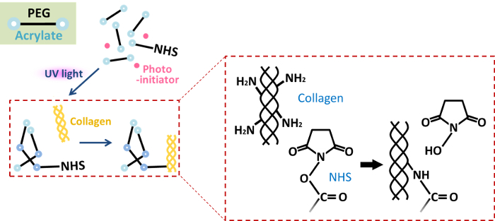
Figure 2. Conjugation of collagen to the PEGDA scaffold via NHS chemistry. Bioactive scaffolds use PEG-NHS that has an amine-reactive succinimidyl (NHS) ester that reacts to the amine group in collagen allowing conjugation to the scaffold. PEG, poly(ethylene glycol); NHS, N-hydroxysuccinimide; UV, ultraviolet. Please click here to view a larger version of this figure.
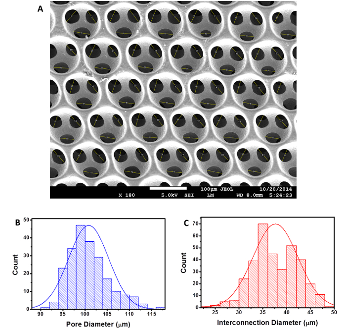
Figure 3. Analysis of the size of the ICC cavities and interconnections. (A) SEM micrographs of the ICC scaffold (scale bar = 100 µm) were analyzed using ImageJ software and quantitatively represented as histograms of (B) pore diameter and (C) interconnection diameter. This figure has been modified and used with permission from Wiley47. Please click here to view a larger version of this figure.
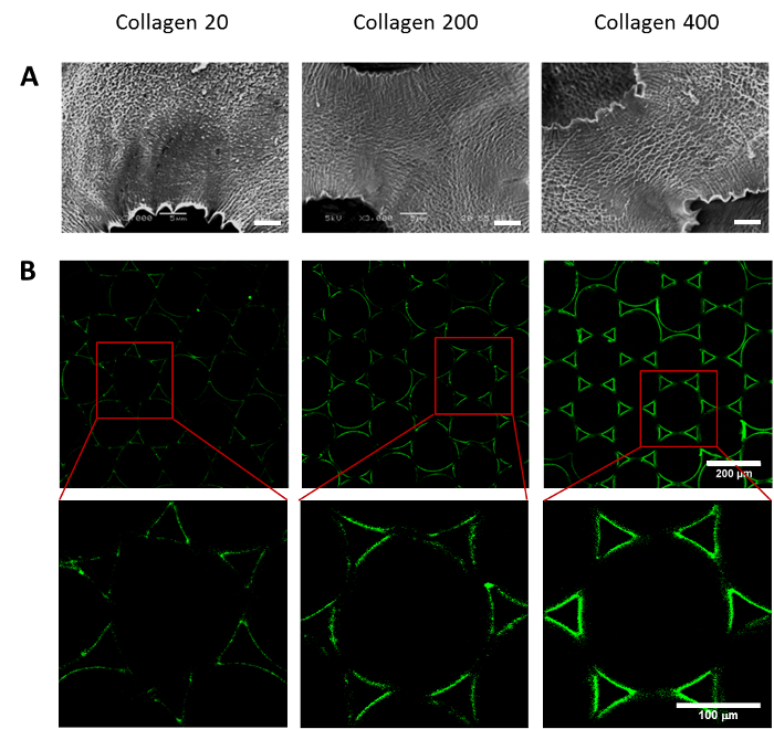
Figure 4. Influence of collagen coating on ICC scaffold topology. (A) SEM images and (B) confocal images were taken to qualitatively assess the surface topology of scaffolds coated with 20 µg/ml, 200 µg/ml, and 400 µg/ml of collagen. Red boxes surround the cavity shown in higher magnification images below. Scale bars are (A) 5 µm, (B) 200 µm and 100 µm (higher magnification). This figure has been modified and used with permission from Wiley47. Please click here to view a larger version of this figure.
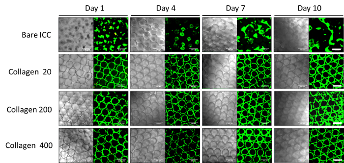
Figure 5. Effect of ICC platform conditions on cell viability and morphology using qualitative Live/Dead assay. Confocal images of the Live/Dead stained Huh-7.5 cells seeded in ICC scaffolds with different collagen concentration coatings (0, 20, 200, and 400 µg/ml) were taken 1, 4, 7, and 10 days after seeding. Green stain (calcein) indicates live cells and red stain (Ethidium homodimer-1) indicates cell death. Scale bars indicate 200 µm. [This figure has been modified and used with permission from Wiley47] Please click here to view a larger version of this figure.
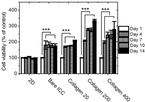
Figure 6. Effect of platform type and ICC platform conditions on cell viability using a quantitative colorimetric cell viability assay. Huh-7.5 cells seeded on a 2D polystyrene culture plate and in ICC scaffolds with different collagen concentration coatings (0, 20, 200, and 400 µg/ml) were subjected to the colorimetric MSR viability assay 1, 4, 7, 10, and 14 days after seeding. Absorbance was measured at 450 nm and the data were normalized to Day 1 absorbance values. (n = 3, mean ± SD; ***: P < 0.001 compared to MSR solution absorbance reading for Day 1 of each group.) [This figure has been modified and used with permission from Wiley47] Please click here to view a larger version of this figure.
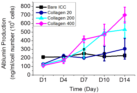
Figure 7. Effect of ICC platform condition on Huh-7.5 function using secreted albumin concentration ELISA analysis. Albumin ELISA was used to quantify the serum albumin secretion in media collected from cell-laden ICC scaffolds with different collagen concentration coatings (0, 20, 200, and 400 µg/ml) on Days 1, 4, 7, 10, and 14 after seeding. Each data point represents an average of 3 samples and is normalized to the number of cells found using the colorimetric MSR cell viability assay and the created standard curve (n = 3, mean ± SD). [This figure has been modified and used with permission from Wiley47] Please click here to view a larger version of this figure.
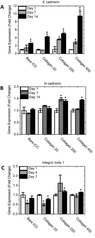
Figure 8. Effect of ICC platform condition on Huh-7.5 function using gene expression analysis. Real-time quantitative PCR was used to profile gene expression of cells cultured in ICC scaffolds with different collagen concentration coatings (0, 20, 200, and 400 µg/ml) on Days 1, 4, and 7 after seeding. Three junction proteins were chosen, namely (A) E-cadherin, (B) N-cadherin, and (C) Integrin Beta 1. The mRNA expression levels were normalized by GAPDH. (n = 3, mean ± SD. *: P < 0.05. **: P < 0.01 compared to gene expression of Day 1 each group.) [This figure has been modified and used with permission from Wiley47] Please click here to view a larger version of this figure.
