Tissue-engineered renal constructs provide a solution for the organ shortage and deleterious effects of dialysis. Here, we describe a protocol to micro dissect murine kidneys for isolation of cortico-medullary segments. These segments are implanted into scaffold-free cellular constructs, forming renal organoids.
Kidney transplantation is now a mainstream therapy for end-stage renal disease. However, with approximately 96,000 people on the waiting list and only one-fourth of these patients achieving transplantation, there is a dire need for alternatives for those with failing organs. In order to decrease the harmful consequences of dialysis along with the overall healthcare costs it incurs, active investigation is ongoing in search of alternative solutions to organ transplantation. Implantable tissue-engineered renal cellular constructs are one such feasible approach to replacing lost renal functionality. Here, described for the first time, is the microdissection of murine kidneys for isolation of living corticomedullary renal segments. These segments are capable of rapid incorporation within scaffold-free endothelial-fibroblast constructs which may enable rapid connection with host vasculature once implanted. Adult mouse kidneys were procured from living donors, followed by stereoscope microdissection to obtain renal segments 200 – 300 µm in diameter. Multiple renal constructs were fabricated using primary renal segments harvested from only one kidney. This method demonstrates a procedure which could salvage functional renal tissue from organs that would otherwise be discarded.
Chronic kidney disease (CKD) is one of the current major public health challenges worldwide1. The prevalence of CKD in the United States is over 14% of the total population, with over 600,000 Americans suffering from the most severe form, end-stage renal disease (ESRD)2. The current treatment options available for those with ESRD include dialysis and kidney transplantation. Although approximately 25,000 patients undergo renal transplantation each year, a significant number of patients are added annually leading to a large disparity between those awaiting a life-saving organ and those receiving transplantation3. In addition to its serious negative effects on longevity and quality of life, dialysis is associated with an astonishing financial burden. In 2014, Medicare paid claims totaled over $30 billion for ESRD patients2. With a limited organ supply and no apparent downtrend in patients requiring dialysis, research efforts aimed at identifying alternative solutions to dialysis and transplantation are ever important. Even a relatively short delay in the need for dialysis increases a patient's number of quality-adjusted life years and productivity substantially while postponing dialysis-related costs4,5,6.
Solutions for functional tissue loss, like that in ESRD, are currently being studied in tissue engineering and regenerative medicine laboratories, with widely varied approaches ranging from scaffold-based organoid fabrication to whole organ engineering using decellularized organ structures for cellular implantation7,8,9,10,11. Recapitulating complex renal structures from marginal or discarded kidneys has only partially been investigated. In fact, nearly 20% of kidneys procured for transplantation are discarded for various reasons12,13. The functional renal tissue from these putative grafts could be utilized and incorporated into one or many tissue-engineered constructs. Prior studies have demonstrated the feasibility of working with these discarded organs, utilizing kidneys for the extra-cellular matrix for tissue engineering purposes14,15. However, few have used primary nephronal tissue from healthy kidneys for tissue-engineering purposes16,17,18.
One method previously described by Kim et al. involves isolation of renal "segments" from healthy rat kidneys, which were then seeded on polyglycolic acid (PGA) scaffolds for construct fabrication16. However, little information is given regarding exact dissection methodology, and segments were obtained from a combination of fine mincing and filtration. We describe a modification of this protocol, which similarly produces discrete renal segments with intact nephronal architecture, but instead relies on microdissection techniques. Nephrectomies are performed on living adult mice, after which the kidneys are transferred to the dissection microscope where the renal capsule is removed, and the tissue is further dissected. Small-bore 30½ G needles are used as cutting instruments and also as guides aiding in dissection, as the needle diameter is equal to the target diameter of the renal segments. The isolated, in this case murine, renal segments maintain viability in culture and incorporate with scaffold-free endothelial-fibroblast cellular constructs19. These constructs have previously been used to engineer other organs, including a bio-artificial pancreas20.
All animal surgical procedures described below were approved by the Institutional Animal Care and Use Committee (IACUC) at the Medical University of South Carolina prior to any animal surgeries or use of any animal tissues.
1. Murine Nephrectomy
2. Murine Kidney Microdissection for Renal Segment Isolation
3. Renal Segment Cellular Construct Fabrication
The protocol described produces approximately 50 renal segments per pyramidal 2 mm3 section of renal tissue. The renal segments that have been processed and imaged have tubular and glomerular components in differing proportions (see Figure 2). The intact segments were subjected to an assay in order to determine the viability of different segments once every 24 h for three days. Green-fluorescent calcein-AM is present with intracellular esterase activity, indicative of living cells. Red-fluorescent ethidium homodimer-1 is seen with loss of the integrity of the plasma membrane. Though these segments are intact, three-dimensional structures, significant assay penetration is appreciable with confocal microscopy (see Figure 3).
Segments were further characterized for size uniformity using the same cell viability assay. Using glass slides with depressions, segments were placed under cover slips without mechanical force placed on any dimension of the segments. Free floating, the segments were imaged 30 minutes after placement in the cell viability/toxicity assay. A z-projection with 10-µm step size was taken through the entire segment. The largest 'X' and 'Y' dimensions were obtained and recorded. The 'Z' dimension was obtained by calculating the distance from the first to the last visible fluorophore. Given that the segments are roughly cubic, a simple cubic volume measurement was obtained and plotted to compare 10 randomly selected segments from one microdissection experiment. Target volume of (300 µm)3 = 0.027 mm3 is shown with a red marker on the histogram (Figure 4A). Representative 'X' and 'Y' measurements are shown in Figure 4B. Although there are outliers, there are many segments close to the target volume. Repeat experimentation has shown consistent results (n = 20). The raw measurements (not shown here) show that in the vast majority of segments, there is at least one dimension measuring 200 – 300 µm. This has important implications for diffusion limitations.
The renal segments are embedded in scaffold-free endothelial-fibroblast constructs and cultured for 3 days. The renal segments incorporate with the cellular constructs to form intact structures by day 3 (see Figure 5A). The constructs maintain their pre-vascular endothelial network, shown by labeling with von Willebrand Factor (Figure 5E). However, it does not appear that the network invades the renal cellular material. Renal epithelial cells are labeled with Cytokeratin-18 (Figure 5B, green). These constructs were incubated with FITC-labeled albumin (Figure 5C, gray) in order to test in vitro renal functionality. While there is residual albumin found away from the segments of embedded renal tissue, there are "hot spots" in the area of the renal tubular epithelial cells, those known to take up albumin that traverses intra-luminally. This is thought to represent albumin reuptake in the renal segment cellular construct.
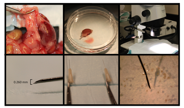
Figure 1: Representative Photographs from Nephrectomy and Renal Segment Microdissection. A murine kidney is removed, rinsed, and placed in a 60-mm dish, and moved to the stereoscope microscope stage. The diameter of the needle tip is used to guide dissection. Hemostats are attached to needle tips for the dissection instruments. Please click here to view a larger version of this figure.
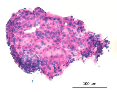
Figure 2: Microdissected Renal Segment. The microdissected renal segments contain varying proportions of glomeruli (lower left corner, basophilic structure) and tubules (remainder of the image) in their native arrangement. 10X image, scale bar as indicated. Please click here to view a larger version of this figure.
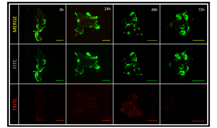
Figure 3: Renal Segment Viability. The microdissected renal segments contain portions of live and dead tissue. Predominantly, the segments are living, and remain so in the culture at 72 h. Note that different segments were used for the different time points, as the viability assay itself is toxic to cells. 10X images, Scale bars = 200 µm. Please click here to view a larger version of this figure.
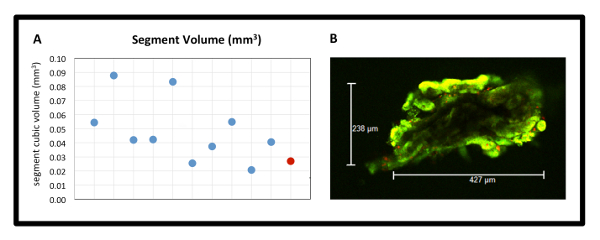
Figure 4: Renal Segment Volume Uniformity. (A) Segment cubic volume in mm3 from one dissection experiment (n = 10). Individual segments are represented by blue points, in comparison to target red point (target volume 0.027 mm3). (B) Representative X and Y dimension measurements from the live-dead assay, 10X image. Please click here to view a larger version of this figure.
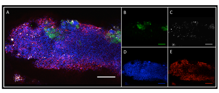
Figure 5: Tissue-Engineered Scaffold-Free Renal Segment Construct. (A) Merged image showing incorporation of renal segments into the SPEC. (B) Cytokeratin-18-positive renal tubular epithelial cells (green). (C) FITC-labeled albumin. (D) Hoescht stain for nuclei. (E) von-Willebrand factor (vWF) positivity highlighting the prevascular network. 10X images, Scale Bar = 200 µm. Please click here to view a larger version of this figure.
Methods used to engineer living renal tissue constructs vary widely with regard to both the type of cells and biomaterials utilized, and in many cases, are outdated or not well-characterized in the literature7. While many are using stem cell approaches or recapitulating individual components of the renal architecture in isolation, the prospect of artificially recreating an entire organ with over 26 different differentiated cell types from cellular suspensions is overwhelming to consider21. This is likely why others have moved to approaches using segments of renal tissue in tissue engineering applications. As described above, previous work has been done in isolation of renal segments for seeding on PGA scaffolds16. However, the methodology used to describe the isolation of segments was described in little detail and the segments were not cultured for any period of time to assess for viability. The method described here, along with the use of the cellular viability assay, shows that at the minimum, portions of the renal segments are maintaining viability to the 72-h mark, which has not been described before. The method also has the advantage of producing segments with fairly predictable dimensions. Additionally, these methods provide a clinically translatable approach to augmenting failing renal function, primarily demonstrating a feasible method for isolation of segments of primary renal tissue from organs that would otherwise be discarded.
In developing the method, the greatest challenges were, first, identifying a sterile object of the appropriate size, and second, maintaining sterility as the procedure involved many steps outside of the cell culture hood. 30½ G needle tips proved to be useful measurement as well as dissection devices. Sterility was a challenge early in experimentation, mostly related to surgical set-up and controlling murine fur.
Limitations of the procedure are related to the three-dimensional nature of the renal tissue segments. In one view under the microscope, the dimensions of the segment may appear to be roughly the size of the needle diameter, 0.260 mm. However, the Z-dimension is not visible on a stereoscope. The microdissection is done by hand, which itself is another limitation. In the future, robotic standardization would be ideal. If the 2 mm3 pyramidal portion of renal tissue was maintained in the correct orientation during dissection, dissecting out intact nephrons would not be unfathomable. Finally, comparing these constructs to the bio-artificial pancreas, in which islet cells were embedded in the SPEC, one must acknowledge key differences20. The kidney is functionally and architecturally unique from the pancreas and arguably more complex. Additionally, islet cells are only one subtype of functional cell within the pancreas, as opposed to the conglomerate of functional cells isolated with renal segments in this method. With architecture and specifically directionality playing such a large role in the function of the kidney and particularly filtration, the lack of uniform orientation of the segments within the construct is an additional weakness of this methodology.
Ongoing research is aimed at assessment of renal segment and renal construct functionality, to clarify the extent to which the albumin uptake is occurring and evaluate other renal functions. Future animal experiments would involve the implantation of the renal constructs in the bladder wall. Embedding the construct in this highly vascular bodily structure would allow rapid anastomosis as well as obviate the need for a collection system, with filtrate excreting directly into the bladder. Considering that in vitro functionality of renal tissues is extremely limited, in vivo testing is vital. The only in vitro tests currently reported in the literature are centered around albumin uptake and erythropoietin expression17,22. These living segments of intact renal tissue in its native architecture embedded in constructs with primitive endothelial networks, which do not rely on any foreign biomaterials, show promise in moving towards functional, biocompatible renal tissue replacement.
The authors have nothing to disclose.
NIH Institutional Postdoctoral Training Grant, NIH-HL-007260
Non-fenestrated Sterile Field | Busse Hospital Disposables | 696 | |
Fenestrated Sterile Field | Busse Hospital Disposables | 697 | |
Halsted Mosquito Forceps 5 Curved | Miltex | Mil-7-4 | "Hemostat" in manuscript |
Extra Fine Graefe Forceps, Curved with teeth | Fine Science Tools | 11155-10 | Fine forceps with teeth |
Extra Fine Graefe Forceps, Serrated (without teeth) | Fine Science Tools | 11152-10 | Fine forceps without teeth |
Fine Scissors – Tungsten Carbide | Fine Science Tools | 14568-09 | Iris Scissors |
Betadine Surgical Scrub with Pump, Povidone-iodine 7.5% | Purdue Products L.P. | 67618-151-17 | |
Sterile Cotton Gauze Pad (4" x 4") | Fisher Healthcare | 22-415-469 | |
Dulbecco's Phosphate Buffered Solution | Corning | 21-030-CV | |
Penicillin/Streptomycin Solution, 100X | Corning | 30-002-Cl | |
Isoflurane, USP | Manufacturer: Piramal, Distributor: McKesson | 2254845 | |
Nair Hair Remover | Nair | 22600-23307 | Hair Removal Cream in text |
200 Proof Ethanol | Decon Laboratories | 2705 | Diluted to 70% Ethanol Solution |
BioLite 60mm Tissue Culture Dish | Themo-Scientific | 130181 | |
Press'n Seal | Glad | 12587-70441 | Applied to Stereoscope |
SZX16 Stereo Microscope | Olympus | SZX16 | |
Fiber Optic Illuminator | Cole Parmer | 41720-20 | |
Self-Supporting Dual-Light Pipe, 23" L Gooseneck | Cole Parmer | EW-41720-60 | |
Scalpel Handle #3 | Miltex | Mil-4-7 | |
Sterile Rib-Back Carbon Steel Blade, Blade Size 15 | Bard-Parker | 371115 | |
31 1/2 Gauge Needle | ThermoFisher Scientific | 14-826F | Becton Dickinson 305106 |
Dulbecco's Modified Eagle's Medium | Corning | 10-017-CV | |
Fetal Select 100% Bovine Serum | Atlas Biologicals | FS-0500-AD | |
Normal Human Dermal Fibroblasts | Lonza | CC-2511 | |
Human Adipose Microvascular Endothelial Cells | Sciencell Research Laboratories | 7200 | |
Surgical Loupes (2.5x) | Orascoptic | (N/A) Custom Order | |
FGM-2 (Fibroblast Basal Medium with FGM-2 SingleQuots Added) | Lonza | CC-3131, CC-4126 | |
EGM-2 (Endothelial Basal Medium with EGM-2 SingleQuots Added) | Lonza | CC-3156, CC-4176 | |
Live/Dead Viability/Cytotoxicity Kit for Mammalian Cells | ThermoFisher Scientific | L3224 | |
Anti-Cytokeratin-18 Antibody | Abcam | ab668 | |
Goat anti-Mouse IgG, Alexa Fluor 633 | ThermoFisher Scientific | A-21052 | |
Goat anti-Rabbit IgG, Alexa Fluor 546 | ThermoFisher Scientific | A-11010 | |
Anti-Von Willebrand Factor Antibody | Abcam | ab6994 | |
Albumin, Fluorescein isothiocyanate Conjugate | Sigma Aldrich | A9771-50MG | |
Hoescht 33342 | BD Pharmingen | 561908 | |
Background Buster | Innovex Biosciences | NB306 |