Summary

Combining Microfluidics and Microrheology to Determine Rheological Properties of Soft Matter during Repeated Phase Transitions

Published: April 19, 2018
doi:

Summary

We demonstrate the fabrication and use of a microfluidic device that enables multiple particle tracking microrheology measurements to study the rheological effects of repeated phase transitions on soft matter.

Abstract

The microstructure of soft matter directly impacts macroscopic rheological properties and can be changed by factors including colloidal rearrangement during previous phase changes and applied shear. To determine the extent of these changes, we have developed a microfluidic device that enables repeated phase transitions induced by exchange of the surrounding fluid and microrheological characterization while limiting shear on the sample. This technique is µ2rheology, the combination of microfluidics and microrheology. The microfluidic device is a two-layer design with symmetric inlet streams entering a sample chamber that traps the gel sample in place during fluid exchange. Suction can be applied far away from the sample chamber to pull fluids into the sample chamber. Material rheological properties are characterized using multiple particle tracking microrheology (MPT). In MPT, fluorescent probe particles are embedded into the material and the Brownian motion of the probes is recorded using video microscopy. The movement of the particles is tracked and the mean-squared displacement (MSD) is calculated. The MSD is related to macroscopic rheological properties, using the Generalized Stokes-Einstein Relation. The phase of the material is identified by comparison to the critical relaxation exponent, determined using time-cure superposition. Measurements of a fibrous colloidal gel illustrate the utility of the technique. This gel has a delicate structure that can be irreversibly changed when shear is applied. µ2rheology data shows that the material repeatedly equilibrates to the same rheological properties after each phase transition, indicating that phase transitions do not play a role in microstructural changes. To determine the role of shear, samples can be sheared prior to injection into our microfluidic device. µ2rheology is a widely applicable technique for the characterization of soft matter enabling the determination of rheological properties of delicate microstructures in a single sample during phase transitions in response to repeated changes in the surrounding environmental conditions.

Introduction

Phase transitions in soft matter can change the scaffold structure, which has implications in the processing and final stability of the material1,2,3. The characterization of soft materials during dynamic phase transitions provides essential information about the relationship between structural evolution and equilibrium structure and rheological properties. For example, many home care products require a phase change during consumer use. Also, during manufacturing, processing steps, including dilution and mixing, can impart shear affecting the rheological properties and final microstructure of the product. Understanding the rheological properties throughout a phase change ensures that the product performs as designed. Additionally, if forces alter the starting rheology of the material during manufacturing, phase transitions can yield unexpected and undesired results, changing the intended function and effectiveness.At the critical gelation point, defined as the point where the material transitions from a solution of associated colloids or polymers to a sample-spanning gel network, material properties change drastically with slight changes to association. Any modification to the structure at the critical gel point can impact the end product4. During these dynamic transitions, soft materials have weak mechanical properties and measurements that use classical experimental techniques can be within the measurement noise limit5,6,7. To account for this, techniques such as microrheology, which is sensitive in the low moduli range (10-3 – 4 Pa), are used to characterize the weak incipient gel during dynamic evolution. Some materials are susceptible to changes in microstructure due to external forces, which presents a challenge during characterization, as any transfer of material or fluid can affect the structure and, ultimately, the final material properties. To avoid altering the material microstructure, we have developed a microfluidic device that can exchange the environmental fluid around a sample while minimizing shear. By exchanging the fluid environment, changes in rheological properties and microstructure are measured during phase transitions with minimal contributions from shear. The device is combined with multiple particle tracking microrheology (MPT) in a technique called µ2rheology. This technique is used to quantify material properties during consecutive phase changes of a gel in response to an external driving force. The technique will be illustrated using a fibrous colloidal gel, hydrogenated castor oil (HCO)9,10,11.

Gel scaffolds can undergo changes in association and dissociation due to their sample environment12,13,14,15. The driving force for gelation and degradation are material specific and must be tailored for each material of interest. µ2rheology can be used to characterize gel systems that respond to external stimuli, including colloidal and polymeric networks. Altering pH, osmotic pressure or salt concentration are examples of driving forces that can induce changes in the material microstructure. For example, HCO undergoes controlled phase transitions by creating an osmotic pressure gradient. When a concentrated HCO gel sample (4 wt% HCO) is submerged in water, the attractive forces between colloidal particles weaken, causing degradation. Alternatively, when a dilute solution of HCO (0.125 wt% HCO) is contacted with a hydrophilic material (referred to as the gelling agent and composed of mostly glycerin and surfactant), the attractive forces return, causing gelation. This gel system will be used to show the operation of the device as a tool for measuring consecutive phase transitions on a single sample9,10. To characterize these gel scaffolds during dynamic transitions and the delicate incipient gel structure at the critical phase transition, we use MPT to characterize these materials with high spatio-temporal resolution.

Microrheology is used to determine gel properties and structure, especially at the critical transition, of an array of soft materials, including colloidal and polymeric gels5,6,9,16. MPT is a passive microrheological technique that uses video microscopy to record the Brownian motion of fluorescent probe particles embedded within a sample. The particle positions throughout the videos are precisely determined to within 1/10th of a pixel using classical tracking algorithms17,18. The ensemble averaged mean-squared displacement (MSD, (Δr2(t))) is calculated from these particle trajectories. The MSD is related to material properties, such as the creep compliance, using the Generalized Stokes-Einstein Relation17,19,20,21,22,23. The state of the material is determined by calculating the logarithmic slope of the MSD curve as a function of lag time, α,

Equation 1

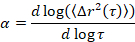
where t is the lag time, and comparing it to the critical relaxation exponent, n. n is determined using time-cure superposition, a well-documented technique that was modified to analyze MPT data by Larsen and Furst6. By comparison of n to α the state of the material is quantitatively determined. When α > n the material is a sol, and when α < n the material is a gel. Previous work has characterized the HCO system using microrheology to determine the critical relaxation exponent9. Using this information, we precisely determine when the material transitions from a gel to a sol during an experiment. Additionally, the non-Gaussian parameter, αNG, can be calculated to determine the extent of structural heterogeneity of a system,

Equation 2

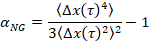
where Δx(t) is the one-dimensional particle movement in the x direction. Using MPT, we can characterize a single phase transition, but by characterizing materials with MPT in a microfluidic device, we are able to manipulate the surrounding fluid environment and collect data of several phase transitions on a single gel sample.

This microfluidic device is designed to investigate the critical transitions of a single gel sample that undergoes phase changes in response to changes in the surrounding fluid environment. The device exchanges fluid surrounding the sample when it is either in the gel or sol state by locking the sample in place to induce a phase transition while minimizing shear. A solvent basin is located directly above the sample chamber, which are connected by six symmetrically spaced inlet channels. This symmetry allows for the exchange of fluid from the solvent basin to the sample chamber while creating equal pressure around the sample, locking it in place. There have been several studies that use this technique for single particle and DNA trapping, but this work scales up the volume from single molecules to samples that are approximately 10 µL24,25,26. This unique design also enables real-time microrheological characterization during phase transitions.

µ2rheology is a robust technique that is applicable to many soft matter systems. The technique described in this paper was designed for colloidal gels, but it can be easily adapted to other materials such as polymer or micellar solutions. With this technique, we determine not just how phase transitions affect the equilibrium material properties, but also how different processing steps can have lasting effects on the rheological evolution of the material and the final scaffold structure and properties.

Protocol

1. Fabrication of the Microfluidic Device Microfluidic stamp fabrication. Note: This step requires the use of volatile materials and should be done in a chemical fume hood. Use a negative printed design with the same dimensions as the glass slide (75×50 mm), the channels colored white, and the background colored black (See Figure 1). Print this design on a clear acetate sheet (transparency) with a resolution of 1200 dpi. If the …

Representative Results

A two-layered microfluidic device is constructed with PDMS (Figure 1a,b), which is patterned on a microfluidic stamp. The design of the stamp is shown in Figure 1c. Improper experimental setup can result both in errors in passive microrheology and microfluidic flows during surrounding fluid exchange (Figure 2). Examples of improper experimental setup are detailed in …

Discussion

The two-layer microfluidic device (Figure 1) can be easily made by following well-documented microfluidic fabrication techniques29. Glass supports are added to the bottom of the device to decrease vibrational effects on probe movement. The glass slide is very thin (0.10 mm) in order to accommodate the working distance of the microscope objective. This makes the device susceptible to small vibrations in the building and sample environment that are then measured with th…

Divulgazioni

The authors have nothing to disclose.

Acknowledgements

Funding for this work was provided by the Procter & Gamble Co. and the American Chemical Society Petroleum Research Fund (54462-DNI7). Acknowledgment is made to the Donors of the American Chemical Society Petroleum Research Fund for partial support of this research. The authors would like to acknowledge Dr. Marco Caggioni for helpful discussions.

Materials

150 x 15 mm Petri Dish Corning, Inc. 351058
75 x 50 x 0.15 mm glass slide Fisher Scientific Custom
75 x 50 x 1.0 mm glass slide Fisher Scientific 12-550-C
75 x 25 x 1.0 mm glass Slide Fisher Scientific 12-550-A3
22 x 22 Glass cover slips Fisher Scientific 12-542-B
Acetone, 99.5% VWR Analytical 67-64-1
Low intensity UV source UVP UVL-56
Chloroform, 99.9% Fisher Chemical C298-500
Cotton Swabs Q-tips 83289205
Ethanol, 90% Fisher Chemical A962-4
Fluoresbrite® YG Carboxylate Microspheres 0.50µm Polysciences, Inc.  15700-10
High-Intensity UV Lamp Spectroline Corp. SB-100P
Hot plate Corning, Inc. PC-420
Hydrochloric Acid, 6N Ricca Chemical Company 3750-32
Methyltriethyoxysilane, 98% Acros Organics 174622500
Microcentrifuge Eppendorf 5424
Plasma cleaner Harrick Plasma, Inc. PDC-32G
Polydimethylsiloxane (PDMS) Robert McKwown Company 2065622
Sonicator Branson, Emerson Electric 1800
Steel connectors, ID 0.023 inch New England Small Tube Corp. Custom
Tetraethoxysilane, 98% Alfa Aesar A14965
Thiol-ene Resin (UV curable) Norland Products, Inc.  NOA81
Transparency Staples Inc.  21828
Tygon tubing, ID 1/32 inch McMaster-Carr E-3603
Vacuum oven Fisher Scientific 282A
Biopsy punch 8 mm World Precision Instruments 504535
Bioposy punch 0.5 mm World Precision Instruments 504528
Syringe, 30 mL BD 309659
Syringe, 3 mL BD 309651
Needle, 18 gauge BD 305195
Microcentrifuge tube, 1.5 mL Eppendorf 22-36-320-4
High-speed Camera Vision Research Miro M120 
Microscope Carl Zeiss AG Zeiss Observer, Z1
Syringe pump New Era Pump Systems NE-300
Hydrogenated castor oil Procter & Gamble N/A
Afício MP 6002 Printer Ricoh Company, Ltd. 415877

Riferimenti

  1. Mitchell, P. Microfluidics-downsizing large-scale biology. Nat. Biotech. 19, 717-721 (2001).
  2. Haber, C. Microfluidics in commercial applications; an industry perspective. Lab Chip. 6, 1118-1121 (2006).
  3. Whitesides, G. M. The origins and the future of microfluidics. Nature. 442, 368-373 (2006).
  4. Huang, X., Raghavan, S. R., Terech, P., Weiss, R. G. Distinct kinetic pathways generate organogel networks with contrasting fractality and thixotropic properties. J. Am. Chem. Soc. 128, 15341-15352 (2006).
  5. Larsen, T. H., Schultz, K. M., Furst, E. M. Hydrogel microrheology near the liquid-solid transition. Korea-Aust. Rheol. J. 20, 165-173 (2008).
  6. Larsen, T. H., Furst, E. M. Microrheology of the liquid-solid transition during gelation. Phys. Rev. Lett. 100, 146001 (2008).
  7. Schultz, K. M., Baldwin, A. D., Kiick, K. L., Furst, E. M. Rapid rheological screening to identify conditions of biomaterial hydrogelation. Soft Matter. 5, 740-742 (2009).
  8. Switzer, L. H., Klingenberg, D. J. Flocculation in simulations of sheared fiber suspensions. Int. J. Multiph. Flow. 30, 67-87 (2004).
  9. Wehrman, M. D., Lindberg, S., Schultz, K. M. Quantifying the dynamic transition of hydrogenated castor oil gels measured via multiple particle tracking microrheology. Soft Matter. 12, 6463-6472 (2016).
  10. Wehrman, M. D., Milstrey, M. J., Lindberg, S., Schultz, K. M. Using µ2rheology to quantify rheological properties during repeated reversible phase transitions of soft matter. Lab Chip. 17, 2085-2094 (2017).
  11. Wehrman, M. D., Lindberg, S. E., Schultz, K. M. Impact of shear on the structure and rheological properties of a hydrogenated castor oil colloidal gel during dynamic phase transitions. J. Rheol. , (2018).
  12. Loh, X. J. Dual-responsive “reversible micelles”. J. Appl. Polym. Sci. 127, 992-1000 (2013).
  13. Kern, F., Zana, R., Candau, S. J. Rheological properties of semidilute and concentrated aqueous solutions of cetyltrimethylammonium chloride in the presence of sodium salicylate and sodium chloride. Langmuir. 7, 1344-1351 (1991).
  14. Trappe, V., Prasad, V., Cipelletti, L., Segre, P. N., Weitz, D. A. Jamming phase diagram for attractive particles. Nature. 411, 772-775 (2001).
  15. Philipse, A. P., Wierenga, A. M. On the density and structure formation in gels and clusters of colloidal rods and fibers. Langmuir. 14, 49-54 (1998).
  16. Schultz, K. M., Bayles, A. V., Baldwin, A. D., Kiick, K. L., Furst, E. M. Rapid, high resolution screening of biomaterial hydrogelators by mu2rheology. Biomacromolecules. 12, 4178-4182 (2011).
  17. Crocker, J. C., Grier, D. G. Methods of digital video microscopy for colloidal studies. J. Colloid Interface Sci. 179, 298-310 (1996).
  18. Mason, T. G. Estimating the viscoelastic moduli of complex fluids using the generalized Stokes–Einstein equation. Rheol. Actac. 39, 371-378 (2000).
  19. Mason, T. G., Ganesan, K., van Zanten, J. H., Wirtz, D., Kuo, S. C. Particle tracking microrheology of complex fluids. Phys. Rev. Lett. 79, 3282-3285 (1997).
  20. Mason, T. G., Weitz, D. A. Optical measurements of frequency-dependent linear viscoelastic moduli of complex fluids. Phys. Rev. Lett. 74, 1250-1253 (1995).
  21. Squires, T. M., Mason, T. G. Fluid mechanics of microrheology. Annu. Rev. Fluid Mech. 42, 413-438 (2010).
  22. Gittes, F., Schnurr, B., Olmsted, P. D., MacKintosh, F. C., Schmidt, C. F. Microscopic viscoelasticity: shear moduli of soft materials determined from thermal fluctuations. Phys. Rev. Lett. 79, 3286-3289 (1997).
  23. Mai, D. J., Brockman, C., Schroeder, C. M. Microfluidic systems for single DNA dynamics. Soft Matter. 8 (41), 10560-10572 (2012).
  24. Tanyeri, M., Ranka, M., Sittipolkul, N., Schroeder, C. M. A microfluidic-based hydrodynamic trap: design and implementation. Lab Chip. 11, 1786-1794 (2011).
  25. Lee, J. S., Dylla-Spears, R., Teclemariam, N. P., Muller, S. J. Microfluidic four-roll mill for all flow types. Appl. Phys. Lett. 90, 074103 (2007).
  26. Crocker, J. C., Grier, D. G. Methods of digital video microscopy for colloidal studies. J. Colloid Interface Sci. 179 (1), 298-310 (1996).
  27. Mason, T. G., Weitz, D. Optical measurements of frequency-dependent linear viscoelastic moduli of complex fluids. Phys. Rev. Lett. 74 (7), 1250 (1995).
  28. Schultz, K. M., Furst, E. M. High-throughput rheology in a microfluidic device. Lab on a chip. 11, 3802-3809 (2011).
  29. Abate, A. R., Lee, D., Do, T., Holtze, C., Weitz, D. A. Glass coating for PDMS microfluidic channels by sol-gel methods. Lab Chip. 8, 516-518 (2008).
  30. Happel, J., Brenner, H. . Low Reynolds Number Hydrodynamics: with special applications to particulate media. , (1965).
check_url/it/57429?article_type=t

Play Video

Citazione di questo articolo
Wehrman, M. D., Milstrey, M. J., Lindberg, S., Schultz, K. M. Combining Microfluidics and Microrheology to Determine Rheological Properties of Soft Matter during Repeated Phase Transitions. J. Vis. Exp. (134), e57429, doi:10.3791/57429 (2018).

View Video