Presented here are protocols for the creation of peptide-based small unilamellar vesicles capable of growth. To facilitate in vesiculo production of the membrane peptide, these vesicles are equipped with a transcription-translation system and the peptide-encoding plasmid.
Compartmentalization of biochemical reactions is a central aspect of synthetic cells. For this purpose, peptide-based reaction compartments serve as an attractive alternative to liposomes or fatty acid-based vesicles. Externally or within the vesicles, peptides can be easily expressed and simplify the synthesis of membrane precursors. Provided here is a protocol for the creation of vesicles with diameters of ~200 nm based on the amphiphilic elastin-like polypeptides (ELP) utilizing dehydration-rehydration from glass beads. Also presented are protocols for bacterial ELP expression and purification via inverse temperature cycling, as well as their covalent functionalization with fluorescent dyes. Furthermore, this report describes a protocol to enable the transcription of RNA aptamer dBroccoli inside ELP vesicles as a less complex example for a biochemical reaction. Finally, a protocol is provided, which allows in vesiculo expression of fluorescent proteins and the membrane peptide, whereas synthesis of the latter results in vesicle growth.
The creation of synthetic living cellular systems is usually approached from two different directions. In the top-down method, the genome of a bacterium is reduced to its essential components, ultimately leading to a minimal cell. In the bottom-up approach, artificial cells are assembled de novo from molecular components or cellular subsystems, which need to be functionally integrated into a consistent cell-like system.
In the de novo approach, compartmentalization of the necessary biochemical components is usually achieved using membranes made from phospholipids or fatty acids1,2,3,4. This is because "modern" cell membranes mainly consist of phospholipids, while fatty acids are regarded plausible candidates of prebiotic membrane enclosures5,6. For the formation of new membranes or to facilitate membrane growth, amphiphilic building blocks must be provided from the exterior7 or ideally through production within a membranous compartment using the corresponding anabolic processes4,8.
While lipid synthesis is a relatively complex metabolic process, peptides can be produced quite readily using cell-free gene expression reactions9,10. Hence, peptide membranes formed by amphiphilic peptides represent an interesting alternative to lipid membranes as enclosures for artificial cell mimics that are able to grow11.
Amphiphilic elastin-like di-block copolymers (ELPs) are an attractive class of peptides, which can serve as the building block for such membranes12. The basic amino acid sequence motif of ELPs is (GaGVP)n, where “a” can be any amino acid except for proline and “n” is the number of motif repeats13,14,15,16,17. ELPs have been created with a hydrophobic block containing mainly phenylalanine for a and a hydrophilic block mainly composed of glutamic acid11. Depending on a and solution parameters, such as pH and salt concentration, ELPs exhibit a so-called inverse temperature transition at temperature Tt, where the peptides undergo a fully reversible phase transition from a hydrophilic to hydrophobic state. The synthesis of the peptides can be easily implemented inside vesicles using the “TX-TL” bacterial cell extract11,18,19,20,21, which provides all necessary components for coupled transcription and translation reactions.
The TX-TL system was encapsulated together, with the DNA template encoding the ELPs into ELP vesicles utilizing dehydration-rehydration from glass beads as a solid support. The formation of vesicles occurs through rehydration of the dried peptides from the bead surface11. Other methods22 for vesicle formation can be used, which potentially show lower polydispersity and larger vesicle sizes (e.g., electro-formation, emulsion phase transfer, or microfluidics-based methods). To test the viability of the encapsulation method, transcription of the fluorogenic aptamer dBroccoli23 can alternatively be used11, which is less complex than gene expression with the TX-TL system.
Due to the expression of the membrane building blocks in vesiculo and their subsequent incorporation into the membrane, the vesicles start to grow11. Membrane incorporation of the ELPs can be demonstrated through a FRET assay. To this end, the ELPs used for formation of the initial vesicle population are be conjugated with fluorescent dyes in equal shares constituting a FRET pair. Upon expression of non-labeled ELPs in vesiculo and their incorporation into the membrane, the labeled ELPs in the membrane are diluted and consequently the FRET signal decreases11. As a versatile and common method for conjugation, copper catalyzed azide-alkyne cycloaddition is used. With the use of a stabilizing ligand such as tris(benzyltriazolylmethyl)-amine, the reaction can be carried out in an aqueous solution at a physiological pH without the hydrolysis of reactants11, which is appropriate for conjugation reactions involving peptides.
The following protocol presents a detailed description of the preparation for growing ELP-based peptidosomes. The expression of the peptides and vesicle formation using the glass beads method are described. Furthermore, it is described how to implement transcription of the fluorogenic dBroccoli aptamer and the transcription-translation reaction for protein expression inside the ELP vesicles. Finally, provided is a procedure for the conjugation of ELPs with fluorophores, which can be used to prove vesicle growth through a FRET assay11.
1. Expression of Elastin-like Polypeptides
2. Vesicle Production Using the Glass Beads Method
3. Transcription of RNA Aptamer dBroccoli Inside the Vesicles
4. Transcription-translation (TX-TL) Reaction
NOTE: For the transcription-translation reaction, a crude cell extract is required as well as reaction buffer and DNA. The crude cell extract is prepared as described in Sun et al.18. For a TX-TL reaction, use the following: 33% (v/v) of the crude E. coli extract, 42% (v/v) reaction buffer, and 25% (v/v) phenol-chloroform purified DNA plus additives. The final concentrations are approximately 9 mg/mL protein, 50 mM HEPES (pH = 8), 1.5 mM ATP, 1.5 mM GTP, 0.9 mM CTP, 0.9 mM UTP, 0.2 mg/mL tRNA, 26 mM coenzyme A, 0.33 mM NAD, 0.75 mM cAMP, 68 mM folinic acid, 1 mM spermidine, 30 mM PEP, 1 mM DTT, 2% PEG-8000, 13.3 mM maltose, 1 U of T7 RNA polymerase, and 50 nM plasmid DNA in ultrapure water.
5. Conjugation of Elastin-like Polypeptides with Fluorophores via Copper Catalyzed Azide-alkyne Huisgen Cycloaddition
Vesicle production
Figure 1 shows transmission electron microscopy (TEM) images of vesicles prepared with different swelling solutions and the glass beads method (also see Vogele et al.11). For the sample in Figure 1A, only PBS was used as swelling solution to prove the formation of vesicles and to determine their size. When TX-TL was used as swelling solution (Figure 1B), the vesicles also formed. Dynamic light scattering (DLS) measurements were performed to show that the glass beads method has an effect on vesicle formation. Figure 2 depicts the measured intensity autocorrelation curves of an EF sample prepared without glass beads in PBS (blue) and an EF sample prepared with the glass beads method with PBS as the swelling solution (red). The sizes were calculated from a cumulant fit25 and resulted in a diameter of 134 nm with a polydispersity of 25% when the vesicles were prepared without the glass beads method. When the glass beads were used, the cumulant fit resulted in a diameter of 168 nm with a polydispersity of 21%.
In vesiculo transcription11
Figure 3 shows the fluorescence intensity profile over time for the transcription of the dBroccoli aptamer inside the EF vesicles (green). As a negative control, DNase I was added to the swelling solution and thus also incorporated during vesicle formation (blue). The measurements were performed in fluorescence plate reader with excitation at 480 nm and emission at 520 nm.
In vesiculo protein expression11
Figure 4 shows the fluorescence intensity of two fluorescent proteins which were expressed inside the ELP vesicles using a fluorescence plate reader. Excitation was carried out 500 nm and emission at 520 nm. Transcription-translation of mVenus (Figure 4A) was performed to investigate expression dynamics and expression level of proteins in the ELP vesicle. It is important to note that after vesicle formation, the contents of the vesicles and outer solution are the same. Hence, to suppress protein expression outside of the vesicle, the antibiotic kanamycin was added to the exterior solution. As a control, kanamycin was also added to the swelling solution, in which case protein expression inside of the vesicle was suppressed. This further indicates that the small molecule kanamycin stays inside the vesicle and suggests that the membrane is not permeable for this molecule over the time scale of these experiments. If kanamycin diffused through the membrane, the mVenus expression would not have been suppressed and at 5 h, the fluorescence would be higher. In the second experiment, the expression of YPet (Figure 4B) was carried out. Both proteins were chosen because they exhibit a faster maturation time than, for instance, GFP. Furthermore, the T7 promoter was used for mVenus transcription and a constitutive promoter used for YPet transcription to show that both inducible and continuous expression are possible.
FRET assay11
Figure 5A shows the results of the FRET assay performed to demonstrate ELP incorporation into the membrane. Therefore, the vesicles were produced using a mixture of two equally concentrated fluorophore-peptide constructs. These were Cy3-EF (donor) and Cy5-EF (acceptor). At time t = 0, the starting FRET level was measured. The signals of the donor and the FRET signal depend on the mean distance between the dyes in the membrane. Upon expression of the membrane ELPs, additional peptides incorporate into the membrane, which increases the average distance between the FRET pairs. The latter was measured through the increase in donor fluorescence and a decrease of the FRET signal at time t = 2.5 h. Figure 5B shows the negative control. Here, kanamycin was added to the swelling solution before vesicle formation. Kanamycin suppresses protein expression; therefore, no change in FRET was visible. It is important to note that this assay only shows EF incorporation.
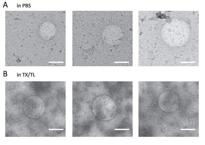
Figure 1: Representative TEM images. (A) EF vesicles formed in the swelling solution PBS. (B) EF vesicles formed in the swelling solution TX-TL. Scale bars = 200 nm. Please click here to view a larger version of this figure.
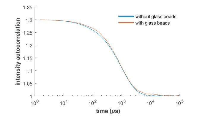
Figure 2: Representative DLS data. Intensity autocorrelation curves measured by DLS. The blue curve depicts EF peptides in PBS prepared without the glass beads method. The red curve depicts an EF solution produced with the glass beads method and PBS as the swelling solution. Please click here to view a larger version of this figure.
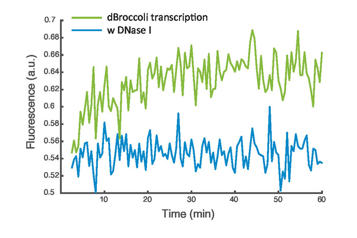
Figure 3: dBroccoli transcription. Representative data of the transcription of the dBroccoli aptamer inside EF vesicles. The green curve depicts the fluorescence signal and the blue curve the control measurement with encapsulated DNase I. Please click here to view a larger version of this figure.
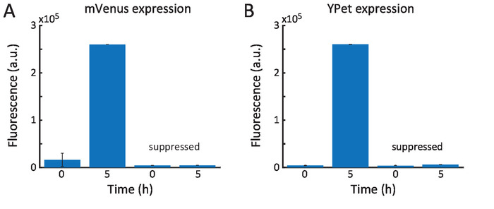
Figure 4: Protein expression inside peptide vesicles. (A) A plasmid encoding mVenus and the TX-TL expression mix were encapsulated in the ELP vesicles. After approximately 5 h, the fluorescence saturated. When the antibiotic kanamycin was encapsulated as well, no fluorescence was observed. (B) Similar results were obtained upon encapsulation of a YPet plasmid. Error bars represent the standard deviation of the measured values at the indicated time during a time frame of 15 min. Please click here to view a larger version of this figure.
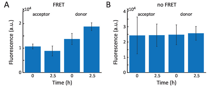
Figure 5: FRET assay. (A) Starting fluorescence levels at time t = 0 for donor emission and FRET signal (acceptor emission). At t = 2.5 h, the donor showed increased emission, and the FRET signal decreased. (B) When only hydrophilic ELP were expressed inside the vesicles, the FRET signal and donor emission stayed constant. Error bars represent the standard deviation of the measured values at the indicated time during a time frame of 15 min. Please click here to view a larger version of this figure.
Supplemental File: Contains all plasmid sequences resp. gene sequences. Please click here to download this file.
Film rehydration is a common procedure for the creation of small unilamellar vesicles. The main source of failure is the wrong handling of the materials used in the procedure.
Initially, the ELPs are produced by E. coli cells. The yield after ELP purification can vary significantly depending on how carefully the protocol is conducted during its crucial steps. These are the inverse temperature cycling (ITC) step and the resuspension of the ELPs after precipitation. For purification, we triggered the hydrophobic collapse of the peptide through the addition of phosphoric acid, which changes the pH to acidic and leads to protonation of the glutamic acid side chains in the hydrophilic block. After this step, both blocks are hydrophobic at RT. It is preferable to use an acid whose salt is already in the solution to keep the ionic strength constant, but other acids such as hydrochloric acid can be used, as well. To increase the yield of ELP production, additional salts can be added to enhance the coacervation process, because salt decreases the transition temperature Tt and makes the ELP more hydrophobic. Usually sodium chloride is used, but kosmotropic salts such as sodium sulphate are far better, whereas the salt concentration can be close to its solution limit. Additional heating using a water bath can increase the yield even further.
Another critical step is the redissolution of the ELP pellet. For maximum ELP solubility, the solution should be pre-cooled, and a quick pH adjustment can help to dissolve the pellet faster. The pH may need to be readjusted during this process several times. For further improvement, the sample can be stored in the fridge at 4 °C and shaking of the sample should be avoided. To exchange salts, the described dialysis step at the end of the purification protocol should be preferred. In principle, ITC can be used to separate the peptides from the salt-containing supernatant. But depending on the previous salt concentration, residual salt can be still found in this pellet. Furthermore, dialysis can be used to concentrate the peptides needed for the experiments without losses as mentioned before.
The next crucial step is the evaporation of the chloroform/methanol mixture. During evaporation at reduced pressures, a delay in boiling can cause splatters or turbulences, which may result in large loss of glass beads. This can be prevented using either filters or tissues. The same problem arises when the desiccator is used, but aluminum foil at the opening of the round bottom flask can be used to prevent this. For the handling of the ELP-coated glass beads, it is recommended to use disposable antistatic micro-spatula, which also reduces losses and simplifies the procedure.
The swelling of the vesicles using various swelling solutions such as PBS and transcription mix is straightforward and less error prone. However, for the TX-TL system, variations from batch-to-batch of up to 10% can occur. To minimize this problem, all experiments should be performed with one batch of cell-free extract only, which can be used for up to 3,000 reactions. After rehydration, the vesicle content and outer solution are the swelling solution. A purification of the outside solution should not be performed, because this may change the osmotic pressure difference between vesicle inside and outside. The result would be an uncontrolled size change or even bursting of the vesicles. Therefore, possible reactions at the outside should be only suppressed. Depending on the reaction, this can be done, for instance, by the addition DNase I to digest present DNA or by adding antibiotics such as kanamycin, which inhibits translation.
Film rehydration from glass beads has several advantages when compared to other preparation methods. In contrast to solvent injection, emulsion phase transfer or microfluidics displacement of solvent is not required, since the initial solvent is completely evaporated, and the peptides are rehydrated in aqueous solution. Therefore, the glass beads method is particularly suitable for sensitive samples such as TX-TL, which can be significantly affected or even destroyed by residual organic solvents used in other methods. Furthermore, the protocol is straightforward, less error prone, and able to be easily scaled up. Because of the large surface to volume ratio only small amounts of glass beads are needed and allow a high degree of parallelization with high throughput.
As its main disadvantage, the glass beads method is only useful for the creation of small unilamellar vesicles, which cannot be observed via fluorescence microscopy. The described method is somewhat limited when microscopic observation of the dynamics of single vesicles is desired, which is sometimes necessary (e.g., when observing potential vesicle division processes). Furthermore, the small size of SUVs limits the encapsulation of low-concentrated components such as the peptide-encoding plasmid, which explains the relatively low expression of the peptides. The underlying encapsulation process is a Poisson process, and thus the concentration of any particular component must be at least 150 nM to guarantee an encapsulation probability of 95%. Therefore, it is quite remarkable that it is possible to observe such a large increase in vesicle size in these experiments.
The protocol presented here will enable researchers to create peptide-based vesicles from elastin-like polypeptides. It also opens up opportunities to produce artificial cell-like structures encapsulated by peptide membranes, which represent attractive alternatives to classical compartments made from fatty acids or phospholipids. Peptides are advantageous in that they can be easily expressed in a cell-free environment (for instance, in the TX-TL system used here), which allows coupling of membrane growth directly to a transcription-translation process inside the vesicle. Furthermore, ELPs can be designed and adjusted to specific physicochemical parameters such as contour length, hydrophobicity/hydrophilicity of the used deblock, and sensitivity to stimuli such as pH or ionic strength.
The authors have nothing to disclose.
We gratefully acknowledge financial support through the DFG TRR 235 (Emergence of Life, project P15), the European Research Council (grant agreement no. 694410 AEDNA), and the TUM International Graduate School for Science and Engineering IGSSE (project no. 9.05). We thank E. Falgenhauer for her help with sample preparation. We thank A. Dupin and M. Schwarz-Schilling for their help with the TX-TL system and useful discussions. We thank N. B. Holland for useful discussions.
2xYT | MP biomedicals | 3012-032 | |
3-PGA | Sigma-Aldrich | P8877 | |
5PRIME Phase Lock GelTM tube | VWR | 733-2478 | |
alkine-conjugated Cy3 | Sigma-Aldrich | 777331 | |
alkine-conjugated Cy5 | Sigma-Aldrich | 777358 | |
ATP | Sigma-Aldrich | A8937 | |
benzamidin | Carl Roth | CN38.2 | |
BL21 Rosetta 2 E. coli strain | Novagen | 71402 | |
Bradford BSA Protein Assay Kit | Bio-rad | 500-0201 | |
cAMP | Sigma-Aldrich | A9501 | |
carbenicillin | Carl Roth | 6344.2 | |
Chloramphenicol | Sigma-Aldrich | C1919 | |
chloramphenicol | Carl Roth | 3886.3 | |
chloroform | Carl Roth | 4432.1 | |
CoA | Sigma-Aldrich | C4282 | |
CTP | USB | 14121 | |
CuSO4 | Carl Roth | P024.1 | |
DFHBI | Lucerna Technologies | 410 | |
DMSO | Carl Roth | A994.1 | |
DNase I | NEB | M0303S | |
DTT | Sigma-Aldrich | D0632 | |
Ethanol | Carl Roth | 9065.2 | |
Folinic acid | Sigma-Aldrich | F7878 | |
Glass beads, acid-washed | Sigma-Aldrich | G1277 | |
GTP | USB | 16800 | |
HEPES | Sigma-Aldrich | H6147 | |
IPTG (β-isopropyl thiogalactoside ) | Sigma-Aldrich | I6758 | |
KCl | Carl Roth | P017.1 | |
K-glutamate | Sigma-Aldrich | G1149 | |
LB Broth | Carl Roth | X968.2 | |
lysozyme | Sigma-Aldrich | L6876 | |
methanol | Carl Roth | 82.2 | |
MgCl2 | Carl Roth | KK36.3 | |
Mg-glutamate | Sigma-Aldrich | 49605 | |
Micro Bio-Spin Chromatography Columns | Bio-Rad | 732-6204 | |
NAD | Sigma-Aldrich | N6522 | |
NHS-azide linker (y-azidobutyric acid oxysuccinimide ester) | Baseclick | BCL-033-5 | |
PEG-8000 | Carl Roth | 263.2 | |
pH stripes | Carl Roth | 549.2 | |
phenylmethylsulfonyl fluoride | Carl Roth | 6367.2 | |
phosphate-buffered saline | VWR | 76180-684 | |
phosphoric acid | Sigma-Aldrich | W290017 | |
polyethyleneimine | Sigma-Aldrich | 408727 | |
Potassium phosphate dibasic solution | Sigma-Aldrich | P8584 | |
Potassium phosphate monobasic solution | Sigma-Aldrich | P8709 | |
Qiagen Miniprep Kit | Qiagen | 27106 | |
RNAPol reaction buffer | NEB | B9012 | |
RNase inhibitor murine | NEB | M0314S | |
RNaseZap Wipes | ThermoFisher | AM9788 | |
rNTP | NEB | N0466S | |
Roti-Phenol/Chloroform/Isoamyl alcohol | Carlroth | A156.1 | |
RTS Amino Acid Sampler | 5 Prime | 2401530 | |
Slide-A-Lyzer Dialysis Cassettes, 10k MWCO (Kit) | Thermo-Scientific | 66382 | |
sodium chloride | Carl Roth | 9265.1 | |
sodium hydroxide | Carl Roth | 8655.1 | |
Spermidine | Sigma-Aldrich | 85558 | |
sterile-filtered (0.22 µm filter) | Carl Roth | XH76.1 | |
T7 polymerase | NEB | M0251S | |
TBTA (tris(benzyltriazolylmethyl)amine) | Sigma-Aldrich | 678937 | |
TCEP (tris(2-carboxyethyl)-phosphine hydrochloride) | Sigma-Aldrich | C4706 | |
Tris base | Fischer | BP1521 | |
tRNA (from E. coli) | Roche Applied Science | MRE600 | |
UTP | USB | 23160 |