Dry powder formulations for inhalation have great potential in treating respiratory diseases. Before entering human studies, it is necessary to evaluate the efficacy of the dry powder formulation in preclinical studies. A simple and noninvasive method of the administration of dry powder in mice through the intratracheal route is presented.
In the development of inhalable dry powder formulations, it is essential to evaluate their biological activities in preclinical animal models. This paper introduces a noninvasive method of intratracheal delivery of dry powder formulation in mice. A dry powder loading device that consists of a 200 µL gel loading pipette tip connected to an 1 mL syringe via a three-way stopcock is presented. A small amount of dry powder (1-2 mg) is loaded into the pipette tip and dispersed by 0.6 mL of air in the syringe. Because pipette tips are disposable and inexpensive, different dry powder formulations can be loaded into different tips in advance. Various formulations can be evaluated in the same animal experiment without device cleaning and dose refilling, thereby saving time and eliminating the risk of cross-contamination from residual powder. The extent of powder dispersion can be inspected by the amount of powder remaining in the pipette tip. A protocol of intubation in mouse with a custom-made light source and a guiding cannula is included. Proper intubation is one of the key factors that influences the intratracheal delivery of dry powder formulation to the deep lung region of the mouse.
The pulmonary route of administration offers various benefits in delivering therapeutics for both local and systemic actions. For the treatment of lung diseases, high local drug concentration can be achieved by pulmonary delivery, thereby reducing the required dose and lowering the incidence of systemic side effects. Moreover, the relatively low enzymatic activities in the lung can reduce premature drug metabolism. The lungs are also efficient for drug absorption for systemic action due to the large and well-perfused surface area, the extremely thin epithelial cell layer and the high blood volume in pulmonary capillaries1.
Inhaled dry powder formulations have been widely investigated for the prevention and treatment of various diseases such as asthma, chronic obstructive pulmonary disease, diabetes mellitus and pulmonary vaccination2,3,4. Drugs in the solid state are generally more stable than in the liquid form, and dry powder inhalers are more portable and user-friendly than nebulizers5,6. In the development of inhaled dry powder formulations, the safety, the pharmacokinetic profile and the therapeutic efficacy need to be evaluated in preclinical animal models following pulmonary administration7. Unlike humans who can inhale dry powder actively, pulmonary delivery of dry powder to small animals is challenging. It is necessary to establish an efficient protocol of delivering dry powder to the lungs of animals.
Mice are widely used as research animal models because they are economical and they breed well. They are also easy to handle and many disease models are well-established. There are two major approaches to administer dry powder to the lung of mouse: inhalation and intratracheal administration. For inhalation, the mouse is placed in a whole-body or nose-only chamber where dry powder is aerosolized and the animals breathe in the aerosol without sedation8,9. Expensive equipment is required and the drug delivery efficiency is low. While the whole-body chamber may be technically less challenging, the nose-only exposure chamber could minimize exposure of drugs to the body surface. Regardless, it is still difficult to precisely control and determine the dose delivered to the lungs. The dry powder is mainly deposited in the nasopharynx region where mucociliary clearance is prominent10. Moreover, mice inside the chamber are under significant stress during the administration process because they are constrained and deprived of food and water supply11. For intratracheal administration, it generally refers to the introduction of the substance directly into the trachea. There are two different techniques to achieve this: tracheotomy and orotracheal intubation. The former requires a surgical procedure that makes an incision in the trachea, which is invasive and seldom used for powder administration. Only the second technique is described here. Compared to the inhalation method, intratracheal administration is the more commonly used method for pulmonary delivery in the mouse because of its high delivery efficiency with minimal drug loss12,13. It is a simple and fast method to precisely deliver a small amount of powder within a few milligrams to the mouse. Although the mouse is anatomically and physiologically distinct to humans and anesthetization is required during the intubation process, intratracheal administration bypasses the upper respiratory tract and offers a more effective way to assess the biological activities of the dry powder formulation such as the pulmonary absorption, bioavailability and therapeutic effects14,15.
To administer dry powder intratracheally, the mouse has to be intubated, which could be challenging. In this paper, the fabrication of a custom-made dry powder insufflator and an intubation device is described. The procedures of intubation and insufflation of dry powder in the lung of the mouse are demonstrated.
The experiments conducted in this study have been approved by the Committee on the Use of Live Animals for Teaching and Research (CULATR), The University of Hong Kong. Dry powder formulations prepared by spray freeze drying (SFD) containing 0.5% of luciferase messenger RNA (mRNA), 5% synthetic peptide PEG12KL4 and 94.5% of mannitol (w/w) are used in this study to demonstrate mRNA expression in the lung16. The mass median aerodynamic diameter (MMAD) of SFD powder is 2.4 μm. Spray dried (SD) mannitol powder are used to investigate the effect of air volume used in powder dispersion16. The MMAD of SD powder is 1.5 μm.
1. Fabrication of dry powder insufflator and loading of dry powder
2. Fabrication of intubation device
3. Intubation
4. Powder administration
When a dry powder insufflator is used to deliver powder aerosol to the lung of an animal, the volume of air used is critical as it affects the safety as well as the powder dispersion efficiency. To optimize the method, different volumes of air (0.3 mL, 0.6 mL and 1.0 mL) were used to disperse the dry powder (1 mg of spray dried mannitol) and the weight of mice was monitored for 48 hours after administration (Figure 6). The use of 0.3 mL and 0.6 mL of air did not cause weight loss of the mice up to 48 h post-administration. Dispersing the powder with 1 mL of air resulted in over 5% of weight loss within 24 h, which was not fully recovered after 48 h. In this protocol, BALB/c mice of 7-9 weeks old were used. Depending on the species, the strain and age of animal, the powder properties (e.g., particle size distribution, cohesiveness and density) and the mass of powder to be administered, the volume of air to be used for efficient powder dispersion and animal tolerance may require optimization by investigators for different animal models.
Dry powder formulation prepared by spray freeze drying (SFD) was delivered to the mice using the method described above. The SFD formulation contained 0.5% of mRNA expressing luciferase protein, 5% of synthetic peptide as delivery vector and 94.5% of mannitol16. BALB/c mice were intratracheally administered with 1 mg of SFD powder containing 5 µg of mRNA and the luciferase expression in the lungs was evaluated at 24 h post-administration using in vivo imaging system (IVIS) (Figure 7). The SFD powder were dispersed in the deep lung and luciferase expression was observed. As a comparison, the SFD powder were reconstituted in PBS (to a final volume of 75 µL) and administered to mice as liquid with the same intubation procedure but a microsprayer was used instead to generate liquid aerosol16. The luciferase expression of the reconstituted formulation was significantly higher than the dry powder formulation, which could be due to the powder dissolution issue or different pharmacokinetic profile between powder and liquid form. The histological characteristics of the lungs treated with mRNA dry powder aerosol were compared with untreated control and lipopolysaccharide (LPS) treated groups (Figure 8). The lungs without any treatment illustrated a healthy presentation while the lung treated with 10 µg of LPS intratracheally showed irregular distribution of air space and inflammatory cell infiltration into the interstitial and alveolar spaces. The lungs treated with SFD powder did not show any signs of inflammation.
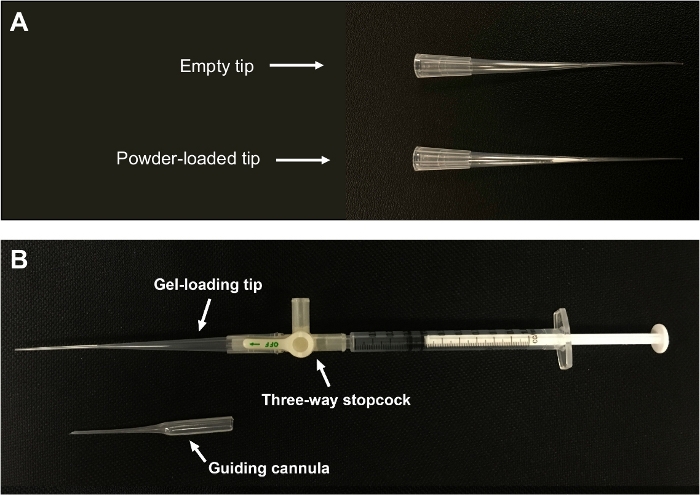
Figure 1: Custom-made dry powder insufflator.
(A) Powder is packed near the narrow end of the tip. (B) A gel-loading pipette tip is connected to a 1 mL syringe via a three-way stopcock. The figure is adapted from Liao et al.21. Please click here to view a larger version of this figure.
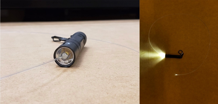
Figure 2: Custom-made light source for intubation.
A flexible optical fiber is connected to a LED torch by creating a small hole on the lens. Please click here to view a larger version of this figure.
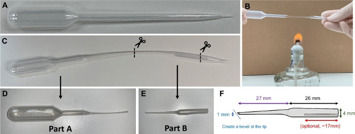
Figure 3: Guiding cannula.
(A) A 1 mL plastic Pasteur pipette is used to make a guiding cannula. (B) The pipette is softened by heating. (C) The heated pipette is stretched and cut. (D) Part A of the pipette is used as fine-tip pipette. (E&F) Part B of the pipette is used as a guiding cannula. A bevel is created to facilitate intubation procedure. A small opening (optional) can be made to increase the flexibility of the cannula. Please click here to view a larger version of this figure.
Figure 4: Intubation platform.
(A) The platform for intubation consists of a Plexiglass plate which is mounted to a stand. (B) An anaesthetized mouse is placed on the platform in a supine position, suspended by hooking its incisors with a nylon floss. Please click here to view a larger version of this figure.
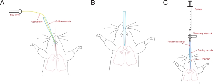
Figure 5: Schematic diagram illustrating the intubation procedure.
(A) The bevel of the guiding cannula is aligned with the midline of the tracheal opening. (B) The guiding cannula is inserted into the trachea and ready for powder administration. (C) The powder-loaded tip (connected to the syringe through a three-way stopcock) is inserted into the guiding cannula which has already been placed in the trachea of the mouse. Please click here to view a larger version of this figure.
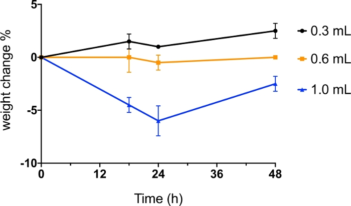
Figure 6: Intratracheal administration of dry powder with different volume of air.
BALB/c mice were administered intratracheally with spray dried (SD) mannitol powder dispersed by 0.3 mL, 0.6 mL and 1.0 mL of air. Body weight of the mice was monitored before administration and at 18 h, 24 h and 48 h post-administration. The data was presented as mean value of percentage of weight change (n=2). Please click here to view a larger version of this figure.
Figure 7: Intratracheal administration of mRNA formulation as dry powder and reconstituted liquid aerosol.
BALB/c mice were administered intratracheally with spray freeze dried (SFD) 0.5% mRNA (luciferase) formulation as powder aerosol (1 mg) using custom-made dry powder insufflator or reconstituted liquid aerosol (1 mg in 75 μL PBS) using microsprayer. Each mouse received a dose of 5 μg of mRNA. PBS (75 µL) was used as control. At 24 h post-administration (A) the lungs were isolated for bioluminescence imaging; (B) luciferase protein expression of the lung tissues were measured. The data was expressed as the mean value of relative light unit (RLU) per mg of protein, analyzed by one-way ANOVA followed by Tukey’s post-hoc test, ***p < 0.001 (n=4). The figure is adapted from Qiu et al.16. Please click here to view a larger version of this figure.
Figure 8: Histology of the lungs of mice following intratracheal administration of mRNA dry powder formulation.
(A) untreated control; mice were intratracheally administered with (B) LPS (10 mg in 25 μL PBS), and (C) spray freeze dried mRNA powder (1 mg). Slides were viewed using an upright microscope at 20x magnification (scale bar = 100 mm). The figure is adapted from Qiu et al.16. Please click here to view a larger version of this figure.
In this paper, custom-made devices for dry powder insufflation and intratracheal intubation are presented. In the powder loading step, dry powder are loaded into a 200 µL gel-loading pipette tip. It is important to gently tap the tip to allow the loose packing of powder at the narrow end of the tip. However, if the powder are packed too tightly, they will get stuck in the tip and cannot be properly dispersed. It is recommended to neutralize the static charges of the powder and the pipette tip in order to facilitate powder loading, particularly for powder with low density and in low relative humidity. The guiding cannula is a critical component of the device. It is used to facilitate the intubation of powder-loaded pipette tip into the trachea of the mouse. The diameter of the guiding cannula should not be too wide; otherwise it will be difficult to insert it into the trachea and may injure the mouse. The diameter of the guiding cannula should be just wide enough to fit the optical fiber and the powder-loaded pipette tip, and the pipette tip should protrude the guiding cannula by approximately 1-2 mm.
The ability to visualize the opening of the trachea is crucial in the intubation process, allowing the guiding cannula to be correctly inserted. The tracheal opening consists of white arytenoid cartilage with regular opening and closing motion at the back of the throat. With the fiber optic illumination, the opening of trachea could be easily visualized. By puffing a tiny volume of air through the fine tip plastic pipette, an inflation at the chest indicates a proper intubation. If inflation at the chest is not observed or resistance is felt during insertion, retract the guiding cannula swiftly and repeat the steps again.
There was a widely used commercially available dry powder insufflator12,17,18 (Table of Materials; this device is now discontinued). The dry powder is loaded into the sample chamber of the device and dispersed by air from a 3 mL plastic air syringe or an air pump. To measure the emitted dose, the device has to be weighed before and after powder administration, which leads to inaccuracy considering the dose of powder is usually very small (relative to the mass of the device). Compared to the commercial insufflator, the biggest advantage of the custom-made device is that the success of powder dispersion could be observed by the absence of powder in the transparent gel-loading pipette tips. Since the pipette tip is light, it can also be weighed accurately before and after administration to measure the emitted dose. The pipette tip is inserted into the guiding cannula rather than being exposed to the trachea of the animal. There is minimal risk of contaminating the tip with the moisture or secretion in the trachea (which may affect the accuracy of emitted dose measurement). As the pipette tips are disposable and inexpensive, different dry powder formulations can be loaded into different tips in advance. Various formulations can be evaluated in the same animal experiment without the need of device cleaning and dose refilling, thereby saving time and eliminating the risk of cross-contamination from residual powder. Moreover, the powder dispersion pattern generated by the commercial insufflator varied among different formulations. A number of studies reported that dry powder dispersed by the commercial insufflator were easily agglomerated and failed to reach the deep lung upon administration19,20. In contrast, other formulations dispersed by devices similar to ours are reported to have a high lung deposition15,21,22.
There are other similar custom-made devices reported in the literature for the administration of powder aerosol to the lung of animal. For instance, Chaurasiya et al. described the use of a cannula tube for intubation as well as powder loading, with a syringe connected to the cannula tube after intubation for powder dispersion23. While their approach uses standardized equipment and material (e.g., otoscope, cannula and syringe) with less customization, the method here offers some distinct advantages. Firstly, it allows confirmation of correct intubation before drug administration. This step is particularly helpful for less experienced user. Secondly, the guiding cannula can act as a protecting shield to prevent any secretion or moisture in the trachea from contaminating the gel-loading pipette tip, allowing a more accurate emitted dose measurement by weighing. Lastly, the more flexible guiding cannula together with the optical fiber may enable easier intubation.
In summary, a custom-made dry powder insufflator which is inexpensive, disposable, reproducible and efficient in dispersing small amount of powder precisely is introduced in this paper. The intubation process mentioned is noninvasive, quick and could deliver powder formulations to the mice safely and accurately. It can also be adopted to administer liquid formulation for pulmonary delivery.
The authors have nothing to disclose.
The authors would like to thank Mr. Ray Lee, Mr. HC Leung and Mr. Wallace So for their kind assistance in making the light source and powder insufflator; and the Faculty Core Facility for the assistance in animal imaging. The work was supported by the Research Grant Council, Hong Kong (17300319).
BALB/c mouse | Female; 7-9 weeks old; Body weight 20-25 g | ||
CleanCap Firefly Luciferase mRNA | TriLink Biotechnology | L-7602 | |
Dry Powder Insufflator | PennCentury | Model DP-4M | |
Ketamine 10% | Alfasan International B.V. | NA | |
Light emitting diode (LED) torch | Unilite Internation | PS-K1 | |
Mannitol (Pearlitol 160C) | Roquette | 450001 | |
Non-filter round gel loading pipette tip (200 µL) | Labcon | 1034-800-000 | |
Nylon floss | Reach | 30017050 | |
One milliliter syringe without needle | Terumo | SS-01T | |
Optical fibre | Fibre Data | OMPF1000 | |
PEG12KL4 peptide | EZ Biolab | (PEG12)-KLLLLKLLLLKLLLLKLLLLK-NH2 | |
Plastic Pasteur fine tip pipette | Alpha Labotatories | LW4061 | |
Three-way stopcock | Braun | D201 | |
Xylazine 2% | Alfasan International B.V. | NA | |
Zerostat 3 anti-static gun | MILTY | 5036694022153 |