This protocol presents an improved method to obtain transient myocardial hypertrophy with absorbable suture, simulating left ventricular hypertrophy decrease after removing pressure overload. It could be valuable for the studies on myocardial hypertrophic preconditioning.
Based on twice transverse aortic constrictions (TACs) in mice, it is proved that myocardial hypertrophic preconditioning (MHP) could attenuate cardiomyocyte hypertrophy and slow down progression to heart failure. For novices, however, the MHP model is usually quite difficult to establish because of the technical obstacles in ventilator operation, opening the chest repeatedly, and bleeding caused by debanding. To facilitate this model, to increase the surgical success rate and to reduce the incidence of bleeding, we switched to absorbable sutures for the first TAC combing with a ventilator-free technique. Using a 2-week absorbable suture, we demonstrated that this procedure could cause significant myocardial hypertrophy in 2 weeks; and 4 weeks after surgery, myocardial hypertrophy was almost completely regressed to the baseline. Using this protocol, the operators could master the MHP model easily with a lower operation mortality.
Ischemic preconditioning is a phenomenon that induces brief non-lethal episodes of ischemia and reperfusion to the heart and has the capacity to dramatically reduce myocardial injury1. Given the obvious clinical implications of ischemic preconditioning, such as limiting myocardial infarct size2 and suppressing ventricular tachyarrhythmias after myocardial revascularization3, there have been lots of research to dissect the mechanisms underlying cardio-protective effects induced by preconditioning4,5. In contrast, other non-ischemic types of preconditioning have received relatively little attention. Cardiac hypertrophy may be blunted in patients with aortic stenosis undergoing aortic valve replacement6. Wherever the state of pathological myocardial hypertrophy exists, the principle of preconditioning is rarely reported.
In 1991, Rockman et al. firstly established a mouse model of left ventricular hypertrophy by transverse aortic constriction (TAC)7. By operating TAC twice in mice, we have previously proved that myocardial hypertrophic preconditioning (MHP) leads to transient hypertrophic stimulation in the heart thereby making the heart more resistant to sustained hypertrophic stress in the future8. The characteristics of the MHP model have been validated by ultrasound biomicroscope and hemodynamic assessment9. Key points in constructing the model was to perform thoracotomy three times, TAC for a week, debanding for a week, and secondary TAC for 6 weeks. However, debanding could cause bleeding, which made it difficult to be mastered by novices and difficult to be popularized. In addition, it is also a technical challenge to intubate mice. Improper intubation could cause tracheal injury, pneumothorax, and even death in mice. So, it is necessary and valuable to improve some procedures while constructing the MHP model.
To reduce the difficulty of the model and increase its success, we switched to absorbable sutures for the first TAC and monitored the model's success by measuring pressure gradient across the aortic constriction under echocardiography10. Based on our preliminary experiment, it would be difficult to induce sufficient myocardial hypertrophy in mice with too low-pressure gradient, while mice with too high-pressure gradient would develop acute heart failure or even die. The ideal pressure gradient for the model ranges from 40–80 mmHg11. In addition, this experiment did not rely on a ventilator, which could effectively avoid ventilator-associated technical manipulation and injury12.
All procedures were carried out following the guidelines of the Care and Use of Laboratory Animals published by the US National Institutes of Health (NIH Publication No. 85-23, revised in 1996). C57BL/6J male mice (8–10 weeks, 20–25 g) were provided by the Animal Center of South Medical University.
1. Preoperative preparation
2. Induction of anesthesia and shaving
3. Surgery
4. Echocardiographic assessment of successful ligation and measurements
In this study, we randomly divided 45 mice into three groups, the sham, the silk suture group, and the absorbable suture group (the number of each group on D0 (baseline), D14, and D28 after TAC was 15, 10, and 5, respectively). On D7, D14, D21, and D28 after the surgery, the constricted peak velocity was determined by echocardiography. We found that the blood flow velocity at the constriction was still greater than 3,000 mm/s in the second week after TAC even though an absorbable suture had been used to constrict the aortic arch (Figure 1A). Moreover, the pressure gradient at the constriction of the absorbable suture group was maintained above 40 mmHg in 2 weeks (Figure 1B). Interestingly, there was no constriction in the fourth week after the surgery, indicating that the absorbable suture had been completely absorbed.
We also found that the left ventricular posterior wall thickness at end-diastole increased and left ventricular internal diameter at end-diastole decreased slightly on D14 after TAC (Figure 2A-C). It is interesting that the left ventricular posterior wall thickness at end-diastole in the absorbable suture group regressed substantially on D28 after TAC, which had no significant difference from the baseline level. In addition, the use of absorbable suture to make the model did not affect the ejection fraction of the left ventricle (Figure 2D).
HW / BW ratios of the silk suture group and the absorbable suture group had increased by 30% compared to the sham group. On D28 after TAC, myocardial hypertrophy regressed and the ratio fell back to the baseline level (Figure 3A) in the absorbable suture group, while the ratio increased by 64% in the silk suture group. The results of H&E staining also supported cardiac hypertrophy (Figure 3B). In conclusion, absorbable suture was suitable to cause transient pathological hypertrophy stimulation, which met the requirements of the myocardial hypertrophic preconditioning model.
Quantitative data has been presented as the mean ± SD. Comparisons among the sham, the silk suture, and the absorbable suture were performed using one-way ANOVA followed by Bonferroni’s post-hoc.
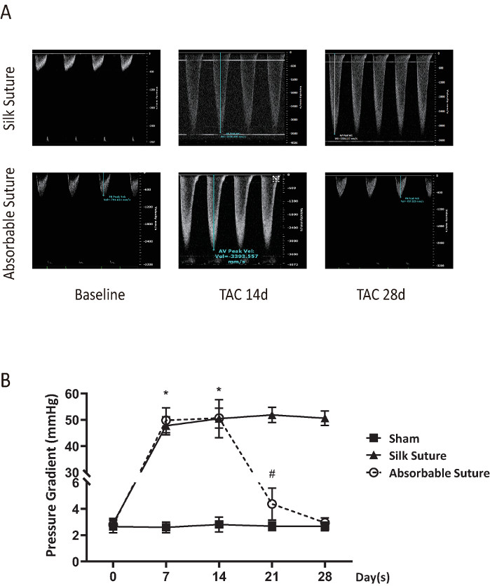
Figure 1: Pulsed-wave Doppler imaging and results from peak velocity and pressure gradient of the constriction. On D0 (baseline), D7, D14, D21, and D28 after TAC using silk suture or absorbable suture, aortic arch or the constriction was measured by a pulsed-wave Doppler. (A) Representative imaging of peak velocity on D0 (baseline), D14, and D28 after surgery in the silk suture group and the absorbable suture group. The blood flow velocity of the absorbable suture group returned to baseline while the velocity of the silk suture group was still greater than 3,000 mm/s on D28 after surgery. (B) The pressure gradient was calculated according to the modified Bernoulli's equation: pressure gradient = 4 x Vmax2 (V= maximum peak velocity). *: p < 0.05 vs sham. #: p < 0.05 vs the silk suture group (the number of each group on D0 (baseline), D14, and D28 after TAC was 15, 10, and 5, respectively). Data presented as mean ± SD. Please click here to view a larger version of this figure.
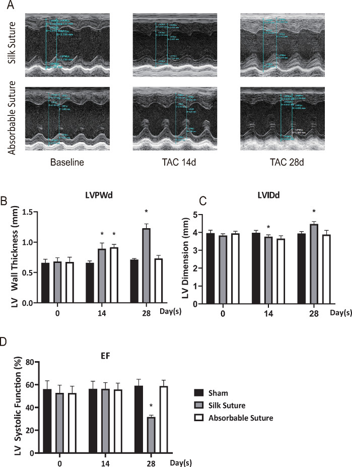
Figure 2: Left ventricular parameters of structure and systolic function. (A) M-mode imaging from the left ventricle (LV) in the silk suture group and the absorbable suture group on D0 (baseline), D14, and D28 after TAC. The representative image of LV wall thickness at end-diastole was about 0.70 mm (D0) to 1.089 mm (D28) in the silk suture group. As for the absorbable suture group, the wall thickness was about 0.658 mm (D28), which returned almost to the baseline. (B) Left ventricular posterior wall thickness at end-diastole (LVPWd). (C) Left ventricular internal diameter at end-diastole (LVIDd). (D) Left ventricular ejection fraction (EF). *: p < 0.05 vs the sham at the same time (the number of each group on D0 (baseline), D14, and D28 after TAC was 15, 10, and 5, respectively). Data presented as mean ± SD. Please click here to view a larger version of this figure.
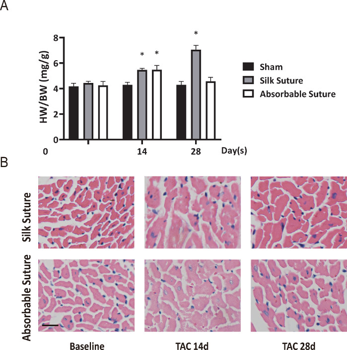
Figure 3: Reversible cardiac hypertrophy in the model of myocardial hypertrophic preconditioning. (A) Heart weight (HW) to body weight (BW) ratio. (B) Histological slices of heart stained with H&E (scale bar = 50 μm). Cardiomyocytes in the silk suture group were significantly enlarged from D14 to D28 while the size of cells mostly regressed on D28 in the absorbable suture group. *: p < 0.05 vs the sham at the same time (the number of each group on D0 (baseline), D14, and D28 after TAC was 15, 10, and 5, respectively). Data presented as mean ± SD. Please click here to view a larger version of this figure.
There is still a vastly underexplored area in cardiac non-ischemic preconditioning. Based on our previous studies, we switched to using absorbable sutures to improve the myocardial hypertrophic preconditioning model.
In previous reports, many investigators used silk suture to constrict the aortic arch8,14,15. Silk suture was easily available and was often used for surgical wound suture, tissue ligation, and tissue fixation. In this protocol, we replaced silk suture with absorbable suture for the first TAC. We found that the blood flow velocity in the constriction still reached 3,000 mm/s or more and lasted for 2 weeks, which was enough to serve as a long-term hypertrophic stimulation. Then, it would return to the baseline on D28. However, it is difficult to control the absorption time of absorbable sutures. The time for absorption is related to the material and batch of absorbable suture, adaptability of tissues, and tightness when constricting. We found earlier that tighter the constriction, less is the absorption time, but at the same time the mortality rate of mice would increase. Therefore, we selected peak velocity at about 3,000 mm/s as the standard, so that the absorption time could be controlled within 2 weeks and the survival rate of mice could also be increased. A more significant point was that the use of absorbable sutures could reduce the chest-opening times to two times, which reduced the difficulty in the surgery and increased the success rate of the model.
A ventilator is usually required for the life support of mice during the surgery. It is reported that aortic arch of mice could be constricted without a ventilator using the suprasternal fossa approach15. However, this required cutting the sternum of mice, which might prolong the surgical recovery time. In this study, we selected this approach: constriction of the first intercostal space close to the sternum, to perform the surgery. This was a minimally invasive approach via lateral thoracotomy14. Surprisingly, we could perform TAC without a ventilator if the incision was as close as possible to the sternum. This approach could also effectively avoid ventilator-associated technical manipulation and injury12. Moreover, entering the mediastinum of mice via the incision, the aortic arch was exposed clearly because there was almost no interference with thymic tissues and other blood vessels in the surgical field.
Although it is convenient not to use a ventilator during the whole operation, the improved method still carries the risk of pneumothorax caused by pleural injury. There are some suggestions, which may be useful to avoid damaging the pleura. Blunt the tip of the needle as much as possible. After entering the mediastinum, keep the tip of the elbow tweezers as vertical as possible during the whole operation. During ligation, place the spacer into the mediastinum along the anterior median line.
In summary, we improved a mouse model of myocardial hypertrophy and regression using absorbable sutures, which could have many potential applications in establishing the model of myocardial hypertrophic preconditioning, exploring the mechanism of hypertrophy regression, and investigating the time of left ventricular reversible remodeling, and so on.
The authors have nothing to disclose.
This work was supported by grants from the National Natural Science Foundation of China (81770271; to Y, Liao), the Joint Funds of the National Natural Science Foundation of China (U1908205; to Y, Liao), and the Municipal Planning Projects of Scientific Technology of Guangzhou (201804020083; to Dr Liao).
Absorbable suture (5-0) | Shandong Kang Lida Medical Products Co., Ltd | 5-0 | Ligation |
Animal ultrasound system VEVO2100 | Visual Sonic | VEVO2100 | Echocardiography |
Cold light illuminator | Olympus | ILD-2 | Light |
Heat pad- thermostatic surgical system (ALC-HTP-S1) | SHANGHAI ALCOTT BIOTECH CO | ALC-HTP-S1 | Heating |
Isoflurane | RWD life science | R510-22 | Inhalant anaesthesia |
Matrx VIP 3000 Isofurane Vaporizer | Midmark Corporation | VIP 3000 | Anesthetization |
Medical nylon suture (5-0) | Ningbo Medical Needle Co. | 5-0 | Close the skin |
Pentobarbital sodium salt | Merck | 25MG | Anesthetization |
Precision electronic balance | Denver Instrument | TB-114 | Weighing sensor |
Self-made spacer | 25-gauge needle | ||
Silk suture (5-0) | Yangzhou Yuankang Medical Devices Co., Ltd. | 5-0 | Ligation |
Small animal microsurgery equipment | Napox | MA-65 | Surgical instruments |
Transmission Gel | Guang Gong pai | 250ML | Echocardiography |
Veet hair removal cream | Reckitt Benchiser | RQ/B 33 Type 2 | Remove hair of mice |
Vertical automatic electrothermal pressure steam sterilizer | Hefei Huatai Medical Equipment Co. | LX-B50L | Auto clean the surgical instruments |