Science Education
>

Measurement of Glutamate Uptake using Radiolabeled L-[3H]-Glutamate in Acute Transverse Slices Obtained from Rodent Resected Hippocampus

PREPARAZIONE ISTRUTTORI
CONCETTI
Student Protocol
JoVE Journal
Biochimica
Author Produced
È necessario avere un abbonamento a JoVE per visualizzare questo.  Accedi o inizia la tua prova gratuita.
JoVE Journal Biochimica
Measurement of Glutamate Uptake using Radiolabeled L-[3H]-Glutamate in Acute Transverse Slices Obtained from Rodent Resected Hippocampus

All procedures were performed in accordance with the NIH Guide for the Care and Use of Laboratory Animals and approved by the local Ethics Committee (project approval # 33732/CEUA-UFRGS). All efforts were made to minimize discomfort and the number of animals used in the experiments.

1. Preparing Hank's Balanced Salt Solution (HBSS)

  1. In a 1 L beaker, add approximately 0.5 L of sterile or double-distilled water and begin stirring vigorously on a magnetic stirrer. Add the following HBSS components (in mM): 137 NaCl, 0.63 Na2HPO4, 4.17 NaHCO3, 5.36 KCl, 0.44 KH2PO4, 0.41 MgSO4, 0.49 MgCl2, 5.55 D-Glucose (see Table 1).
  2. Adjust the pH to 7.4 with NaOH or HCl before adding 1.26 mM CaCl2 to avoid precipitation. Add this salt very slowly and under agitation. Bring it up to a volume of 1 L with sterile or double-distilled water. This buffer will be used to wash the tissue and to maintain the AHS in a viable state during the experiment. It will provide inorganic ions while maintaining a physiological pH and osmotic pressure.
    NOTE: The addition of divalent cations is done after titration of pH to avoid precipitation.
  3. Separate two different fractions of HBSS: one will be kept in a water bath at 37 °C and used during the uptake paradigm, while the other will be kept ice-cold (4 °C) and used for washings before and after the experiment.
    ​NOTE: It is possible to prepare stock solutions of each salt and keep them frozen until use, store it in the freezer (-20 °C) and use it within a few weeks.

2. Preparing Sodium-Free HBSS

  1. Before mixing the components for sodium-free HBSS, prepare in advance two other solutions: Glucamine-HCl (137 mM) and Glucamine-HEPES (4.17 mM). Mix equimolar concentrations of NMDG base and HCl to obtain the Glucamine-HCl solution. Do the same procedure with 2.0 M HEPES free acid to obtain the NMDG-HEPES solution (see Table 2).
  2. In a 0.5 L beaker, add approximately 0.2 L of sterile or double-distilled water and begin stirring vigorously in a magnetic stirrer. Add the following buffer components (in mM): 137 Glucamine-HCl, 4.17 Glucamine-HEPES, 5.36 KCl, 0.44 KH2PO4, 1.26 MgSO4, 0.41 MgCl2, and 5.55 D-Glucose (see Table 2).
  3. Adjust the pH to 7.4 with KOH or HCl and keep stirring. Add 1.26 mM CaCl2 very slowly and under agitation. Bring the solution up to a volume of 0.3 L with sterile or double-distilled water.
    NOTE: This buffer will be used to measure non-sodium-dependent glutamate uptake, which will allow the measurement of passive/non-specific glutamate uptake. After the experiment, this parameter is subtracted from the total glutamate uptake to determine sodium-dependent uptake.
  4. After preparation, this buffer can be stocked in the freezer (-20 °C) and used afterward in subsequent experiments. The same goes for the NMDG solutions.
  5. During the experiment, always keep the sodium-free HBSS ice cold.

3. Organizing the material for hippocampus dissection from rats

  1. Organize the dissection area next to the sink before starting the experiment. Position a specific guillotine for rats (for mice, it will be possible to use sharp scissors) into the sink to keep the bench clean.
  2. Arrange the surgical instruments and other materials for brain removal and hippocampal dissection in the order of use and as close as possible to where they are needed (see Table of Materials). Place the Petri dish in an ice bucket. Also, keep a plastic bag nearby for disposal of the carcass.
  3. Prepare both HBSS solutions at the recommended concentration for use. The sodium-free HBSS can be frozen and thawed before the experiment.
    1. Separate sodium-containing HBSS into two distinct portions, as previously recommended: one heated at 37 °C and another at 4 °C that will be used for dissection. Thaw sodium-free HBSS and keep at 4 °C.
  4. To induce anesthesia, use 3% isoflurane31. Before beginning the experiment, make sure to verify the absence of corneal reflex and nociceptive stimulation of the hind paw.
    ​NOTE: Be careful to choose the adequate anesthetic drug, as it should not interfere with any parameters that will be under investigation. For this protocol, it is recommended the use of inhalant anesthetic (e.g., isoflurane)32.

4. Euthanizing the rat

  1. Euthanize the rodent by using the approved methods by the Institutional Animal Care and Use Committee (IACUC), as well as by the National Institutes of Health Guide for the Care and Use of Laboratory Animals (NIH Publications No. 8023, revised 1978).
  2. Using a guillotine, decapitate the rat immediately prior to the first cervical vertebra.
  3. Place the head on a clean paper towel moistened with cold saline solution. Completely remove the scalp with the surgical scissor size S, as well as the cutaneous muscle aiming to fully exposing the sutures on the superior surface of the skull. Initiate this procedure carefully, starting near the occipital bone and running posteriorly to the nasal bone.
    ​NOTE: In case the experimental model is mice, use sharp scissors to remove the head. Be sure to cut just behind the skull to exclude excess tissue.

5. Removal of the rat brain

  1. Perform brain removal as quickly as possible. With an appropriate bone plier, make a single cut between both eyeholes joining both orifices in the rat's skull. After, place one tip of the surgical scissor size S into the foramen magnum and make a single cut laterally into the skull. Repeat for the other side.
  2. Cut the skull plates from the occipital plate, then along the midline suture of the parietal plates, and finally to the frontal plate towards the nose. Be sure that the rodent's head is held firmly and that the pressure is directed away from the brain to prevent damaging the tissue underneath.
    NOTE: It is critically important that pressure is directed away from the brain to prevent damaging the tissue underneath. In young rats and mice, it is possible to cut the skull with small surgical scissors.
  3. Gently remove the occipital and parietal plates from both hemispheres using a spatula or forceps. Carefully inspect for any dura matter that may be attached to the skull bones and stretched across the surface of the brain.
  4. Remove the brain immediately with the thin double-ended spatula and gently place the whole brain on an ice-cold Petri dish covered with filter paper. With the plastic Pasteur pipette dampen it with ice-cold saline solution. Immediately start to dissect the hippocampi from both brain hemispheres as explained below.
  5. Carefully resect the cerebellum from the whole brain with the #11 scalpel blade. In order to completely separate the cerebral hemispheres run a scalpel blade throughout the intrahemispheric fissure. With the plastic Pasteur pipette, dampen it with ice-cold HBSS buffer frequently to keep the tissue wet and preserved.
  6. Take each hemisphere separately, resect brain stem and midbrain from the overlying cortex with an Iris scissors. Carefully, with the surgical scissor cut the brainstem/midbrain/thalamus, while with a thin brush push down and pull these brain structures. Below it will be possible to observe the hippocampus (medial surface) and the lateral ventricle. The cortex with the hippocampus should now be free from the brainstem.

6. Hippocampi dissection from rats

  1. To resect the hippocampus, put one brain hemisphere in the coronal plane with the parietal cortex facing down. At this moment, it is possible to observe the white fimbria fibers that form a shallow hyperbola at the bottom of the hippocampus.
  2. Very carefully, with the non-dominant hand, anchor the brain tissue with thin tweezers. With your dominant hand, place other tweezers under the caudal tip of the hippocampus.
  3. Anchoring the medial white matter tracts, position the tweezers completely under the hippocampus to separate it from the rest of the brain gently. Apply pressure while moving the second tip slightly anteriorly and laterally. If done properly, the hippocampus should land on the filter paper.
  4. To finish the hippocampus dissection, remove the spare tissue with the same tweezers (usually any remaining cortex, blood vessels, and white matter). Repeat the same methodology for the second hemisphere.
  5. Once both hippocampi are removed, transfer it to a Petri dish with HBSS placed on the ice bucket and transfer to the cutting table area above the blade in the tissue chopper. Be sure to properly identify the dorsal and ventral areas if the investigation requires analyzing them separately.
    NOTE: It is important to always distinguish the ventral and dorsal parts of the hippocampus during dissection. By doing so, the anterior end will be facing upwards33. This part of the hippocampus is slightly thinner than the posterior end.

7. Slices preparation

NOTE: While arranging the surgical material on the bench (session 2), prepare the tissue chopper to obtain the transverse slice from the resected hippocampus.

  1. Set the parameters in the tissue chopper before its use. The Chopper can cut slices from 100 to 400 µm; set it to 300 µm. Additionally, it is recommended a range of 60-80 strokes per minute, as it maintains the tissue integrity during the slicing procedure.
  2. After all the procedures performed in session 2, once the hippocampi were resected from the brain and placed on the filter paper humidified with cold saline solution on top of a smooth polypropylene support, align each hippocampus on the filter paper before submitting them to the chopping procedure.
  3. Place the hippocampus at a time on the circular stainless-steel cutting table from tissue chopper, perpendicular to the cutting blades to obtain the transverse hippocampus slices along the septo-temporal axis. Before starting the chopping process, lightly humidify the blade using isopropyl alcohol. This procedure keeps the tissue from sticking on the blade, allowing a more smooth and precise slicing.
  4. After turning the equipment on, the blade in the chopping arm will be raised and dropped, slicing the tissue in the selected thickness. This is a quick process, and normally the tissue will be properly sliced after a minute. It is possible to chop more than one hippocampus at the same time if they are kept properly separated on the cutting table.
  5. To separate the slices, take each sliced sample from the tissue chopper carefully using the double-ended spatula and plunge it into a Petri dish with ice-cold saline solution. Using two thin brushes, gently pinch the hippocampus and separate the slices. Be careful to maintain slice integrity through this process.
    1. Perform this experiment in triplicates, hence select 6 slices from each sample: 3 slices for the total glutamate uptake and 3 for the unspecific uptake.
  6. With the brush, gently transfer each slice from the Petri dish to a 24-well plate containing: (i) 1 mL of HBSS; and (ii) 1 mL of HBSS sodium-free. Identify the well/plate with the correspondent experimental groups and perform the analysis at least in triplicates.
    NOTE: Due to a time-limited issue, if the experiment is performed in triplicates, it limits the number of animals/groups to only 6 at a time (one hippocampus per animal in 5 min uptake). This means that the L-[3H]-Glutamate will be administered every 15 s until all slices are incubated. To stop the reaction, the same interval should be observed to ensure that all samples are exposed to a 5 min incubation period.

8. Glutamate uptake assay

  1. During the slices stabilization in the respective buffer (HBSS or sodium-free HBSS), prepare the radioactive-containing solution by diluting L-[3H]-Glutamate in sterile water to a final concentration of 4.5 μCi/mL in a final volume that allows 20 μL per slice plus some excess, which will prevent the lack of radioactive input at the end of the batch, since it will be needed to quantify how many disintegrations per minute (DPM) was offered to each slice in the beginning.
    NOTE: For each 24-well plate, it is recommended to prepare 500 μL of 4.5 μCi/mL L-[3H]- Glutamate due to the total incubation time needed for the L-[3H]-Glutamate uptake (5 min) and the time to pipette each 20 µL aliquot (0.33 μCi in each well), as explained below. The stock concentration of L-[3H]-Glutamate is usually 1.0 mCi/mL, thus prepare a 1:200 dilution to apply on the slices.
  2. After 15 min of pre-incubation in HBSS, completely remove the buffer with a Pasteur pipette and replace it by adding to the slices 280 μL of one of the following: (i) regular HBSS (37 °C) for total uptake; or (ii) sodium-free HBSS (4 °C) for unspecific uptake. It is important to notice that total uptake must be performed on a heating plate at 37 °C, while the unspecific uptake is performed on ice.
  3. Prepare one timer and set the beginning of the experiment when adding 20 μL input in the first well containing a slice. Wait for 15 – 20 s (depending on the number of slices in the plate) and add the radioactive input to the next well and so on until the end of the slice-containing wells. Distribute 20 μL of input in all the wells always considering the total incubation time (5 min).
  4. Once each of the wells containing slices in the plate are filled, wait for the first slice to complete 5 min incubation with L-[3H]-Glutamate. Then, immediately remove the incubation media to a proper flask for radioactive waste.
  5. Immediately after the removal, quickly add 300 μL of ice-cold HBSS and gently remove it. Repeat this procedure two more times in each well, completing three washes for each slice. Be sure to use the sodium-free HBSS for washing the slices devoted to the unspecific uptake measurement. After washing all the slices in the plate, add 300 μL 0.5 M NaOH to each well and store the plate protected with parafilm at 4 °C overnight.

9. Counting the radioactive L-[3H]-Glutamate

  1. On the next day, label as many tubes as slices in the experiment and collect the content of the well to each tube. Be sure that the slices are completely solubilized in the lysis solution. If necessary, homogenize the sample using a 26-gauge needle coupled to a 1 mL syringe.
  2. Add 1.2 mL of scintillation liquid to a final volume of approximately 1.5 mL and homogenize all samples in a vortex before reading them in a scintillation counter for 60 s each.
    ​NOTE: Be sure that the detection parameters are set to tritium and the result will be delivered in DPM.

10. Calculations

  1. Convert the results given by the scintillation counter to the radioactivity emitted by the reagent trapped within the slice and represents the amount of glutamate taken up. Deduct the value obtained for the unspecific uptake from the result obtained with the regular HBSS. One μCi is equivalent to 2.22×10⁶ DPM.
  2. By knowing the amount of μCi and the original concentration of [3H]L-Glutamate (information usually provided in the reagent's flask or datasheet), calculate the L-[3H]-Glutamate captured by each slice.

Measurement of Glutamate Uptake using Radiolabeled L-[3H]-Glutamate in Acute Transverse Slices Obtained from Rodent Resected Hippocampus

Learning Objectives

Glutamate uptake is one of the most important mechanisms controlling neurotransmission in the brain. The hippocampus, specifically, is a critical place in glutamate signaling, being an important hub connecting memory, cognition, and emotions in the brain. Following the protocol, adult male Wistar rats were used to generate representative results. Animals were anesthetized using isoflurane 3% until unconscious. After dissecting the brain, hippocampi were removed and placed in the chopper table perpendicularly to the blade. Three hundred µm thick slices were obtained at a rate of 60 per minute and collected to a petri dish containing ice-cold saline solution. Slices were separated each in a well of a 24-well plate and L-[3H]-Glutamate uptake was carried out for 5 min (unless stated otherwise) in sodium-free or regular HBSS, at 4 or 37 °C, respectively.

After an overnight period in lysis solution at 4 °C, samples were mixed with scintillation liquid and read in a scintillation counter for 60 s. The results are given in DPM and the value of the uptake carried out in sodium-free HBSS is deducted from the value obtained with regular HBSS. One DPM corresponds to 0.000000000450 or 4,5 x 10⁻¹⁰ Curie (Ci). By knowing the specific radioactivity of the L-[3H]-Glutamate, is possible to establish a relationship between the result in DPM and the amount of L-[3H]-Glutamate captured by each sample.

First, we carry out the uptake assay using 200 and 300 μm slices to demonstrate that the total protein amount does not influence the uptake parameter. No differences in glutamate uptake in 5 min were observed comparing slices with 200 and 300 μm (data not shown). Accordingly, the researcher has to use slices obtained from the same hippocampal region of all animals to prevent size bias. In summary, this protocol highlights the usual thickness (300 µm) used in different experiments with slices from hippocampus, striatum or cortex10,34,35. Moreover, in order to clarify if there was any difference in L-[3H]-Glutamate uptake in the three main hippocampal regions, dorsal, intermediate and ventral regions were assayed separately. There was no difference in L-[3H]-Glutamate between the three regions (Figure 3A, p = 0.638, one-way ANOVA of repeated measures). This is not surprising since the experiments were performed with naïve animals. However, the results may vary between regions when considering different experimental models or pharmacological challenges.

To make sure that the experiment was not carried out in a saturation range, the uptake was performed for 2.5, 5, 10 or 15 min. The results demonstrate that in a 10 min incubation time the amount of L-[3H]-Glutamate taken up was significantly higher when compared with 5 min (Figure 3B, p = 0.0061, F = 6.864, one-way ANOVA of repeated measures). Conversely, 10 and 15 min uptake results were not statistically different, suggesting a plateau was reached. This data shows that a 5 min incubation is adequate to measure the uptake in most conditions.

To demonstrate that the method is sensitive enough to respond to a blockage in glutamate uptake, 100 µM dihydrokainic acid (DHK), a specific GLT-1 blocker, was administered to the AHS, 5 min prior the uptake. DHK was supplied in stabilization media during 5 min previous to media replacement and [3H]L-Glutamate uptake start36, according to the following timeline: t= 0: slice stabilization in 37 °C HBSS, t= 10 min: addition of DHK to specific slices, t= 15 min: substitution of stabilization media by fresh 37 °C HBSS and beginning of [3H]L-Glutamate uptake. As expected, DHK was able to decrease glutamate uptake (Figure 3C, p = 0.0034, r squared = 0.9069, paired Student's t test) in the protocol conditions, indicating that other pharmacological or genetic manipulations can be used to modulate glutamate uptake.

As a way to determine if the samples are stable enough to withstand a delay in the measurement, some replicates were kept with no scintillation liquid at 4 °C (Figure 3D) or frozen for 7 days (Figure 3E), having the scintillation of their counterparts measured in the next day of the experiment. The data shows that there was no difference between the DPM values of the samples tested 24 h after the experiment and the samples kept refrigerated or frozen for 7 days (p = 0.229 and p = 0.9623, respectively, paired Student's t test).

D-[3H]-Aspartate is also a substrate to glutamate transporters and represents an index of glutamate uptake, since it is metabolized in a lesser extent than L-[3H]-Glutamate. The accumulation of D-[3H]-Aspartate in the slice would provide another important index of transporters activity. Therefore, the uptake of D-[3H]-Aspartate in 300 μm hippocampal slices was estimated for 2.5 and 5 min in the same conditions than L-[3H]-Glutamate. Different time points for the uptake of L-[3H]-Glutamate and D-[3H]-Aspartate under basal conditions did not present significant differences comparing both groups (Figure 3D; p = 0.0632 for radiolabeled substrate, 2-way ANOVA followed by Tukey).

Finally, to demonstrate that unlabeled glutamate in incubation media could hamper the radiolabeled transport activity the uptake buffer was supplemented with different concentrations of unlabeled glutamate (0, 50, 100 and 200 µM). Here, in Figure 3G it is demonstrated that the presence of unlabeled glutamate in incubation media significantly impact the L-[3H]-Glutamate uptake in a concentration-dependent manner in 300 μm hippocampal slices (p = 0.02, repeated measures ANOVA).

Reagent Final concentration (mM)
CaCl2 1.26
D-Glucose 5.55
KCl 5.36
KH2PO4 0.44
MgCl2 0.49
MgSO4 0.41
NaCl 137
NaHCO3 4.17
Na2HPO4 0.63

Table 1. Hank's Balanced Salt Solution composition

Reagent Final concentration (mM)
CaCl2 1.26
D-Glucose 5.55
Glucamine-HCl 137
pH 7.4
(adjust with concentrated HCl)
Glucamine-HEPES 4.17
pH 7.4
(adjust pH with HEPES free acid 2 M)
KCl 5.36
KH2PO4 0.44
MgCl2 0.49
MgSO4 0.41

Table 2. Sodium-Free HBSS composition

Figure 1
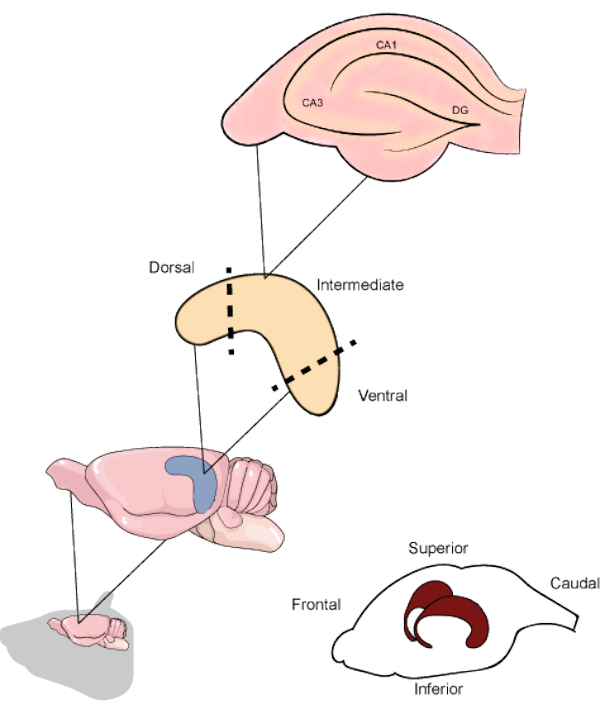
Figure 1. Schematic representation of the rodent brain and hippocampi location beneath the temporoparietal and occipital cortices. When resected, the hippocampus can be divided into the dorsal, intermediate, and ventral parts, the first being oriented in the upper direction of the brain. The hippocampal slices along the transverse axis contain the main regions (CA1, CA3 and DG). CA1: Cornu ammonis 1, CA3: Cornu ammonis 3 and DG: dentate gyrus. Please click here to view a larger version of this figure.

Figure 2
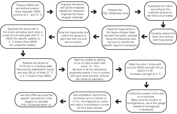
Figure 2: Step-by-step flow of the experiment, from buffer preparation to result calculations. Please click here to view a larger version of this figure.

Figure 3
Figure 3: Representative results of L-[3H]-Glutamate uptake performed in AHS of adult male Wistar rats. A) Different regions take up the same amount of L-[3H]-Glutamate under basal conditions (p = 0.638, one-way ANOVA of repeated measures). B) The contact of L-[3H]-Glutamate with the slices for 10 min increases the amount of L-[3H]-Glutamate captured, compared with 5 min (p = 0.02, F = 6.864, one-way ANOVA). C) Dihydrokainic acid (DHK), a specific GLT-1 blocker, decreases glutamate uptake (p = 0.0034, r squared = 0.9069, paired Student's t test). Sample stability was measured after 7 days. Replicates were kept D) at 4 °C (p = 0.229, paired Student's t test) and E) -20 °C (p = 0.9623, paired Student's t test) and compared to its counterparts measured 24 h after the experiment. F) Different time points for the uptake of L-[3H]-Glutamate and D-[3H]-Aspartate under basal conditions (p = 0.0632 for radiolabeled substrate, 2-way ANOVA followed by Tukey); G) Different concentrations of unlabeled glutamate (0, 50, 100 and 200 µM) supplemented in uptake buffer (p = 0/02, one-way ANOVA of repeated measures). GLT-1: Glutamate transporter type 1. DPM: disintegrations per minute. The data in A and B are expressed as minimum value, median and maximum value. The data in C, D, and E represent paired slices, therefore they are unique values.Please click here to view a larger version of this figure.

List of Materials

#11 scalpel blade Swann-Morton 525
100 mm glass petri dish Common suppliers
110 mm diameter Whatman Filter Sigma Aldrich WHA1001110
42.5 mm diameter Whatman Filter Sigma Aldrich WHA1001042
24-well cell culture plate Falcon 353047
Becker Common suppliers
Blades for the tissue chopper Wilkinson 3241
Bone rongeur Erwin Guth 9,00,005
CaCl2 Sigma Aldrich C4901
D-[2,3-3H]-Aspartic acid PerkinElmer NET581001MC 11.3 Ci/mmol
(37 MBq)
D-Glucose Sigma Aldrich G8270
N-Methyl-D- Glucamine Sigma Aldrich M2004
HEPES Sigma Aldrich H3375
Hidex 300 SL Hidex Oy. Super Low Level #425-020
Iris scissors Erwin Guth 8,00,040
Isoflurane Cristalia (São Paulo, Brazil) 4,10,525 1 mL/mL
KCl Sigma Aldrich P3911
KH2PO4 Sigma Aldrich P0662
L-[3,4-3H]-Glutamic Acid PerkinElmer NET490005MC 49.7 Ci/mmol
(185 MBq)
MgCl2 Sigma Aldrich M8266
MgSO4 Sigma Aldrich M7506
Na2HPO4 Sigma Aldrich S9763
NaCl Sigma Aldrich S9888
NaHCO3 Sigma Aldrich S5761
Plastic Pasteur pipette Common suppliers
Scintillation liquid PerkinElmer 1200.437 for 1 x 5 Liter Optiphase HiSafe 3
Small surgical scissors Erwin Guth 8,00,040
Small tweezers Erwin Guth 6,00,131
Spare chopping discs for the chopper Common suppliers
Standard scissors Erwin Guth 8,00,010
Thin brushes (size 0 or 2) Common suppliers
Thin double-ended spatula Erwin Guth 470.260E
Tissue Chopper Ted Pella, Inc. 10180

Lab Prep

Glutamate removal from the extracellular space by high-affinity Na+-dependent transporters is essential to ensure that the brain's intrinsic connectivity mechanisms work properly and homeostasis is maintained. The hippocampus is a unique brain structure that manages higher cognitive functions, and is the subject of several studies regarding neurologic diseases. The investigation of physiological and pathological mechanisms in rodent models can benefit from acute hippocampal slice (AHS) preparations. AHS has the advantage of providing reliable information on cell function since the cytoarchitecture and synaptic circuits are preserved. Although AHS preparations are commonly used in neurochemistry laboratories, it is possible to find some methodological differences in the literature. Considering that distinctive slice preparation protocols might change the hippocampal regions analyzed, this current protocol proposes a standard technique for obtaining transverse AHS from resected hippocampus. This simple-to-perform protocol may be used in mice and rats' experimental models and allow several ex vivo approaches investigating neurochemical dynamics (in dorsal, intermediate and ventral hippocampus) in different backgrounds (e.g., transgenic manipulations) or after in vivo manipulations (e.g., pharmacological treatments or suitable rodent models to study clinical disorders). After dissecting the hippocampus from the rodent brain, transverse slices along the septo-temporal axis (300 µm thick) were obtained. These AHS contain distinct parts of the hippocampus and were subjected to an individual neurochemical investigation (as an example: neurotransmitter transporters using their respective substrates). As the hippocampus presents a high density of excitatory synapses, and glutamate is the most important neurotransmitter in the brain, the glutamatergic system is an interesting target for in vivo observed phenomena. Thus, the current protocol provides detailed steps to explore glutamate uptake in ex vivo AHS using L-[3H]-Glutamate. Using this protocol to investigate hippocampal function may help to better understand the influence of glutamate metabolism on mechanisms of neuroprotection or neurotoxicity.

Glutamate removal from the extracellular space by high-affinity Na+-dependent transporters is essential to ensure that the brain's intrinsic connectivity mechanisms work properly and homeostasis is maintained. The hippocampus is a unique brain structure that manages higher cognitive functions, and is the subject of several studies regarding neurologic diseases. The investigation of physiological and pathological mechanisms in rodent models can benefit from acute hippocampal slice (AHS) preparations. AHS has the advantage of providing reliable information on cell function since the cytoarchitecture and synaptic circuits are preserved. Although AHS preparations are commonly used in neurochemistry laboratories, it is possible to find some methodological differences in the literature. Considering that distinctive slice preparation protocols might change the hippocampal regions analyzed, this current protocol proposes a standard technique for obtaining transverse AHS from resected hippocampus. This simple-to-perform protocol may be used in mice and rats' experimental models and allow several ex vivo approaches investigating neurochemical dynamics (in dorsal, intermediate and ventral hippocampus) in different backgrounds (e.g., transgenic manipulations) or after in vivo manipulations (e.g., pharmacological treatments or suitable rodent models to study clinical disorders). After dissecting the hippocampus from the rodent brain, transverse slices along the septo-temporal axis (300 µm thick) were obtained. These AHS contain distinct parts of the hippocampus and were subjected to an individual neurochemical investigation (as an example: neurotransmitter transporters using their respective substrates). As the hippocampus presents a high density of excitatory synapses, and glutamate is the most important neurotransmitter in the brain, the glutamatergic system is an interesting target for in vivo observed phenomena. Thus, the current protocol provides detailed steps to explore glutamate uptake in ex vivo AHS using L-[3H]-Glutamate. Using this protocol to investigate hippocampal function may help to better understand the influence of glutamate metabolism on mechanisms of neuroprotection or neurotoxicity.

Procedura

Glutamate removal from the extracellular space by high-affinity Na+-dependent transporters is essential to ensure that the brain's intrinsic connectivity mechanisms work properly and homeostasis is maintained. The hippocampus is a unique brain structure that manages higher cognitive functions, and is the subject of several studies regarding neurologic diseases. The investigation of physiological and pathological mechanisms in rodent models can benefit from acute hippocampal slice (AHS) preparations. AHS has the advantage of providing reliable information on cell function since the cytoarchitecture and synaptic circuits are preserved. Although AHS preparations are commonly used in neurochemistry laboratories, it is possible to find some methodological differences in the literature. Considering that distinctive slice preparation protocols might change the hippocampal regions analyzed, this current protocol proposes a standard technique for obtaining transverse AHS from resected hippocampus. This simple-to-perform protocol may be used in mice and rats' experimental models and allow several ex vivo approaches investigating neurochemical dynamics (in dorsal, intermediate and ventral hippocampus) in different backgrounds (e.g., transgenic manipulations) or after in vivo manipulations (e.g., pharmacological treatments or suitable rodent models to study clinical disorders). After dissecting the hippocampus from the rodent brain, transverse slices along the septo-temporal axis (300 µm thick) were obtained. These AHS contain distinct parts of the hippocampus and were subjected to an individual neurochemical investigation (as an example: neurotransmitter transporters using their respective substrates). As the hippocampus presents a high density of excitatory synapses, and glutamate is the most important neurotransmitter in the brain, the glutamatergic system is an interesting target for in vivo observed phenomena. Thus, the current protocol provides detailed steps to explore glutamate uptake in ex vivo AHS using L-[3H]-Glutamate. Using this protocol to investigate hippocampal function may help to better understand the influence of glutamate metabolism on mechanisms of neuroprotection or neurotoxicity.

Tags