The present protocol describes a unique, clinically relevant model of peripheral arterial disease that combines femoral artery and vein electrocoagulation with the administration of a nitric oxide synthase inhibitor to induce hindlimb gangrene in FVB mice. Intracardiac DiI perfusion is then used for high-resolution, three-dimensional imaging of the footpad vasculature.
Peripheral arterial disease (PAD) is a significant cause of morbidity resulting from chronic exposure to atherosclerotic risk factors. Patients suffering from its most severe form, chronic limb-threatening ischemia (CLTI), face substantial impairments to daily living, including chronic pain, limited walking distance without pain, and nonhealing wounds. Preclinical models have been developed in various animals to study PAD, but mouse hindlimb ischemia remains the most widely used. There can be significant variation in response to ischemic insult in these models depending on the mouse strain used and the site, number, and means of arterial disruption. This protocol describes a unique method combining femoral artery and vein electrocoagulation with the administration of a nitric oxide synthase (NOS) inhibitor to reliably induce footpad gangrene in Friend Virus B (FVB) mice that resembles the tissue loss of CLTI. While traditional means of assessing reperfusion such as laser Doppler perfusion imaging (LDPI) are still recommended, intracardiac perfusion of the lipophilic dye 1,1′-dioctadecyl-3,3,3′,3′-tetramethylindocarbocyanine perchlorate (DiI) is used to label the vasculature. Subsequent whole-mount confocal laser scanning microscopy allows for high-resolution, three-dimensional (3D) reconstruction of footpad vascular networks that complements traditional means of assessing reperfusion in hindlimb ischemia models.
Peripheral arterial disease (PAD), characterized by reduced blood flow to the extremities due to atherosclerosis, affects 6.5 million people in the United States and 200 million people worldwide1. Patients with PAD experience reduced limb function and quality of life, and those with CLTI, the most severe form of PAD, are at increased risk for amputation and death with a 5-year mortality rate nearing 50%2. In clinical practice, patients with ankle-brachial indices (ABI) <0.9 are considered to have PAD, and those with ABI <0.4 associated with either rest pain or tissue loss as having CLTI3. Symptoms vary among patients with similar ABIs depending on daily activity, muscle tolerance to ischemia, anatomic variations, and differences in collateral development4. Digit and limb gangrene is the most severe manifestation of all vascular occlusive diseases that result in CLTI. It is a form of dry necrosis that mummifies the soft tissues. In addition to atherosclerotic PAD, it can also be observed in patients with diabetes, vasculitides such as Buerger's disease and Raynaud's phenomenon, or calciphylaxis in the setting of end-stage renal disease5,6.
Several preclinical models have been developed to study the pathogenesis of PAD/CLTI and test the efficacy of potential treatments, the most common of which remains mouse hindlimb ischemia. Inducing hindlimb ischemia in mice is typically accomplished by the obstruction of blood flow from the iliac or femoral arteries, either by suture ligation, electrocoagulation, or other means of constricting the desired vessel7. These techniques drastically reduce perfusion to the hindlimb and stimulate neovascularization in the thigh and calf muscles. However, there are essential murine strain-dependent differences in sensitivity to ischemic insult partially owing to anatomical differences in collateral distribution8,9. For example, C57BL/6 mice are relatively resistant to hindlimb ischemia, demonstrating reduced limb function but generally no evidence of gangrene in the footpad. On the other hand, BALB/c mice have an inherently poor capacity to recover from ischemia and typically develop auto-amputation of the foot or lower leg following femoral artery ligation alone. This severe response to ischemia narrows the therapeutic window and can preclude longitudinal assessment of limb reperfusion and function. Interestingly, genetic differences in a single quantitative trait locus located on murine chromosome 7 have been implicated in these differential susceptibilities of C57BL/6 and BALB/c mice to tissue necrosis and limb reperfusion10.
Compared to C57BL/6 and BALB/c strains, FVB mice demonstrate an intermediate but inconsistent response to femoral artery ligation alone. Some animals develop footpad gangrene in the form of black ischemic nails or mummified digits, yet others without any overt signs of ischemia11. Concomitant administration of Nω-Nitro-L-arginine methyl ester hydrochloride (L-NAME), a nitric oxide synthase (NOS) inhibitor12, prevents compensatory vasodilatory mechanisms and further increases oxidative stress in hindlimb tissue. In combination with femoral artery ligation or coagulation, this approach consistently produces footpad tissue loss in FVB mice that resembles the atrophic changes of CLTI but rarely progresses to limb auto-amputation11. Oxidative stress is one of the hallmarks of PAD/CLTI and is propagated by endothelial dysfunction and diminished bioavailability of nitric oxide (NO)13,14. NO is a pluripotent molecule that usually exerts beneficial effects on arterial and capillary blood flow, platelet adhesion and aggregation, and leukocyte recruitment and activation13. Reduced levels of NOS have also been shown to activate the angiotensin-converting enzyme, which induces oxidative stress and accelerates the progression of atherosclerosis15.
Once a model of hindlimb ischemia is established, monitoring subsequent limb reperfusion and the therapeutic effect of any potential treatments are also needed. In the proposed murine gangrene model, the degree of tissue loss can first be quantified using the Faber score to assess the gross appearance of the foot (0: normal, 1-5: loss of nails where score represents the number of nails affected, 6-10: atrophy of digits where score represents the number of digits affected, 11-12: partial and complete foot atrophy, respectively)9. Quantitative measurements of hindlimb perfusion are then typically made using LDPI, which relies on Doppler interactions between laser light and red blood cells to indicate pixel-level perfusion in a region of interest (ROI)16. While this technique is quantitative, non-invasive, and ideal for repeated measurements, it does not provide granular anatomical detail of the hindlimb vasculature16. Other imaging modalities, such as micro-computed tomography (micro-CT), magnetic resonance angiography (MRA), and X-ray microangiography, prove either costly, requiring sophisticated instrumentation, or otherwise technically challenging16. In 2008, Li et al. described a technique for labeling blood vessels within the retina with the lipophilic carbocyanine dye DiI17. DiI incorporates into endothelial cells and, by direct diffusion, stains vascular membrane structures such as angiogenic sprouts and pseudopodal processes17,18. Due to its direct delivery into endothelial cells and the highly fluorescent nature of the dye, this procedure provides intense and long-lasting labeling of blood vessels. In 2012, Boden et al. adapted the technique of DiI perfusion to the murine hindlimb ischemia model via whole-mount imaging of harvested thigh adductor muscles following femoral artery ligation19.
The current method provides a relatively inexpensive and technically feasible way for assessing neovascularization in response to hindlimb ischemia and gene or cell-based therapeutics. In a further adaptation, this protocol describes the application of DiI perfusion to image the footpad vasculature in high resolution and 3D in a murine model of hindlimb gangrene.
All animal experiments described in the protocol were approved by the University of Miami Institutional Animal Care and Use Committee (IACUC). FVB mice, both male and female, aged 8-12 weeks, were used for the study.
1. Preparation of L-NAME solution
2. Chemical and surgical induction of hindlimb gangrene
3. Postoperative administration of L-NAME and monitoring of hindlimb gangrene
4. Preparation of DiI and working solutions for animal perfusion
5. Equipment setup and DiI perfusion
6. Preparation of footpad tissue for confocal laser scanning microscopy
7. Confocal laser scanning microscopy
8. Quantitative analysis and 3D reconstruction of footpad vascularity
This protocol details a reliable means of inducing ischemia and tissue loss in the murine footpad using a combination of femoral artery and vein coagulation with L-NAME administration, a nitric oxide synthase inhibitor, in susceptible FVB mice. Figure 1 details the anatomy of the murine hindlimb vasculature and indicates the sites of the femoral artery and vein coagulation (yellow X), just proximal to the lateral circumflex femoral artery (LCFA) and proximal to the saphenopopliteal junction. The LCFA needs to be identified, and the coagulation sites respective to this structure are kept consistent throughout all surgical procedures. As described, 2 h before surgical procedures and on postoperative days 1-3, mice were also administered 40 mg/kg IP of L-NAME to maintain elevated tissue levels of oxidative stress. Figure 2 shows the variation in tissue loss that can be expected from this model one week after surgery, with Faber scores9 recorded in the lower right corner of each image.
DiI perfusion was performed in FVB mice at 5 and 20 days after femoral artery and vein coagulation to assess hindlimb reperfusion following induction of ischemia. Figure 3A illustrates the murine anatomy after dissection to expose the thoracic cavity. A butterfly needle is inserted into the left ventricle to begin cardiac perfusion. Note that the left ventricle appears slightly paler in color than the right ventricle. Figure 3B depicts the equipment set up with stopcocks connected in series and three syringes filled with PBS, DiI solution, and fixative. Following DiI perfusion, feet were harvested, skinned, and compressed between microscope slides as shown in Figure 3C,D before imaging with a confocal laser scanning microscope under 5x magnification. Reconstruction microscopy images revealed normal vascular anatomy in non-ligated control footpad (Figure 4A) compared with severely diminished perfusion to the footpad of ligated hindlimb 5 days after surgery (Figure 4B). Twenty days after surgery, perfusion to the footpad significantly improved (Figure 4C,D, and Figure 5B), although not to the extent of non-ligated control (Figure 4A and Figure 5A). Vascularity was quantified as described above using the Vessel Density plugin in Fiji. The vascular fraction for the control footpad was 28%. Five days after surgery, footpad vascular fraction was severely reduced to 2% but gradually recovered to 15% and 18% in two separate mice by 20 days postoperatively. To visualize the footpad vascular anatomy in 3D, we imported a stitched microscopy image into alternate image analysis and processing software to create a surface rendering as described previously (Supplementary Figure 1). A video of the surface rendering was then created using the animation functionality (Video 1).
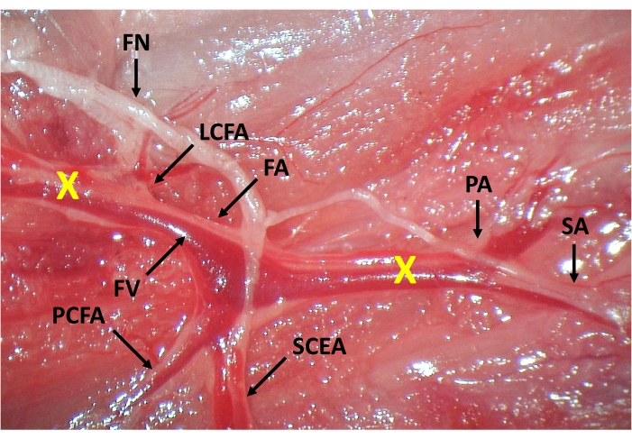
Figure 1: Anatomy of the murine hindlimb vasculature and sites of the femoral artery and vein coagulation. The external iliac artery continues as the femoral artery (FA) distal to the inguinal ligament. The first branches of the femoral artery include the lateral circumflex (LCFA) and deep femoral arteries (not pictured). More distally, the proximal caudal femoral (PCFA) and superficial caudal epigastric arteries (SCEA) branch from the FA proximal to the bifurcation of the saphenous (SA) and popliteal arteries (PA). The femoral nerve (FN) courses alongside the femoral vessels and should be gently isolated before coagulation of the femoral vessels. FA and femoral vein (FV) coagulation sites are also indicated (X). Please click here to view a larger version of this figure.
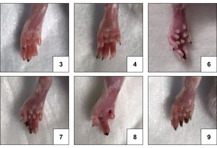
Figure 2: Representative images of hindlimb gangrene in FVB mice with corresponding Faber scores. The degree of ischemic changes induced by this model varies from one or more ischemic nails (Faber scores 1-5) to gangrenous digits (Faber scores 6-10) and partial or complete foot atrophy. Please click here to view a larger version of this figure.
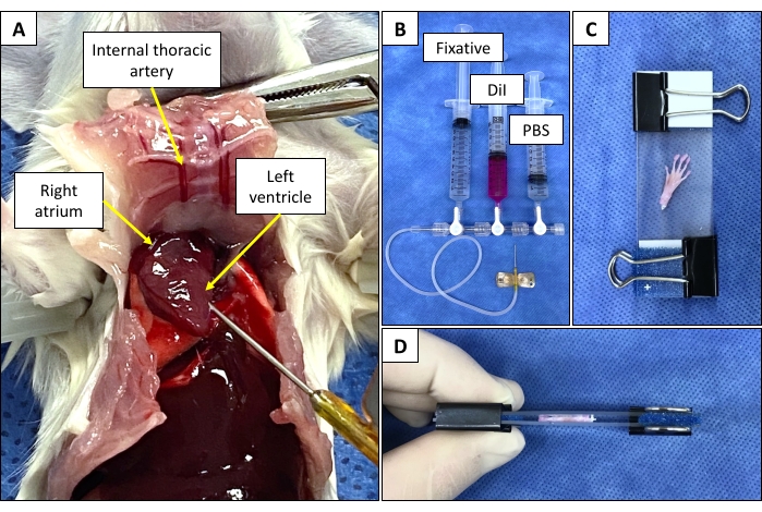
Figure 3: Animal dissection and equipment setup for DiI perfusion and mounting of mouse foot for imaging. (A) Anatomical photograph of the murine anatomy during DiI perfusion. The abdominal and the thoracic cavities are opened, the sternum is reflected, and the ribs are cut on either side of the sternum. A 25 G butterfly needle connected to the stopcock assembly is inserted into the left ventricle. (B) Three 3-way stopcocks are connected in series. Three 10 mL syringes are filled with fixative, DiI, and PBS and connected to the stopcock assembly. A 25 G butterfly needle is connected to the outflow port of the proximal stopcock. (C) Mounting skinned foot between two microscope slides with a folded foam biopsy pad and binder clip at each end to compress the slides together. (D) An alternative view of the skinned foot compressed between microscope slides. Please click here to view a larger version of this figure.
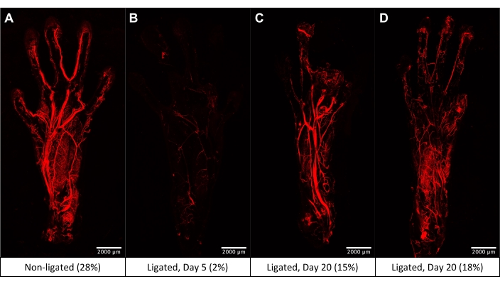
Figure 4: Representative 5x images obtained by confocal laser scanning microscopy of the mouse footpad following DiI perfusion with quantified vessel density expressed as a percent of ROI. (A) Normal footpad vasculature. (B) Footpad vasculature 5 days after femoral artery and vein coagulation shows severely reduced perfusion with minimal vessel opacification. (C) Footpad vasculature 20 days after femoral artery and vein coagulation demonstrates some reconstitution of distal flow to the metatarsal and digital arteries. (D) Image of an additional mouse footpad obtained 20 days after femoral artery and vein coagulation showing minimal large vessel compared to microvascular opacification. Please click here to view a larger version of this figure.
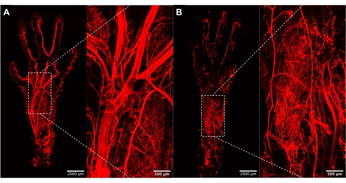
Figure 5: Magnified images of the footpad vasculature. (A) 5x and 20x images of control footpad vasculature demonstrating intact perfusion via the metatarsal and digital arteries. (B) 5x and 20x images of footpad from ligated hindlimb 20 days postoperatively showing reduced perfusion via larger metatarsal arterial branches but the development of an extensive, plush capillary network. Please click here to view a larger version of this figure.
Video 1: Animation of the 3D surface rendering of the footpad vasculature. Video displaying a surface rendering of the footpad vasculature illustrates the 3D resolution achievable with the described protocol. Please click here to download this Video.
Supplementary Figure 1: Steps in the surface rendering of DiI perfusion images. (A) Original DiI perfusion image imported into image analysis and processing software. (B) Surface rendering overlaid onto DiI perfusion image during setting of the threshold intensity. (C) Final 3D surface rendering of DiI perfusion microscopy image. Please click here to download this File.
While mouse hindlimb ischemia is the most widely used preclinical model to study neovascularization in PAD and CLTI, there is significant variation in ischemia severity and recovery depending on the specific mouse strain used and the site, number, and method of arterial disruption. The combination of femoral artery ligation and IP administration of L-NAME can reliably induce hindlimb gangrene in FVB mice11. The same treatment results in hindlimb ischemia without tissue loss in C57BL/6 mice, whereas in BALB/c mice, auto-amputation of the foot or leg can be induced by femoral artery ligation alone. As such, the above-described technique of femoral artery coagulation with concurrent L-NAME administration in FVB mice, which have an intermediate response to ischemia insult, provides a unique and reproducible model of footpad gangrene akin to that seen in the most severe manifestation of diseases that lead to CLTI. The degree of tissue loss observed with this model can vary from a few ischemic nails to multiple gangrenous digits but rarely progresses to auto-amputation of the foot or leg, which allows for longitudinal assessment of limb reperfusion and function. Unlike BALB/c mice, in which the onset of gangrene is rapid with limb auto-amputation typically occurring in less than one week, there is delayed onset of tissue loss in this FVB mouse gangrene model. Femoral artery coagulation acutely restricts blood flow to the hindlimb. Still, accumulation of oxidative stress due to L-NAME administration on postoperative days 0-3 is more gradual, with peak atrophic changes observed between 7-14 days. Therefore, this model offers an improved therapeutic window to evaluate the effects of a particular intervention on gross tissue appearance and rescue of tissue loss in addition to quantifying reperfusion and assessing limb function.
Regarding surgical technique, coagulation or ligation of both the femoral artery and vein is favored due to the relative ease of this operation compared to the isolation of the femoral artery. While this technique can lead to venous thrombosis and insufficiency, it compounds the ischemic insult and helps to induce gangrenous changes more reliably. Additionally, chronic venous insufficiency (CVI) is highly prevalent in the general population, with 10%-30% of adults affected. Consistently, approximately 20% of patients with PAD, especially those with severe arterial insufficiency, also have comorbid CVI21,22. Regardless of whether one decides to ligate or coagulate the femoral vein, it is critically important to maintain the specific site(s) of femoral artery disruption constant across experimental groups. More proximal ligations, such as that of the iliac artery, lead to occlusion of additional downstream collaterals and limit the possibility for arteriogenesis8,16. However, angiogenesis in the distal part of the limb, especially the gastrocnemius muscle, should still be triggered. In FVB mice, double ligation or coagulation of the femoral artery just proximal to the lateral circumflex femoral artery and proximal to the saphenopopliteal bifurcation more consistently induces gangrene than a single ligation or coagulation site.
It should be noted that in PAD and CLTI patients, limb ischemia is caused by atherosclerotic obstruction (a chronic process). In contrast, in mouse models, limb ischemia is induced surgically (an acute process). Although this FVB mouse hindlimb gangrene model has a relatively slower onset of gangrene with delayed peak severity of tissue loss, it is not directly comparable with the chronic, progressive arterial stenosis characteristic of PAD and CLTI. Other groups have developed subacute femoral artery occlusion techniques using ameroid constrictors comprised of an outer metal sleeve and an inner layer of moisture-absorbing material that gradually self-expands. This technique has been shown to result in decreased expression of inflammatory markers, lower blood flow recovery at 4-5 weeks, and reduced muscle necrosis23,24. Other than differences in ischemia acuity, preclinical models using young, healthy animals also fail to replicate risk factors such as diabetes, hypertension, obesity, hyperlipidemia, smoking, and infection that contribute to major adverse limb events and the burden of vascular disease.
In most studies of murine hindlimb ischemia, restoration of blood flow to the ischemic hindlimb is typically assessed via LDPI24,25. This method is non-invasive and repeatable but can be influenced by core body temperature, anesthetic use, hair presence, and positioning of the hindlimbs26. Standardization of these procedures and using the non-ligated hindlimb as an internal control can help mitigate any variations. In contrast to LDPI, micro-CT and MRA provide high-resolution, 3D anatomical information but traditionally require the injection of contrast agent16. X-ray microangiography is also invasive and technically challenging16,27. Like DiI, perfusion with radiopaque silicone casting agents allows for post-mortem 3D reconstruction of the peripheral vasculature28. Intracardiac or tail vein injection of fluorescent lectin has also been described for labeling of tissue vasculature29. Following harvest of tissues of interest, immunohistochemical staining with endothelial-specific markers (e.g., CD31, von Willebrand factor) is often used to quantify capillary density30.
Compared to the techniques mentioned above, DiI perfusion provides several advantages. Firstly, the reagents and materials required are relatively inexpensive, provided access to a confocal laser scanning microscope is available. This method allows for 3D reconstruction of the vasculature, which can be quantified using image analysis software. While this protocol focuses on the footpad vasculature, whole-mount imaging of other murine hindlimb tissues, notably the gastrocnemius and adductor muscles, is also feasible and relevant to angiogenesis and arteriogenesis19 studies. This technique can be modified for larger animal models, including rats and rabbits, by increasing the volume of perfusion solutions. However, imaging constraints regarding tissue size are described below.
Critical portions of DiI perfusion are as follows. Air bubbles in the apparatus may occlude small vessels and hinder the distribution of DiI throughout the vasculature, thereby influencing imaging results. As such, care must be taken to remove any air bubbles in the stopcock apparatus and tubing before perfusion. Filtering all solutions except DiI through a 0.22 µm bottle top filter is also recommended to remove any microparticles. During intracardiac perfusion, carefully monitor the lungs. If they become enlarged and turn pink in color, this is a sign that the butterfly needle has penetrated through to the right ventricle and needs to be retracted slightly.
An important limitation of DiI perfusion is the procedure's terminal nature, which does not allow for repeated measurements. Because poor perfusion results may reflect underlying arterial insufficiency or technical error, harvesting and imaging the non-ligated hindlimb as an internal control is recommended. With regards to imaging, optimal tissue thickness for laser penetration is ~1 mm after compression. Consequently, larger tissues require sectioning into smaller pieces to be mounted on slides and accommodated on the microscope stage and proportionally longer image acquisition times.
In summary, this protocol outlines a unique preclinical model for studying PAD and CLTI. Specifically, femoral artery and vein coagulation with concurrent administration of L-NAME, a NOS inhibitor, reliably induces tissue loss in the footpads of FVB mice. Post-mortem, intracardiac DiI perfusion is then used to label the vasculature fluorescently. Subsequent whole-mount imaging of the harvested feet with confocal laser scanning microscopy allows for high-resolution, 3D reconstruction of the footpad vasculature and visualization of arterial and capillary networks that complements traditional means of assessing reperfusion in hindlimb ischemia models.
The authors have nothing to disclose.
This work was supported by grants to Z-J L and OC V from the National Institutes of Health [R01HL149452 and VITA (NHLBl-CSB-HV-2017-01-JS)]. We also thank the Microscopy and Imaging Facility of the Miami Project to Cure Paralysis at the University of Miami School of Medicine for providing access to their image analysis and processing software.
Binder clips (small) | Office supply store | ||
Buprenorphine (sustained-release) | |||
Butterfly needle (25 G with Luer-Lok) | VWR | 10148-584 | |
Confocal laser scanning microscope | Leica | TCS SP5 | |
DiI (1,1'-Dioctadecyl-3,3,3',3'-tetramethylindocarbocyanine perchlorate) | Invitrogen | D282 | |
Electrocautery device | Gemini Cautery System | 5917 | |
Ethanol (100%) | VWR | 89370-084 | |
Fiji (ImageJ) software | NIH | Used version 2.1.0. Free download, no license required. | |
Foam biopsy pads | Fisher Scientific | 22-038-221 | |
Formalin (neutral buffered, 10%) | VWR | 89370-094 | |
FVB mice | Jackson Laboratory | 001800 | |
Glucose | Sigma-Aldrich | G7528 | Used version 2.1.0. |
HCl (1 M) | Sigma-Aldrich | 13-1700 | |
Imaris software | Oxford Instruments | Used version 9.6.0. | |
Isoflurane | Pivetal | NDC 46066-755-04 | |
KCl | Sigma-Aldrich | P9333 | |
Ketamine | |||
L-NAME (Nω-Nitro-L-arginine methyl ester hydrochloride) | Sigma-Aldrich | N5751 | |
Laser Doppler perfusion imager | MoorLDI | moorLDI2-HIR | Used moorLDI V5 software. |
Microscope slides (25 x 75 x 1 mm) | VWR | 48311-703 | |
Na2HPO4 | Sigma-Aldrich | S7907 | |
NaCl | Sigma-Aldrich | S7653 | |
NaH2PO4 | Sigma-Aldrich | S8282 | |
NaOH | Sigma-Aldrich | S8263 | |
Needles (27 G) | BD | 305109 | |
Povidone-iodine swabstick (10%) | Medline | MDS093901ZZ | |
Surgical instruments | Roboz Surgical | Fine forceps, needle driver, spring scissors, and hemostat are recommended. | |
Suture (5-0 absorbable) | DemeTECH | G275017B0P | |
Syringes (10 mL) | BD | 305482 | |
Three-way stopcocks | Cole-Parmer | 19406-49 | |
Vascular Analysis Plugin | Free download, no license required. See reference: Elfarnawany (2015). | ||
Xylazine |