Summary

살아있는 제브라 피쉬 유충의 눈 제거는 시각 시스템의 신경 의존적 인 성장과 발달을 조사합니다.

Published: February 11, 2022
doi:

Summary

이 기사는 망막 입력이 광학 텍텀의 성장과 발달에 어떻게 영향을 미치는지 조사하기위한 첫 번째 단계로 살아있는 제브라 피쉬 유충에서 눈을 외과 적으로 제거하는 방법을 설명합니다. 또한이 기사는 애벌레 마취, 고정 및 뇌 해부에 대한 정보를 제공하고 면역 조직 화학 및 공초점 이미징을 제공합니다.

Abstract

제브라피쉬는 놀라운 평생 성장과 재생 능력을 발휘합니다. 예를 들어, 배아 발생 중에 확립 된 특수 줄기 세포 틈새 시장은 눈과 뇌 모두에서 전체 시각 시스템의 지속적인 성장을 지원합니다. 망막과 광학 텍텀 사이의 조정 된 성장은 눈과 뇌에 새로운 뉴런이 추가됨에 따라 정확한 망막 토픽 매핑을 보장합니다. 망막 축삭이 생존, 증식 및/또는 분화와 같은 지각 줄기 및 전구 세포 행동을 조절하는 데 중요한 정보를 제공하는지 여부를 해결하기 위해서는 동일한 동물 내의 신경질과 탈핵된 지체 엽을 비교할 수 있어야 합니다.

살아있는 애벌레 제브라 피쉬에서 한쪽 눈을 외과 적으로 제거한 다음 광학 텍텀을 관찰하면이 목표를 달성합니다. 첨부 된 비디오는 애벌레를 마취하고, 전해로 텅스텐 바늘을 날카롭게하고, 한쪽 눈을 제거하는 데 사용하는 방법을 보여줍니다. 다음으로 고정 된 제브라 피쉬 애벌레에서 뇌를 해부하는 방법을 보여줍니다. 마지막으로, 비디오는 면역 조직 화학을위한 프로토콜에 대한 개요와 현미경 검사를 위해 저융점 아가로스에 염색 된 배아를 장착하는 방법에 대한 데모를 제공합니다.

Introduction

이 방법의 목표는 망막 입력이 제브라 피쉬 뇌의 시각 처리 센터 인 광학 텍텀의 성장과 발달에 어떻게 영향을 미치는지 조사하는 것입니다. 한쪽 눈을 제거한 다음 광학 텍텀의 양면을 비교함으로써 동일한 시편 내의 텍탈 변화를 관찰하고 표준화하여 여러 표본에서 비교할 수 있습니다. 이 기술과 결합 된 현대 분자 접근법은 시각적 시스템 성장 및 개발뿐만 아니라 축삭 변성 및 재생의 기초가되는 메커니즘에 대한 통찰력을 얻을 것입니다.

시각, 청각 및 신체 감각 – 감각 시스템은 외부 기관으로부터 정보를 수집하고 그 정보를 중추 신경계로 전달하여 중뇌 1,2에 걸쳐 외부 세계의 “지도”를 생성합니다. 시력은 많은 물고기를 포함한 거의 모든 척추 동물에게 지배적 인 감각 양식입니다. 눈의 신경 조직인 망막은 주로 광수용체, 양극성 세포, 망막의 돌출부 뉴런인 망막 신경절 세포(RGCs)로 구성된 뉴런 회로로 정보를 수집합니다. RGC는 망막의 내부 표면을 가로 질러 시신경 머리로가는 길을 찾는 긴 축삭을 가지고 있으며, 뇌를 통해 매혹되고 함께 여행하며 궁극적으로 등쪽 중뇌의 시각 처리 센터에서 끝납니다. 이 구조는 물고기 및 다른 비 포유류 척추 동물에서 광학 텍텀이라고하며 포유류3에서 우수한 콜리큘러스와 상동성입니다.

광학 텍텀은 등쪽 중뇌의 양측 대칭 다층 구조입니다. 제브라피쉬와 대부분의 다른 물고기에서, 시신경의 각 엽은 대측 눈으로부터 시각 입력만을 수신하여 좌측 시신경이 우측 지골로브에서 종결되고 우측 시신경이 좌측 지골엽(4)에서 종결된다(그림 1). 포유류와 마찬가지로 우수한 콜리큘러스인 광학 텍텀은 오디션과 소마토센세이션을 포함한 다른 감각 입력과 시각 정보를 통합하여 사케이드 1,5,6과 같은 시각적 관심과 안구 움직임의 변화를 제어합니다. 그러나, 포유류의 우수한 콜리큘러스와는 달리, 광학 텍텀은 텍탈 증식 구역7이라고 불리는 텍탈 로브의 내측 및 꼬리 가장자리 근처의 특수 줄기 세포 틈새 시장에서 지속적으로 새로운 뉴런과 글리아교를 생성합니다. 시신경 텍텀 및 중추 신경계의 다른 영역에서 증식성 전구 인자의 유지는 부분적으로 제브라피쉬8에 문서화 된 놀라운 재생 능력에 기여합니다.

맹인 또는 한쪽 눈의 물고기의 두뇌를 조사한 이전 연구에 따르면 광학 텍텀 크기는9,10,11을받는 망막 신경의 양에 직접적으로 비례한다는 것이 밝혀졌습니다. 초기 배아 발생시 눈이 퇴화하는 성인 동굴 물고기에서 광학 텍텀은 밀접하게 관련된, 관찰 된 표면 물고기의 그것보다 눈에 띄게 작습니다9. 동굴 물고기 눈 변성은 배아 발생 동안 내인성 렌즈를 표면 물고기의 렌즈로 대체함으로써 차단 될 수 있습니다. 이 한쪽 눈의 동굴 물고기가 성인기까지 자랄 때, 신경 과민 지체 엽은 비 신경 수축 엽9보다 약 10 % 더 많은 세포를 포함합니다. 마찬가지로, 동일한 개체 내에서 다른 크기의 눈을 생성하기 위해 화학 처리와 함께 배양 된 애벌레 킬리 피쉬에서, 더 많은 신경을 가진 텍텀의 측면은 더 크고 더 많은 뉴런(10)을 포함했다. 성인 금붕어에서 시신경 분쇄 실험의 증거는 신경이 증식을 촉진한다는 것을 나타내며, 신경이 파괴되었을 때 지각 세포 증식이 감소합니다11.

이러한 고전적 연구를 확인하고 확장하면서, 최근의 몇몇 보고서는 신경과민에 대한 반응의 증식이 BDNF-TrkB 경로12,13에 의해 적어도 부분적으로 조절된다는 것을 암시하는 데이터를 제공한다. 발달하는 감각 시스템이 손상과 축삭 변성에 어떻게 대처하는지, 세포 및 분자 신호가 망막 입력을 가능하게 하여 광학 텍텀 성장을 조절할 수 있는지, 이러한 메커니즘이 활성화될 때, 신경 연결된 증식 및 분화가 망막과 표적 조직이 성장 속도를 조정하고 정확한 망막 토픽 매핑을 보장할 수 있는지 여부 등 광학 텍텀의 성장과 발달에 관한 많은 열린 질문이 남아 있습니다. 또한, 아래에 설명 된 것과 같은 외과 적 접근법으로 제브라 피쉬 시각 시스템을 심문함으로써 해결할 수있는 활동 의존적 발달에 관한 훨씬 더 큰 질문이 있습니다.

신경 활동, 특히 시각적 입력으로부터 세포 생존 및 증식을 변화시키는 세포 및 분자 메커니즘을 조사하기 위해 설명 된 접근법은 개별 제브라 피쉬 유충 내의 신경 및 탈질 된 텍탈 엽 (그림 1)을 직접 비교합니다. 이 방법은 광학 구조체에서 RGC 축삭 변성에 대한 문서화와 유사분열 세포의 수가 신경과민과 상관관계가 있음을 확인할 수 있게 한다.

Figure 1
그림 1 : 일방적 인 눈 제거 전후의 제브라 피쉬 유충의 스케치. (A) 해부 현미경으로 볼 때 5 dpf 유충의 그림. 각 유충은 저융점 아가로스에 내장되어 있으며 날카로운 후크 팁이있는 텅스텐 바늘 앞에 옆으로 향하게되어 눈을 위로 향하게합니다 (이 예에서는 왼쪽 눈). (B) A에 묘사 된 수술로 인한 9 dpf 유충의 등쪽 뷰의 그림. 오른쪽 눈에서 고도로 개략화 된 RGC 축삭 돌기는 왼쪽 지체 엽의 뉴런과 수축하고 연결하는 것으로 나타났습니다. 약어 : dpf = 수정 후 일; dps = 수술 후 일수; RGC = 망막 신경절 세포. 이 그림의 더 큰 버전을 보려면 여기를 클릭하십시오.

Protocol

이 논문의 방법은 Reed College 및 University College London의 기관 동물 관리 및 사용위원회의 지침과 승인에 따라 수행되었습니다. 이 연구에 사용된 제브라피쉬 균주에 대한 자세한 내용은 재료 표를 참조하십시오. 1. 재료 및 도구 준비 솔루션을 만드세요. 60x 스톡(300 mM NaCl, 10.2 mM KCl, 20 mM CaCl2-이수화물, 및 20 mMMgCl2-육수화물)을 탈이온?…

Representative Results

눈 제거가 완료되었는지 여부를 확인하고 시신경 텍텀이 어떻게 변하는지를 평가하기 위해, 모든 RGC를 막 표적 RFP로 라벨링하는 Tg[atoh7:RFP] 균주에서 수술을 수행했으며, 따라서 망막으로부터 투사되어 시신경을 형성하는 모든 축삭기(24)를 형성한다. 이 균주를 사용하는 것이 절대적으로 필요한 것은 아니지만, 시신경 텍텀 뉴로필에서 시신경 테르미니를 직접 관찰하고 ?…

Discussion

이 논문에 설명 된 기술은 제브라 피쉬에서 척추 동물 시각 시스템 개발을 연구하기위한 많은 접근법 중 하나를 보여줍니다. 다른 연구자들은 배아 망막을 해부하고 유전자 발현 분석19 를 수행하거나 광학 텍텀30에서 신경 활동을 시각화하는 방법을 발표했습니다. 이 논문은 차등 망막 입력이 광학 텍텀의 세포 행동에 어떻게 영향을 미칠 수 있는지 탐구하기위…

Divulgazioni

The authors have nothing to disclose.

Acknowledgements

이 작업에 대한 기금은 주로 Reed College에서 KLC에 이르는 창업 기금, Helen Stafford Research Fellowship 기금에서 OLH로, Reed College Science Research Fellowship에서 YK에 지원되었습니다. 이 프로젝트는 Steve Wilson의 실험실에서 Wellcome Trust Studentship (2009-2014)의 지원을 받은 HR과의 협력으로 시작되었습니다. 이 프로젝트에 대한 초기 토론을 위해 Máté Varga, Steve Wilson 및 Wilson 연구소의 다른 구성원들에게 감사 드리며, 특히 KLC에 아가로스에 배아를 장착하고 제브라 피쉬 뇌 해부를 수행하는 방법을 처음으로 가르친 Florencia Cavodeassi와 Kate Edwards에게 감사드립니다. 우리는 또한 Greta Glover와 Jay Ewing에게 텅스텐 바늘 연마 장치를 조립하는 데 도움을 주신 것에 감사드립니다.

Materials

Equipment and supplies:
Breeding boxes Aquaneering ZHCT100
Dow Corning high vacuum grease Sigma or equivalent supplier Z273554
Erlenmeyer flasks (125 mL) For making Marc's Modified Ringers (MMR) with antibiotics for post-surgery incubation
Fine forceps – Dumont #5 Fine Science Tools (FST) 11252-20
Glass Pasteur pipettes DWK Lifescience 63A53 & 63A53WT For pipetting embryos and larvae
Glass slides for microscopy VWR or equivalent supplier 48311-703 Standard glass microscope slides can be ordered from many different laboratory suppliers.
Glassware including graduated bottles and graduated cylinders For making and storing solutions
2-part epoxy resin ACE Hardware or other equivalent supplier of Gorilla Glue or equivalent 0.85 oz syringe https://www.acehardware.com/departments/paint-and-supplies/tape-glues-and-adhesives/glues-and-epoxy/1590793
Microcentrifuge tube (1.7 mL) VWR or equivalent supplier 22234-046
Nickel plated pin holder (17 cm length) Fine Science Tools (FST) 26018-17 To hold tungsten wire while sharpening and performing surgeries/dissections.
Nylon mesh tea strainer or equivalent Ali Express or equivalent For harvesting zebrafish eggs after spawning; https://www.aliexpress.com/item/1005002219569756.html
Paper clip For Tungsten needle sharpening device.
Petri dishes 100 mm Fischer Scientific or equivalent supplier 50-190-0267
Petri dishes 35 mm Fischer Scientific or equivalent supplier 08-757-100A
Pipette pump SP Bel-Art or equivalent F37898-0000
Potassium hydroxide (KOH) Sigma 909122 For Tungsten needle sharpening device. Make a 10% w/v solution of KOH in the hood by adding pellets to deionized water.
Power supply (variable voltage) For Tungsten needle sharpening device. Any power supply with variable voltage will work (even one used for gel electrophoresis).
Sylgard 184 Elastomer kit Dow Corning 3097358
Tungsten wire (0.125 mm diameter) World Precision Instruments (WPI) TGW0515 
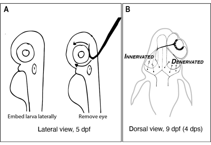
Sharpen to remove eye and dissect larvae.
Variable temperature heat block The Lab Depot or equivalent supplier BSH1001 or BSH1002 Set to 40-42 °C ahead of experiments.
Wide-mouth glass jar with lid (e.g., clean jam or salsa jar) For Tungsten needle sharpening device.
Wires with alligator clip leads For Tungsten needle sharpening device.
Microscopes:
Dissecting microscope Any type will work but having adjustable transmitted light on a mirrored base is preferred.
Laser scanning confocal microscope High NA, 20-25x water dipping objective lens is recommended.
Microscope control and image capture software (Elements) is used here but any confocal microscope will work.
Reagents for surgeries and dissections:
Calcium chloride dihydrate Sigma C7902 For Marc's Modified Ringers (MMR) and embryo medium (E3).
HEPES Sigma H7006 For Marc's Modified Ringers (MMR).
Low melting point agarose Invitrogen 16520-050 Make 1% in embryo medium (E3) or Marc's Modified Ringers (MMR).
Magnesium chloride hexahydrate Sigma 1374248 For embryo medium (E3).
Magnesium sulfate Sigma M7506 For Marc's Modified Ringers (MMR).
Paraformaldehyde Electron Microscopy Sciences 19210 Dilute 8% (w/v) stock with 2x concentrated PBS (diluted from 10x PBS stock).
Penicillin/Streptomycin Sigma P4333-20ML Dilute 1:100 in Marc's Modified Ringers.
Phosphate buffered saline (PBS) tablets Diagnostic BioSystems DMR E404-01 Make 10x stock in deionized water, autoclave and store at room temperature. Dilute to 1x working concentration.
Potassium chloride Sigma P3911 For Marc's Modified Ringers (MMR) and embryo medium (E3).
Sodium chloride Sigma S9888 For Marc's Modified Ringers (MMR) and embryo medium (E3).
Sodium hydroxide Sigma S5881 Make 10 M and use to adjust pH of MMR to 7.4.
Sucrose Sigma S9378
Tricaine-S Pentair 100G #TRS1 Recipe: https://zfin.atlassian.net/wiki/spaces/prot/pages/362220023/TRICAINE
Reagents for immunohistochemistry:
Alexafluor 568 tagged Secondary antibody to detect rabbit IgG Invitrogen A-11011 Use at 1:500 dilution for wholemount immunohistochemistry.
DAPI or ToPro3 Invitrogen 1306 or T3605 Make up 1 mg/mL solutions in DMSO; 1:5,000 dilution for counterstaining.
Dimethyl sulfoxide (DMSO) Sigma D8418 A component of immunoblock buffer.
Methanol (MeOH) Sigma 34860 Mixing MeOH with aqueous solutions like PBST is exothermic. Make the MeOH/PBST solutions at least several hours ahead of time or cool them on ice before using.
Normal goat serum ThermoFisher Scientific 50-062Z A component of immunoblock buffer. Can be aliquoted in 1-10 mL volumes and stored at -20 °C.
Primary antibody to detect phosphohistone H3 Millipore 06-570 Use at 1:300 dilution for wholemount immunohistochemistry.
Primary antibody to detect Red Fluorescent Protein (RFP; detects dsRed derivatives) MBL International PM005 Use at 1:500 dilution for wholemount immunohistochemistry.
Proteinase K (PK) Sigma P2308-10MG Make up 10 mg/mL stock solutions in PBS and use at 10 µg/mL.
Triton X-100 Sigma T8787 Useful to make a 20% (v/v) stock solution in PBS.
Software for data analysis
ImageJ (Fiji) freeware for image analysis; https://imagej.net/software/fiji/
Rstudio freeware for statistical analysis and data visualization; https://www.rstudio.com/products/rstudio/download/
Adobe Photoshop or GIMP Proprietary image processing software (Adobe Photoshop and Illustrator) are often used to compose figures). A freeware alternative is Gnu Image Manipulation Program (GIMP; https://www.gimp.org/)
Zebrafish strains available from the  Zebrafish International Resource Centers in the US (https://zebrafish.org/home/guide.php) or in Europe (https://www.ezrc.kit.edu/). Specialized transgenic strains that have not yet been deposited in either resource center can be requested from individual labs after publication.

Riferimenti

  1. Butler, A. B., Hodos, W. Optic tectum. Comparative Vertebrate Neuroanatomy: Evolution and Adaptation. , 311-340 (2005).
  2. Cang, J., Feldheim, D. A. Developmental mechanisms of topographic map formation and alignment. Annual Review of Neuroscience. 36 (1), 51-77 (2013).
  3. Basso, M. A., Bickford, M. E., Cang, J. Unraveling circuits of visual perception and cognition through the superior colliculus. Neuron. 109 (6), 918-937 (2021).
  4. Burrill, J. D., Easter, S. S. Development of the retinofugal projections in the embryonic and larval zebrafish (Brachydanio rerio). The Journal of Comparative Neurology. 346 (4), 583-600 (1994).
  5. Regeneration in the goldfish visual system. Webvision: The Organization of the Retina and Visual System Available from: https://webvision.med.utah.edu/book/part-x-repair-and-regeneration-in-the-visual-system/regeneration-in-the-goldfish-visual-system/ (2021)
  6. Regeneration in the visual system of adult mammals. Webvision: The Organization of the Retina and Visual System Available from: https://webvision.med.utah.edu/book/part-x-repair-and-regeneration-in-the-visual-system/regeneration-in-the-visual-system-of-adult-mammals/ (2021)
  7. Cerveny, K. L., Varga, M., Wilson, S. W. Continued growth and circuit building in the anamniote visual system. Developmental Neurobiology. 72 (3), 328-345 (2012).
  8. Lindsey, B. W., et al. Midbrain tectal stem cells display diverse regenerative capacities in zebrafish. Scientific Reports. 9 (1), 4420 (2019).
  9. Soares, D., Yamamoto, Y., Strickler, A. G., Jeffery, W. R. The lens has a specific influence on optic nerve and tectum development in the blind cavefish Astyanax. Developmental Neuroscience. 26 (5-6), 308-317 (2004).
  10. White, E. L. An experimental study of the relationship between the size of the eye and the size of the optic tectum in the brain of the developing teleost, Fundulus heteroclitus. Journal of Experimental Zoology. 108 (3), 439-469 (1948).
  11. Raymond, P., Easter, S., Burnham, J., Powers, M. Postembryonic growth of the optic tectum in goldfish. II. Modulation of cell proliferation by retinal fiber input. The Journal of Neuroscience. 3 (5), 1092-1099 (1983).
  12. Sato, Y., Yano, H., Shimizu, Y., Tanaka, H., Ohshima, T. Optic nerve input-dependent regulation of neural stem cell proliferation in the optic tectum of adult zebrafish. Developmental Neurobiology. 77 (4), 474-482 (2017).
  13. Hall, Z. J., Tropepe, V. Visual experience facilitates BDNF-dependent adaptive recruitment of new neurons in the postembryonic optic tectum. The Journal of Neuroscience. 38 (8), 2000-2014 (2018).
  14. Nusslein-Volhard, C., Dahm, R. . Zebrafish. , (2002).
  15. Cold Spring Harbor Protocols. Marc’s modified Ringer’s (MMR) (10X, pH 7.4). Cold Spring Harbor Protocols. , (2009).
  16. Turner, K. J., Bracewell, T. G., Hawkins, T. A. Anatomical dissection of zebrafish brain development. Brain Development. 1082, 197-214 (2014).
  17. Brady, J. A simple technique for making very fine, durable dissecting needles by sharpening tungsten wire electrolytically. Bulletin of the World Health Organization. 32 (1), 143-144 (1965).
  18. . ZFIN Tricaine recipe Available from: https://zfin.atlassian.net/wiki/spaces/prot/pages/362220023/TRICAINE (2018)
  19. Zhang, L., Leung, Y. F. Microdissection of zebrafish embryonic eye tissues. Journal of Visualized Experiments: JoVE. (40), e2028 (2010).
  20. . ZFIN protocols Available from: https://zfin.atlassian.net/wiki/spaces/prot/overview (2021)
  21. Engerer, P., Plucinska, G., Thong, R., Trovò, L., Paquet, D., Godinho, L. Imaging subcellular structures in the living zebrafish embryo. Journal of Visualized Experiments: JoVE. (110), e53456 (2016).
  22. ImageJ with batteries included. Fiji Available from: https://figi.sc/ (2021)
  23. O’Brien, J., Hayder, H., Peng, C. Automated quantification and analysis of cell counting procedures using ImageJ plugins. Journal of Visualized Experiments: JoVE. (117), e54719 (2016).
  24. Poggi, L., Vitorino, M., Masai, I., Harris, W. A. Influences on neural lineage and mode of division in the zebrafish retina in vivo. The Journal of Cell Biology. 171 (6), 991-999 (2005).
  25. Karlstrom, R. O., et al. Zebrafish mutations affecting retinotectal axon pathfinding. Development. 123 (1), 427-438 (1996).
  26. Harvey, B. M., Baxter, M., Granato, M. Optic nerve regeneration in larval zebrafish exhibits spontaneous capacity for retinotopic but not tectum specific axon targeting. PLOS ONE. 14 (6), 0218667 (2019).
  27. Robles, E., Filosa, A., Baier, H. Precise lamination of retinal axons generates multiple parallel input pathways in the tectum. Journal of Neuroscience. 33 (11), 5027-5039 (2013).
  28. Vargas, M. E., Barres, B. A. Why Is Wallerian degeneration in the CNS so slow. Annual Review of Neuroscience. 30 (1), 153-179 (2007).
  29. Hughes, A. N., Appel, B. Microglia phagocytose myelin sheaths to modify developmental myelination. Nature Neuroscience. 23 (9), 1055-1066 (2020).
  30. de Calbiac, H., Dabacan, A., Muresan, R., Kabashi, E., Ciura, S. Behavioral and physiological analysis in a zebrafish model of epilepsy. Journal of Visualized Experiments: JoVE. (176), e58837 (2021).
  31. Adams, S. L., Zhang, T., Rawson, D. M. The effect of external medium composition on membrane water permeability of zebrafish (Danio rerio) embryos. Theriogenology. 64 (7), 1591-1602 (2005).
  32. Fredj, N. B., et al. Synaptic activity and activity-dependent competition regulates axon arbor maturation, growth arrest, and territory in the retinotectal projection. Journal of Neuroscience. 30 (32), 10939-10951 (2010).
  33. Alberio, L., et al. A light-gated potassium channel for sustained neuronal inhibition. Nature Methods. 15 (11), 969-976 (2018).
  34. Kay, J. N., Finger-Baier, K. C., Roeser, T., Staub, W., Baier, H. Retinal ganglion cell genesis requires lakritz, a zebrafish atonal homolog. Neuron. 30 (3), 725-736 (2001).
  35. Gnuegge, L., Schmid, S., Neuhauss, S. C. F. Analysis of the activity-deprived zebrafish mutant macho reveals an essential requirement of neuronal activity for the development of a fine-grained visuotopic map. The Journal of Neuroscience. 21 (10), 3542-3548 (2001).
  36. Jeffery, W. R. Astyanax surface and cave fish morphs. EvoDevo. 11 (1), 14 (2020).
  37. Sieger, D., Peri, F. Animal models for studying microglia: The first, the popular, and the new. Glia. 61 (1), 3-9 (2013).
  38. Svahn, A. J., et al. Development of ramified microglia from early macrophages in the zebrafish optic tectum. Developmental Neurobiology. 73 (1), 60-71 (2013).
  39. Herzog, C., et al. Rapid clearance of cellular debris by microglia limits secondary neuronal cell death after brain injury in vivo. Development. 146 (9), (2019).
  40. Chen, J., Poskanzer, K. E., Freeman, M. R., Monk, K. R. Live-imaging of astrocyte morphogenesis and function in zebrafish neural circuits. Nature Neuroscience. 23 (10), 1297-1306 (2020).
check_url/it/63509?article_type=t

Play Video

Citazione di questo articolo
Hagen, O. L., Kim, Y., Kushkowski, E., Rouse, H., Cerveny, K. L. Eye Removal in Living Zebrafish Larvae to Examine Innervation-dependent Growth and Development of the Visual System. J. Vis. Exp. (180), e63509, doi:10.3791/63509 (2022).

View Video