Summary

Directe vergelijking van hyperspectrale gestimuleerde Raman-verstrooiing en coherente anti-stokes Raman-verstrooiingsmicroscopie voor chemische beeldvorming

Published: April 28, 2022
doi:

Summary

Dit artikel vergelijkt direct de resolutie, gevoeligheid en beeldcontrasten van gestimuleerde Raman-verstrooiing (SRS) en coherente anti-Stokes Raman-verstrooiing (CARS) geïntegreerd in hetzelfde microscoopplatform. De resultaten laten zien dat CARS een betere ruimtelijke resolutie heeft, SRS betere contrasten en spectrale resolutie geeft, en beide methoden hebben een vergelijkbare gevoeligheid.

Abstract

Gestimuleerde Raman-verstrooiing (SRS) en coherente anti-Stokes Raman scattering (CARS) microscopie zijn de meest gebruikte coherente Raman-verstrooiingsbeeldvormingstechnologieën. Hyperspectrale SRS- en CARS-beeldvorming bieden Raman spectrale informatie op elke pixel, wat een betere scheiding van verschillende chemische samenstellingen mogelijk maakt. Hoewel beide technieken twee excitatielasers vereisen, zijn hun signaaldetectieschema’s en spectrale eigenschappen heel verschillend. Het doel van dit protocol is om zowel hyperspectrale SRS- als CARS-beeldvorming op één platform uit te voeren en de twee microscopietechnieken voor het afbeelden van verschillende biologische monsters te vergelijken. De spectrale focusmethode wordt gebruikt om spectrale informatie te verkrijgen met behulp van femtosecondelasers. Door standaard chemische monsters te gebruiken, worden de gevoeligheid, ruimtelijke resolutie en spectrale resolutie van SRS en CARS in dezelfde excitatieomstandigheden (d.w.z. vermogen bij het monster, pixel dwell-tijd, objectieve lens, pulsenergie) vergeleken. De beeldcontrasten van CARS en SRS voor biologische monsters worden naast elkaar geplaatst en vergeleken. De directe vergelijking van CARS- en SRS-prestaties zou een optimale selectie van de modaliteit voor chemische beeldvorming mogelijk maken.

Introduction

Het Raman-verstrooiingsfenomeen werd voor het eerst waargenomen in 1928 door C. V. Raman1. Wanneer een invallend foton interageert met een monster, kan spontaan een inelastische verstrooiingsgebeurtenis optreden, waarbij de energieverandering van het foton overeenkomt met een vibrationele overgang van de geanalyseerde chemische soort. Dit proces vereist geen gebruik van een chemische tag, waardoor het een veelzijdig, labelvrij hulpmiddel is voor chemische analyse en tegelijkertijd de verstoring van monsters minimaliseert. Ondanks de voordelen lijdt spontane Raman-verstrooiing aan een lage verstrooiingsdoorsnede (meestal 1011 lager dan de infrarood [IR] absorptiedoorsnede), wat lange acquisitietijden vereist voor analyse2. De zoektocht naar het verhogen van de gevoeligheid van het Raman-verstrooiingsproces is dus essentieel bij het pushen van Raman-technologieën voor realtime beeldvorming.

Een effectieve manier om de gevoeligheid van Raman-verstrooiing aanzienlijk te verbeteren, is door middel van coherente Raman-verstrooiingsprocessen (CRS), waarvoor meestal twee laserpulsen worden gebruikt om moleculaire trillingsovergangen te exciteren 3,4. Wanneer het fotonenergieverschil tussen de twee lasers overeenkomt met de trillingsmodi van monstermoleculen, worden sterke Raman-signalen gegenereerd. De twee meest gebruikte CRS-processen voor beeldvorming zijn coherente anti-Stokes Raman scattering (CARS) en gestimuleerde Raman scattering (SRS)5. In de afgelopen twee decennia hebben technologische ontwikkelingen CARS- en SRS-microscopietechnieken verbeterd om krachtige hulpmiddelen te worden voor labelvrije kwantificering en opheldering van chemische veranderingen in biologische monsters.

Chemische beeldvorming door CARS-microscopie kan worden gedateerd op 1982 toen laserscanning voor het eerst werd toegepast om CARS-beelden te verkrijgen, gedemonstreerd door Duncan et al6. De modernisering van CARS-microscopie werd sterk versneld na de brede toepassingen van laserscanning multifotonenfluorescentiemicroscopie7. Vroeg werk van de Xie-groep met behulp van lasers met een hoge herhalingssnelheid heeft CARS omgevormd tot een snel, labelvrij, chemisch beeldvormingsplatform voor de karakterisering van moleculen in biologische monsters 8,9,10. Een van de belangrijkste problemen voor CARS-beeldvorming is de aanwezigheid van een niet-resonante achtergrond, die het beeldcontrast vermindert en het Raman-spectrum vervormt. Er zijn veel inspanningen geleverd om ofwel de niet-resonante achtergrond11,12,13,14,15 te verminderen of om resonante Raman-signalen uit de CARS-spectra16,17 te halen. Een andere vooruitgang die het veld enorm heeft verbeterd, is hyperspectrale CARS-beeldvorming, die spectrale mapping mogelijk maakt bij elke beeldpixel met verbeterde chemische selectiviteit 18,19,20,21.

Gestimuleerde Raman-verstrooiing (SRS) is een jongere beeldvormingstechnologie dan CARS, hoewel het eerder werd ontdekt22. In 2007 werd SRS-microscopie gerapporteerd met behulp van een laserbron met lage herhalingssnelheid23. Al snel demonstreerden verschillende groepen high-speed SRS-beeldvorming met behulp van lasers met hoge herhalingssnelheid 24,25,26. Een van de belangrijkste voordelen van SRS-microscopie ten opzichte van CARS is de afwezigheid van de niet-resonante achtergrond27, hoewel andere achtergronden zoals cross-phase modulatie (XPM), transiënte absorptie (TA), twee-fotonenabsorptie (TPA) en fotothermisch (PT) effect, kunnen optreden met SRS28. Bovendien hebben het SRS-signaal en de monsterconcentratie lineaire relaties, in tegenstelling tot CARS, dat een kwadratische signaalconcentratieafhankelijkheid heeft29. Dit vereenvoudigt chemische kwantificering en spectrale ontmenging. Multicolor en hyperspectrale SRS is geëvolueerd in verschillende vormen 30,31,32,33,34,35,36, waarbij spectrale focus een van de meest populaire benaderingen is voor chemische beeldvorming 37,38.

Zowel CARS als SRS vereisen de focus van de pomp en Stokes laserstralen op het monster om de trillingsovergang van de moleculen voor signaalexcitatie te matchen. CARS en SRS microscopen hebben ook veel gemeen. De fysica die ten grondslag ligt aan deze twee processen en signaaldetecties die betrokken zijn bij deze microscopietechnologieën hebben echter verschillen 3,39. CARS is een parametrisch proces dat geen netto foton-molecuul energiekoppelingheeft 3. SRS is echter een niet-parametrisch proces en draagt bij aan de energieoverdracht tussen fotonen en moleculaire systemen27. In CARS wordt een nieuw signaal met anti-Stokes-frequentie gegenereerd, terwijl SRS zich manifesteert als de energieoverdracht tussen de pomp en Stokes-laserstralen.

CARS-signaal voldoet aan Eq (1)28.
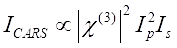
Equation 1 (1)

Ondertussen kan het SRS-signaal worden geschreven als Eq (2) 28.
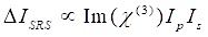
Equation 1(2)

Hier zijn Ip, Is, ICARS en ΔISRS de intensiteiten van respectievelijk de pompbundel, Stokes-bundel, CARS-signaal en SRS-signalen. χ(3) is de derde-orde niet-lineaire optische gevoeligheid van het monster en is een complexe waarde die bestaat uit reële en imaginaire delen.

Deze vergelijkingen drukken de spectrale profielen en signaalconcentratieafhankelijkheid van CARS en SRS uit. Verschillen in fysica resulteren in ongelijksoortige detectieschema’s voor deze twee microscopietechnologieën. Signaaldetectie in CARS omvat meestal spectrale scheiding van nieuw gegenereerde fotonen en detectie met behulp van een fotomultiplicatorbuis (PMT) of charge-coupled device (CCD); voor SRS wordt de energie-uitwisseling tussen de pomp en stokesbundels meestal gemeten door intensiteitsmodulatie met hoge snelheid met behulp van een optische modulator en demodulatie met behulp van een fotodiode (PD) in combinatie met een lock-in versterker.

Hoewel er de afgelopen jaren veel technologische ontwikkelingen en toepassingen zijn gepubliceerd op zowel CARS- als SRS-gebieden, zijn er geen systematische vergelijkingen van de twee CRS-technieken op hetzelfde platform uitgevoerd, vooral voor hyperspectrale CARS- en SRS-microscopie. Directe vergelijkingen in gevoeligheid, ruimtelijke resolutie, spectrale resolutie en chemische scheidingsmogelijkheden zouden biologen in staat stellen om de beste modaliteit voor chemische kwantificering te selecteren. In dit protocol worden gedetailleerde stappen beschreven om een multimodaal beeldvormingsplatform te bouwen met zowel hyperspectrale CARS- als SRS-modaliteiten op basis van een femtosecondelasersysteem en spectrale focus. De twee technieken zijn vergeleken in de voorwaartse richting voor spectrale resolutie, detectiegevoeligheid, ruimtelijke resolutie en beeldcontrasten van cellen.

Protocol

1. Instrumentele setup voor hyperspectrale CRS-beeldvorming OPMERKING: Het genereren van CRS-signaal vereist het gebruik van krachtige (d.w.z. klasse 3B of klasse 4) lasers. Veiligheidsprotocollen moeten worden aangepakt en de juiste persoonlijke beschermingsmiddelen (PBM’ s) moeten te allen tijde worden gedragen bij het werken met dergelijke hoge piekvermogens. Raadpleeg de juiste documentatie voordat u gaat experimenteren. Dit protocol richt zich op het ontwerpen van het bunde…

Representative Results

Vergelijkingen van de spectrale resolutieFiguur 2 vergelijkt de spectrale resolutie van hyperspectrale SRS (figuur 2A) en CARS (figuur 2B) microscopie met behulp van een DMSO-monster. Voor het SRS-spectrum werden twee Lorentziaanse functies (zie protocol stap 2.3) toegepast om in het spectrum te passen, en een resolutie van 14,6 cm-1 werd verkregen met behulp van de 2.913 cm-1 piek. Voor CA…

Discussion

Het hier gepresenteerde protocol beschrijft de constructie van een multimodale CRS-microscoop en de directe vergelijking tussen CARS- en SRS-beeldvorming. Voor de microscoopconstructie zijn de kritieke stappen ruimtelijke en temporele bundeloverlapping en optimalisatie van de bundelgrootte. Het wordt aanbevolen om een standaardmonster zoals DMSO te gebruiken vóór de biologische beeldvorming voor het optimaliseren van SNR en het kalibreren van Raman-verschuivingen. Directe vergelijking tussen CARS- en SRS-beelden laat z…

Divulgazioni

The authors have nothing to disclose.

Acknowledgements

Dit onderzoek werd ondersteund door het start-upfonds van de Purdue University Department of Chemistry.

Materials

2D galvo scanner set Thorlabs GVS002
Acousto-optic modulator Isomet M1205-P80L-0.5
AOM driver Isomet 532B-2
Data acquisition card National Instruments PCle 6363 Custom ordered filter (980 sp)
Delay stage Zaber X-LSM050A
Deuterium oxide Millipore Sigma 151882-100G
Dichroic mirror for beam combination Thorlabs DMLP1000
Dichroic mirror for signal separation Semrock FF776-Di01-25×36
DMSO MiliporeSigma 200-664-3
MIA PaCa 2 Cells ATCC CRL-1420
Femtosecond laser system Spectral Physics InSightX3+
Filter for CARS Chroma AT655/30m
Filter for SRS Chroma ET980sp
Function generator Rigol DG1022Z
Glass rods Lattice Electro Optics SF-57
Half-wave plate Newport 10RP02-51; 10RP02-46
LabVIEW 2020 National Instruments This is the image acquisition software
Lock-in amplifier Zurich Instrument HF2LI
Microscope housing Olympus BX51W1
Objective lens Olympus UPLSAPO60XW
Origin Pro 2019b OriginLab Corporation This is the spectral fitting software
Oscilloscope Tektronix TBS2204B
Photodiode Hamamatsu S3994-01
PMT detector Hamamatsu H7422P-40
PMT voltage amplifier Advanced Research Instrument Corp. PMT4V3
Polarizing beamsplitter cube Thorlabs PBS255
Terminal block National Instruments BNC-2110

Riferimenti

  1. Raman, C. V. A change of wave-length in light scattering. Nature. 121 (3051), 619 (1928).
  2. Li, S., Li, Y., Yi, R., Liu, L., Qu, J. Coherent anti-Stokes Raman scattering microscopy and its applications. Frontiers in Physics. 8, 515 (2020).
  3. Evans, C. L., Xie, X. S. Coherent anti-Stokes Raman scattering microscopy: chemical imaging for biology and medicine. Annual Review of Analytical Chemistry. 1 (1), 883-909 (2008).
  4. Min, W., Freudiger, C. W., Lu, S., Xie, X. S. Coherent nonlinear optical imaging: beyond fluorescence microscopy. Annual Review of Physical Chemistry. 62, 507-530 (2011).
  5. Suhalim, J. L., Boik, J. C., Tromberg, B. J., Potma, E. O. The need for speed. Journal of Biophotonics. 5 (5-6), 387-395 (2012).
  6. Duncan, M. D., Reintjes, J., Manuccia, T. J. Scanning coherent anti-Stokes Raman microscope. Optics Letters. 7 (8), 350-352 (1982).
  7. Denk, W., Strickler, J. H., Webb, W. W. Two-photon laser scanning fluorescence microscopy. Science. 248 (4951), 73-76 (1990).
  8. Zumbusch, A., Holtom, G. R., Xie, X. S. Three-dimensional vibrational imaging by coherent anti-Stokes Raman scattering. Physical Review Letters. 82 (20), 4142-4145 (1999).
  9. Cheng, J. -. X., Xie, X. S. Coherent anti-Stokes Raman scattering microscopy: instrumentation, theory, and applications. The Journal of Physical Chemistry B. 108 (3), 827-840 (2004).
  10. Evans, C. L., et al. Chemical imaging of tissue in vivo with video-rate coherent anti-Stokes Raman scattering microscopy. Proceedings of the National Academy of Sciences of the United States of America. 102 (46), 16807 (2005).
  11. Cheng, J. -. X., Volkmer, A., Book, L. D., Xie, X. S. An epi-detected coherent anti-Stokes Raman scattering (E-CARS) microscope with high spectral resolution and high sensitivity. The Journal of Physical Chemistry B. 105 (7), 1277-1280 (2001).
  12. Volkmer, A., Book, L. D., Xie, X. S. Time-resolved coherent anti-Stokes Raman scattering microscopy: Imaging based on Raman free induction decay. Applied Physics Letters. 80 (9), 1505-1507 (2002).
  13. Marks, D. L., Boppart, S. A. Nonlinear interferometric vibrational imaging. Physical Review Letters. 92 (12), 123905 (2004).
  14. Ganikhanov, F., Evans, C. L., Saar, B. G., Xie, X. S. High-sensitivity vibrational imaging with frequency modulation coherent anti-Stokes Raman scattering (FM CARS) microscopy. Optics Letters. 31 (12), 1872-1874 (2006).
  15. Potma, E. O., Evans, C. L., Xie, X. S. Heterodyne coherent anti-Stokes Raman scattering (CARS) imaging. Optics Letters. 31 (2), 241-243 (2006).
  16. Liu, Y., Lee, Y. J., Cicerone, M. T. Broadband CARS spectral phase retrieval using a time-domain Kramers-Kronig transform. Optics Letters. 34 (9), 1363-1365 (2009).
  17. Masia, F., Karuna, A., Borri, P., Langbein, W. Hyperspectral image analysis for CARS, SRS, and Raman data. Journal of Raman Spectroscopy. 46 (8), 727-734 (2015).
  18. Knutsen, K. P., Johnson, J. C., Miller, A. E., Petersen, P. B., Saykally, R. J. High spectral resolution multiplex CARS spectroscopy using chirped pulses. Chemical Physics Letters. 387 (4-6), 436-441 (2004).
  19. Okuno, M., Kano, H., Leproux, P., Couderc, V., Hamaguchi, H. -. o. Ultrabroadband multiplex CARS microspectroscopy and imaging using a subnanosecond supercontinuum light source in the deep near infrared. Optics Letters. 33 (9), 923-925 (2008).
  20. Masia, F., Glen, A., Stephens, P., Borri, P., Langbein, W. Quantitative chemical imaging and unsupervised analysis using hyperspectral coherent anti-Stokes Raman scattering microscopy. Analytical Chemistry. 85 (22), 10820-10828 (2013).
  21. Pegoraro, A. F., Slepkov, A. D., Ridsdale, A., Moffatt, D. J., Stolow, A. Hyperspectral multimodal CARS microscopy in the fingerprint region. Journal of Biophotonics. 7 (1-2), 49-58 (2014).
  22. Eckhardt, G., et al. Stimulated Raman scattering from organic liquids. Physical Review Letters. 9 (11), 455-457 (1962).
  23. Ploetz, E., Laimgruber, S., Berner, S., Zinth, W., Gilch, P. Femtosecond stimulated Raman microscopy. Applied Physics B. 87 (3), 389-393 (2007).
  24. Freudiger, C. W., et al. Label-free biomedical imaging with high sensitivity by stimulated Raman scattering microscopy. Science. 322 (5909), 1857-1861 (2008).
  25. Nandakumar, P., Kovalev, A., Volkmer, A. Vibrational imaging based on stimulated Raman scattering microscopy. New Journal of Physics. 11 (3), 033026 (2009).
  26. Slipchenko, M. N., Le, T. T., Chen, H., Cheng, J. -. X. High-speed vibrational imaging and spectral analysis of lipid bodies by compound Raman microscopy. The Journal of Physical Chemistry B. 113 (21), 7681-7686 (2009).
  27. Min, W., Freudiger, C. W., Lu, S., Xie, X. S. Coherent nonlinear optical imaging: beyond fluorescence microscopy. Annual Review of Physical Chemistry. 62 (1), 507-530 (2011).
  28. Zhang, C., Zhang, D., Cheng, J. -. X. Coherent Raman scattering microscopy in biology and medicine. Annual Review of Biomedical Engineering. 17 (1), 415-445 (2015).
  29. Prince, R. C., Frontiera, R. R., Potma, E. O. Stimulated Raman scattering: from bulk to nano. Chemical Reviews. 117 (7), 5070-5094 (2017).
  30. Lu, F. -. K., et al. Multicolor stimulated Raman scattering microscopy. Molecular Physics. 110 (15-16), 1927-1932 (2012).
  31. Ozeki, Y., et al. High-speed molecular spectral imaging of tissue with stimulated Raman scattering. Nature Photonics. 6 (12), 845-851 (2012).
  32. Wang, P., et al. Label-free quantitative imaging of cholesterol in intact tissues by hyperspectral stimulated raman scattering microscopy. Angewandte Chemie International Edition. 125 (49), 13280-13284 (2013).
  33. Freudiger, C. W., et al. Stimulated Raman scattering microscopy with a robust fibre laser source. Nature Photonics. 8 (2), 153-159 (2014).
  34. Liao, C. -. S., et al. Microsecond scale vibrational spectroscopic imaging by multiplex stimulated Raman scattering microscopy. Light: Science & Applications. 4 (3), 265 (2015).
  35. Liao, C. -. S., et al. Spectrometer-free vibrational imaging by retrieving stimulated Raman signal from highly scattered photons. Science Advances. 1 (9), 1500738 (2015).
  36. He, R., et al. Dual-phase stimulated Raman scattering microscopy for real-time two-color imaging. Optica. 4 (1), 44-47 (2017).
  37. Andresen, E. R., Berto, P., Rigneault, H. Stimulated Raman scattering microscopy by spectral focusing and fiber-generated soliton as Stokes pulse. Optics Letters. 36 (13), 2387-2389 (2011).
  38. Fu, D., Holtom, G., Freudiger, C., Zhang, X., Xie, X. S. Hyperspectral imaging with stimulated Raman scattering by chirped femtosecond lasers. The Journal of Physical Chemistry B. 117 (16), 4634-4640 (2013).
  39. Zhang, C., Aldana-Mendoza, J. A. Coherent Raman scattering microscopy for chemical imaging of biological systems. Journal of Physics: Photonics. , (2021).
  40. Martens, W. N., Frost, R. L., Kristof, J., Theo Kloprogge, J. Raman spectroscopy of dimethyl sulphoxide and deuterated dimethyl sulphoxide at 298 and 77 k. Journal of Raman Spectroscopy. 33 (2), 84-91 (2002).
  41. Gill, G. W., Gill, G. W. . Cytopreparation: Principles & Practice. , 309-323 (2013).
  42. Fu, D., et al. Imaging the intracellular distribution of tyrosine kinase inhibitors in living cells with quantitative hyperspectral stimulated Raman scattering. Nature Chemistry. 6 (7), 614-622 (2014).
  43. Wei, L., Yu, Y., Shen, Y., Wang, M. C., Min, W. Vibrational imaging of newly synthesized proteins in live cells by stimulated Raman scattering microscopy. Proceedings of the National Academy of Sciences. 110 (28), 11226-11231 (2013).
  44. Lu, F. -. K., et al. Label-free DNA imaging in vivo with stimulated Raman scattering microscopy. Proceedings of the National Academy of Sciences. 112 (37), 11624-11629 (2015).
  45. Slipchenko, M. N., et al. Vibrational imaging of tablets by epi-detected stimulated Raman scattering microscopy. Analyst. 135 (10), 2613-2619 (2010).
  46. Slipchenko, M. N., Zhou, B., Pinal, R., Teresa Carvajal, M., Cheng, J. -. X. RAMAN-chemical imaging of solid dosage forms based on stimulated Raman scattering. American Pharmaceutical Review. 15 (3), 66 (2012).
  47. Sarri, B., et al. Discriminating polymorph distributions in pharmaceutical tablets using stimulated Raman scattering microscopy. Journal of Raman Spectroscopy. 50 (12), 1896-1904 (2019).
  48. Fussell, A. L., Kleinebudde, P., Herek, J., Strachan, C. J., Offerhaus, H. L. Coherent anti-Stokes Raman scattering (CARS) microscopy visualizes pharmaceutical tablets during dissolution. JoVE (Journal of Visualized Experiments). (89), e51847 (2014).
  49. Freudiger, C. W., et al. Multicolored stain-free histopathology with coherent Raman imaging). Laboratory Investigation. 92 (10), 1492-1502 (2012).
  50. Lim, R. S., et al. Multimodal CARS microscopy determination of the impact of diet on macrophage infiltration and lipid accumulation on plaque formation in ApoE-deficient mice [S]. Journal of Lipid Research. 51 (7), 1729-1737 (2010).
  51. Ji, M., et al. label-free detection of brain tumors with stimulated Raman scattering microscopy. Science Translational Medicine. 5 (201), (2013).
  52. Tabish, T. A., Narayan, R. J., Edirisinghe, M. Rapid and label-free detection of COVID-19 using coherent anti-Stokes Raman scattering microscopy. Mrs Communications. 10 (4), 566-572 (2020).
  53. Camp, C. H., et al. High-speed coherent Raman fingerprint imaging of biological tissues. Nature Photonics. 8 (8), 627-634 (2014).
  54. Wei, L., et al. Live-cell bioorthogonal chemical imaging: stimulated Raman scattering microscopy of vibrational probes. Accounts of Chemical Research. 49 (8), 1494-1502 (2016).
  55. Hu, F., Shi, L., Min, W. Biological imaging of chemical bonds by stimulated Raman scattering microscopy. Nature Methods. 16 (9), 830-842 (2019).
  56. Nie, S., Emory, S. R. Probing single molecules and single nanoparticles by surface-enhanced Raman scattering. Science. 275 (5303), 1102-1106 (1997).
  57. Steuwe, C., Kaminski, C. F., Baumberg, J. J., Mahajan, S. Surface enhanced coherent anti-Stokes Raman scattering on nanostructured gold surfaces. Nano Letters. 11 (12), 5339-5343 (2011).
  58. Fast, A., Kenison, J. P., Syme, C. D., Potma, E. O. Surface-enhanced coherent anti-Stokes Raman imaging of lipids. Applied Optics. 55 (22), 5994-6000 (2016).
  59. Zong, C., et al. Plasmon-enhanced stimulated Raman scattering microscopy with single-molecule detection sensitivity. Nature Communications. 10 (1), 1-11 (2019).
  60. Yampolsky, S., et al. Seeing a single molecule vibrate through time-resolved coherent anti-Stokes Raman scattering. Nature Photonics. 8 (8), 650-656 (2014).
check_url/it/63677?article_type=t

Play Video

Citazione di questo articolo
Clark, M. G., Brasseale III, K. A., Gonzalez, G. A., Eakins, G., Zhang, C. Direct Comparison of Hyperspectral Stimulated Raman Scattering and Coherent Anti-Stokes Raman Scattering Microscopy for Chemical Imaging. J. Vis. Exp. (182), e63677, doi:10.3791/63677 (2022).

View Video