Human induced pluripotent stem cell-derived cardiomyocytes (hiPSC-CMs) offer an alternative to using animals for preclinical cardiotoxicity screening. A limitation to the widespread adoption of hiPSC-CMs in preclinical toxicity screening is the immature, fetal-like phenotype of the cells. Presented here are protocols for robust and rapid maturation of hiPSC-CMs.
Human induced stem cell-derived cardiomyocytes (hiPSC-CMs) are used to replace and reduce the dependence on animals and animal cells for preclinical cardiotoxicity testing. In two-dimensional monolayer formats, hiPSC-CMs recapitulate the structure and function of the adult human heart muscle cells when cultured on an optimal extracellular matrix (ECM). A human perinatal stem cell-derived ECM (maturation-inducing extracellular matrix-MECM) matures the hiPSC-CM structure, function, and metabolic state in 7 days after plating.
Mature hiPSC-CM monolayers also respond as expected to clinically relevant medications, with a known risk of causing arrhythmias and cardiotoxicity. The maturation of hiPSC-CM monolayers was an obstacle to the widespread adoption of these valuable cells for regulatory science and safety screening, until now. This article presents validated methods for the plating, maturation, and high-throughput, functional phenotyping of hiPSC-CM electrophysiological and contractile function. These methods apply to commercially available purified cardiomyocytes, as well as stem cell-derived cardiomyocytes generated in-house using highly efficient, chamber-specific differentiation protocols.
High-throughput electrophysiological function is measured using either voltage-sensitive dyes (VSDs; emission: 488 nm), calcium-sensitive fluorophores (CSFs), or genetically encoded calcium sensors (GCaMP6). A high-throughput optical mapping device is used for optical recordings of each functional parameter, and custom dedicated software is used for electrophysiological data analysis. MECM protocols are applied for medication screening using a positive inotrope (isoprenaline) and human Ether-a-go-go-related gene (hERG) channel-specific blockers. These resources will enable other investigators to successfully utilize mature hiPSC-CMs for high-throughput, preclinical cardiotoxicity screening, cardiac medication efficacy testing, and cardiovascular research.
Human induced pluripotent stem cell-derived cardiomyocytes (hiPSC-CMs) have been validated on an international scale, and are available for in vitro cardiotoxicity screening1. Highly pure hiPSC-CMs can be generated in virtually unlimited numbers, cryopreserved, and thawed. Upon replating, they also reanimate and begin contracting with a rhythm reminiscent of the human heart2,3. Remarkably, individual hiPSC-CMs couple to each other and form functional syncytia that beat as a single tissue. Nowadays, hiPSCs are routinely derived from patient blood samples, so any person can be represented using in vitro hiPSC-CM cardiotoxicity screening assays4,5. This creates the opportunity to perform "Clinical Trials in a Dish", with significant representation from diverse populations6.
One critical advantage over existing animal and animal cell cardiotoxicity screening approaches is that hiPSC-CMs utilize the full human genome and offer an in vitro system with genetic similarities to the human heart. This is especially attractive for pharmacogenomics and personalized medicine – the use of hiPSC-CMs for medication and other therapy development is predicted to provide more accurate, precise, and safe medication prescriptions. Indeed, two-dimensional (2D) hiPSC-CM monolayer assays have proven to be predictive of medication cardiotoxicity, using a panel of clinically used medications with a known risk of causing arrhythmias1,7,8,9. Despite the vast potential of hiPSC-CMs and the promise to streamline and make drug development cheaper, there has been a reluctance to use these novel assays10,11,12.
Until now, one major limitation of widespread adoption and acceptance of hiPSC-CM screening assays is their immature, fetal-like appearance, as well as their function. The critical issue of hiPSC-CM maturation has been reviewed and debated in the scientific literature ad nauseum13,14,15,16. Likewise, many approaches have been employed to promote hiPSC-CM maturation, including extracellular matrix (ECM) manipulations in 2D monolayers and the development of 3D engineered heart tissues (EHTs)17,18. At the moment, there is a widely held belief that the use of 3D EHTs will provide superior maturation relative to 2D monolayer-based approaches. However, 2D monolayers provide a higher efficiency of cell utilization and increased success in plating compared to 3D EHTs; 3D EHTs utilize greater numbers of cells, and often require the inclusion of other cell types that can confound results. Therefore, in this article, the focus is on using a simple method to mature hiPSC-CMs cultured as 2D monolayers of electrically and mechanically coupled cells.
Advanced hiPSC-CM maturation can be achieved in 2D monolayers using an ECM. The 2D monolayers of hiPSC-CMs can be matured using a soft, flexible polydimethylsiloxane coverslip, coated with basement membrane matrix secreted by an Engelbreth-Holm-Swarm mouse sarcoma cell (mouse ECM). In 2016, reports showed that hiPSC-CMs cultured on this soft ECM condition matured functionally, displaying action potential conduction velocities near adult heart values (~50 cm/s)18. Further, these mature hiPSC-CMs displayed many other electrophysiological characteristics reminiscent of the adult heart, including hyperpolarized resting membrane potential and expression of Kir2.1. More recently, reports identified a human perinatal stem cell-derived ECM coating that promotes the structural maturation of 2D hiPSC-CMs19. Here, easy-to-use methods are presented to structurally mature 2D hiPSC-CM monolayers for use in high-throughput electrophysiological screens. Further, we provide validation of an optical mapping instrument for the automated acquisition and analysis of 2D hiPSC-CM monolayer electrophysiological function, using voltage-sensitive dyes (VSDs) and calcium-sensitive probes and proteins.
hiPSC usage in this protocol was approved by the University of Michigan HPSCRO Committee (Human Pluripotent Stem Cell Oversight Committee). See the Table of Materials for a list of materials and equipment. See Table 1 for media and their compositions.
1. Thawing and plating commercially available cryopreserved hiPSC-CMs for maturation on a maturation-inducing extracellular matrix (MECM)
2. hiPSC cardiac-directed differentiation and hiPSC-CM purification
3. hiPSC-CM purification via MACS (magnetic-activated cell sorting)
4. Optical mapping using voltage-sensitive dyes (VSDs) and calcium-sensitive fluorophores (CSFs)
5. Optical mapping using genetically encoded calcium indicator (GECI)
6. Acquisition of optical mapping data and analysis
hiPSC-CM maturation characterized by phase contrast and immunofluorescent confocal imaging
The timeline for ECM-mediated maturation of commercially available hiPSC-CMs using MECM coated 96-well plates is presented in Figure 1A. These data are collected using commercially available cardiomyocytes that arrive in the laboratory as cryopreserved vials of cells. Each vial contains >5 × 10⁶ viable cardiomyocytes. The cells are ~98% pure and rigorously tested for quality control (certificate of analysis is provided with each vial). The high number of CMs enables thawing and plating the CMs onto different ECM combinations using the same batch of cells. In Figure 1, hiPSC-CMs are plated on either mouse ECM- or MECM-coated plates. The hiPSC-CMs plated on the MECM mature and become structurally distinct from the same batch of hiPSC-CMs replated on the mouse ECM. Namely, mature cells become rod-shaped, while immature cells retain a circular shape. This can be seen in phase contrast imaging and upon staining the cardiac myofilaments (Figure 1B; troponin I [TnI], red). A more extensive validation of the structural maturation of hiPSC-CMs is presented in Figure 2. A large field of view (20x objective) of CMs stained with α-actinin antibody shows the typical shape of the cells cultured on each ECM condition. α-actinin is a critical structural protein arranged with regular spacing in the cardiac myofilaments. Consistent with the TnI staining in Figure 1, the α-actinin staining further indicates the maturation of hiPSC-CMs cultured on the MECM. Besides promoting a rod-shaped mature phenotype, the MECM also induces greater sarcomere organization (60x images). Mitochondrial content and activity are also distinct between cells cultured on the mouse ECM and the MECM (Figure 2B). Fetal-immature hiPSC-CM mitochondrial content is limited to the perinuclear space, with little mitochondria being found in the cytosol. In contrast, mature hiPSC-CMs mitochondrial content is distributed throughout the cell. Mitochondrial assessment uses an established protocol19.
hiPSC cardiac-directed differentiation and cardiac chamber specification
Provided here is a protocol for the in-house production and maturation of purified, chamber-specific hiPSC-CMs (Figure 3A). This is based on a previously published report20. Presented are detailed procedures for hiPSC-CM purification using a commercially available, magnet-activated cell sorting (MACS) kit. We recently validated the use of MACS purification and showed the benefits of using MACS compared to metabolic-based hiPSC-CM purification; typically, hiPSC-CM purity above 95% is anticipated21. It is important to point out that if the initial CM content is <50%, MACS purification may reach only ~85%. In these cases, CM enrichment may be necessary following the depletion of non-CMs. If the initial CM content from differentiation is >50%, depletion of non-CMs from the cell population using the MACS kit can achieve purity >95%; in this case, the further enrichment or positive selection of CMs is not necessary. The chamber-specific hiPSC-CMs can also be matured using MECM-coated 96-well plates, as outlined above and shown in Figure 1 and Figure 2. It should be expected that the atrial-specific cells (hiPSC-ACM) have a significantly faster spontaneous beat rate and shorter action potential duration 80 (APD80) than the ventricular-specific cells (hiPSC-VCM). These are typical electrophysiological data for action potentials recorded using VSDs and the optical mapping system (Figure 3B–D).
High-throughput cardiac electrophysiological optical mapping
Scientific rigor is dramatically increased for any assay if it can be carried out in a high-throughput way. Cardiotoxicity screening data are presented in Figure 4, Figure 5, Figure 6, and Figure 7, showing high-throughput electrophysiological screening using mature hiPSC-CM monolayers in a 96-well plate. Whole plate heatmaps for parameters such as APD80 (Figure 4A) reveal the reproducibility of a given parameter within a plate from well to well. Further, whole-plate heatmaps provide a quick examination of any outliers in the data set. For example, in well E4 of the plate presented in Figure 4A, it is clear that this well has a much greater APD80 value, indicated by the well appearing yellow, while the other wells are indigo-blue. Typical action potentials of mature 2D hiPSC-CM monolayers (Figure 4B) are reminiscent of the action potential morphology of adult cardiomyocytes isolated and tested in culture. Moreover, a typical action potential spontaneous rhythm is displayed in Figure 4C. The data in Figure 4C is a time-space plot of row A, columns 1-12. The white line across the plate map in Figure 4A depict this. Each bright fluorescent flash over time in each well represents a single spontaneous activation. Figure 5 and Figure 6 show the utility of using GCaMP6m genetically encoded calcium indicator (GECI) to measure intracellular calcium transients; Figure 6 also shows the expected response to isoproterenol-the classical cardiac positive inotrope. In response to isoproterenol, activation of the β1-adrenergic receptors cause positive chronotropy (Figure 6A), positive inotropy (Figure 6B), and positive lusitropy (Figure 6C). These responses to isoproterenol indicate the significant maturation of hiPSC-CM β1-adrenergic receptors and intracellular signaling cascades.
In Figure 7, hiPSC-CM response to human Ether-a-go-go-related gene (hERG) channel blockers is shown, using GCaMP6m calcium fluorescence to monitor rhythm and to serve as a surrogate marker for contractility. E-4031 is a hERG-specific channel blocker, that slows the spontaneous beat rate and increases the calcium transient duration (CaTD80) and triangulation (CaT triangulation). Figure 7A shows the detection of early after-depolarizations caused by the E-4031 hERG channel blockade. Other hERG channel blockers, including domperidone, vandetanib, and sotalol, were also tested, and results are shown in Figure 7E-G. These compounds and doses were selected based on the recent hiPSC-CM validation study1,7,9.
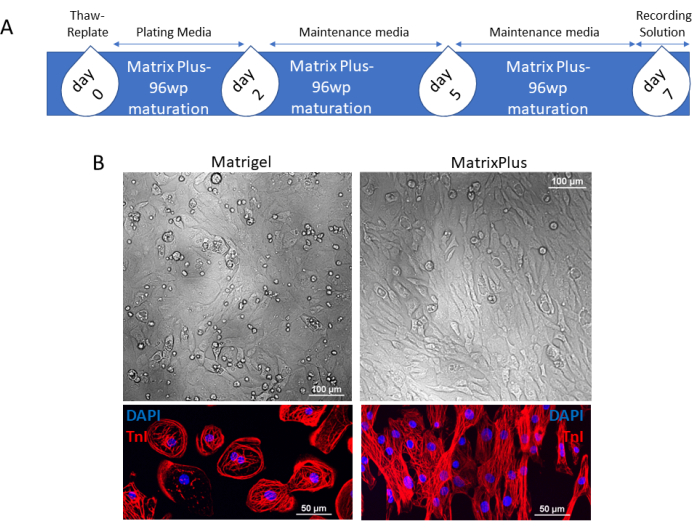
Figure 1: Timeline for fast maturation of commercially available or other-source cryopreserved hiPSC-CMs. (A) Thawed cardiomyocytes suspended in plating medium are applied to the MECM on day 0. On day 2, the medium is replaced with maintenance medium, and the spent medium is changed on day 5. Cells are cultured for an additional 2 days, and on day 7, the mature syncytia of hiPSC-CMs may be loaded with recording solution for downstream applications or cultured for longer periods. (B) Contrast phase of syncytia of cardiomyocytes plated on the mouse ECM or the MECM show that cardiomyocytes plated on the mouse ECM have greater circularity in comparison to cardiomyocytes plated on the MECM; furthermore, immunostaining for TnI indicates that cardiomyocytes plated on the mouse ECM retain a radial symmetry morphology and disorganized sarcomeres in contrast to dolichomorphic and well-structured hiPSC-CMs plated on the MECM. Scale bars = 100 µm (B, upper); 50 µm (B, lower). Abbreviations: hiPSC-CMs = human induced pluripotent stem cell-derived cardiomyocytes; ECM = extracellular matrix; MECM = maturation-inducing ECM; 96wp = 96-well plate; DAPI = 4',6-diamidino-2-phenylindole; TnI = troponin I. Please click here to view a larger version of this figure.
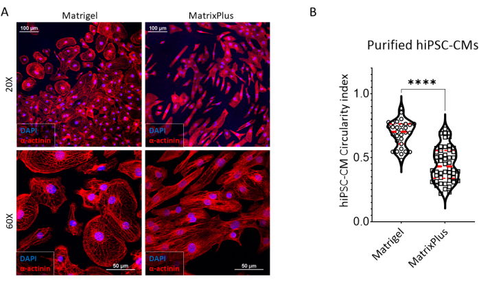
Figure 2: Comparison of sarcomere organization of hiPSC-CMs plated on a mouse ECM or an MECM. (A) Mouse ECM-cultured hiPSC-CMs immunostained against α-actinin indicate radial morphology, with a lower density of sarcomeres dispersed through the cardiomyocyte in contrast to hiPSC-CMs from the same batch plated on the matrix and presenting rod-shaped morphology (20x). (60x) Observation of hiPSC-CMs with confocal microscopy shows that hiPSC-CMs cultured on a mouse ECM have radial symmetry morphology, with a denser perimetral distribution of sarcomeres and a low density of radial sarcomeres in contrast to hiPSC-CMs from the same batch that were cultured on the MECM. They present a homogeneous distribution of sarcomeres organized along the longer axis of the cells. Scale bars = 100 µm (upper); 50 µm (lower). (B) Staining of hiPSC-CMs cultured on the mouse ECM or the MECM with a mitochondrial dye that stains mitochondria with high transmembrane potential show a lower intensity of staining in cardiomyocytes cultured on the mouse ECM in comparison to the MECM. Furthermore, hiPSC-CMs cultured on the MECM have mitochondria homogenously distributed in the cardiomyocytes, in contrast to hiPSC-CMs cultured on the mouse ECM that present a perinuclear accumulation of mitochondria. Scale bars = 200 µm. Abbreviations: hiPSC-CMs = human induced pluripotent stem cell-derived cardiomyocytes; ECM = extracellular matrix; MECM = maturation-inducing ECM; DAPI = 4',6-diamidino-2-phenylindole. Please click here to view a larger version of this figure.
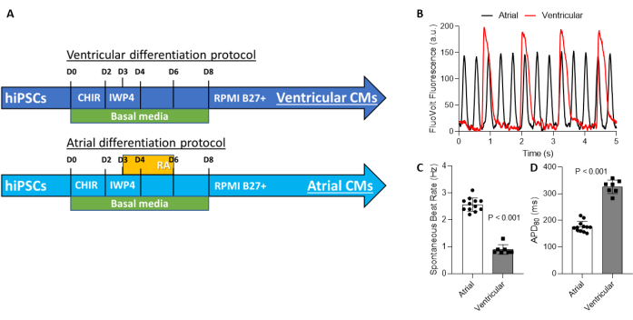
Figure 3: Production of chamber specific cardiomyocytes. (A) Protocols for production of chamber-specific cardiomyocytes share identical Wnt signalizing pathway manipulation, with thr stimulation of Wnt signaling by inhibition of GSK3 from day 0 to 2, and the inhibition of this pathway between day 2 and 4. Chamber specification is achieved with activation of the retinoic acid pathway and Wnt signaling manipulation between days 3 and 6. (B) As a result of chamber specification, atrial cardiomyocytes present a faster rate of spontaneous depolarization in comparison to ventricular cardiomyocytes. (C) Ventricular hiPSC-CMs have slower beat rates in comparison to atrial hiPSC-CMs; therefore, the action potential duration at 80% of repolarization is shorter in hiPSC-ACMs in comparison to hiPSC-VCMs. Abbreviations: hiPSC-CMs = human induced pluripotent stem cell-derived cardiomyocytes; hiPSC-ACM = atrial human induced pluripotent stem cell-derived cardiomyocytes; hiPSC-VCM = ventricular human induced pluripotent stem cell-derived cardiomyocytes. Please click here to view a larger version of this figure.
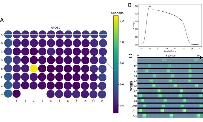
Figure 4: Optical mapping acquired with an optical mapping device and analyzed with Pulse. (A) Example of a heatmap for the holistic observation of parameters assessed in a 96-well plate after movie filtration and determination of regions of interest in 96-well plates, mapped with the optical mapping device. In this example, a APD80% heatmap that indicates an outlier well (E4) and wells that failed to produce data (wells H3, 4, and 5). (B) Furthermore, the user-friendly interface allows easy plotting of average action potential morphology from the selected wells. (C) Additional data visualization tools are available; in this example, a time-space plot generated from the horizontal line crossing the wells on row A (panel A) shows activation across a horizontal section of each well (white line across row A) over a period of 10 s. Please click here to view a larger version of this figure.
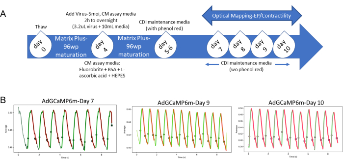
Figure 5: Timeline for mapping of intracellular calcium transient changes with a genetically encoded calcium indicator. On day 4 after plating commercially available cardiomyocytes in an MECM-coated 96-well plate, as indicated in Figure 1A, the cells should be transduced with 5 MOI of virus in CM assay medium overnight. The medium is replaced with CDI maintenance medium until day 6, and changed to CDI maintenance medium without phenol red between days 7 and 11 to allow for prompt or continuous monitoring of intracellular calcium transient changes with Nautilus. (B) hiPSC-CMstransduced with AdGCaMP6f assessed with optical mapping on days 7, 9, and 10 post-thaw indicate the presence of stable, intracellular calcium-mediated fluorescence changes, that allow for daily optical mapping of the same plate over extended period of times without the need for reapplication of calcium-sensitive dyes. Abbreviations: GECI = genetically encoded calcium indicator; CM = cardiomyocyte; MOI = multiplicity of infection; 96wp = 96-well plate; BSA = bovine serum albumin. Please click here to view a larger version of this figure.
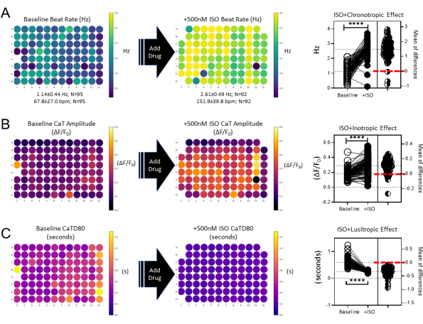
Figure 6: Quick and easy utilization of heatmaps for visual comparison of data acquired from mature functional syncytia of hiPSC-CMs with Nautilus and analyzed with Pulse. (A) hiPSC-CMs treated with isoproterenol show an increase in beat rate, as observed by the inspection of heatmaps and confirmed with paired t-test. (B) Similarly, mapping of cells before and after isoproterenol treatment shows the inotropic effect of β-adrenergic stimulation by visual comparison of heatmaps and with paired t-test. (C) Lastly, utilization of heatmaps for visual comparison of data show lusitropy, another canonical effect of β-adrenergic stimulation, confirmed with paired t-test (p < 0.0001). Absence of a circle indicates failure in data acquisition/analysis for that specific well. Abbreviations: hiPSC-CMs = human induced pluripotent stem cell-derived cardiomyocytes; ISO = isoproterenol. Please click here to view a larger version of this figure.
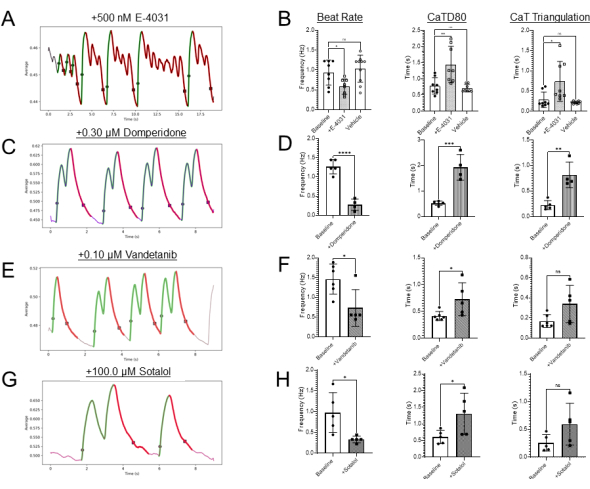
Figure 7: Validation of GECI cardiotoxicity screening assay using hERG channel blockers. (A) Representative spontaneous calcium flux traces from baseline wells in HBSS and in the presence of 500 nM E-4031. (B–D) Quantification of baseline and +E-4031 effects on beat rate, calcium transient duration 80 (CaTD80), and calcium transient triangulation (CaT triangulation) respectively. *,** denotes significant difference; unpaired t-test; p < 0.01; n = 8 in each group. (E) GECI detection of another hERG blocker, domperidone. (F) GECI detection of hERG block induced by vandetanib. (G) GECI detection of hERG block by high dose of sotalol. Abbreviations: GECI = genetically encoded calcium indicator; hERG = human Ether-a-go-go-related gene. Please click here to view a larger version of this figure.
Table 1: Media and their compositions Please click here to download this Table.
There are several different approaches to in vitro cardiotoxicity screening using hiPSC-CMs. A recent "Best Practices" paper on the use of hiPSC-CMs presented the various in vitro assays, their primary readouts, and importantly, each assay's granularity to quantify human cardiac electrophysiological function20. In addition to using membrane-piercing electrodes, the most direct measure of human cardiac electrophysiological function is provided by VSDs. VSD assay readouts enable direct visualization and quantification of critical electrophysiological parameters, including action potential duration, action potential propagation velocity, action potential upstroke, action potential triangulation, beat rate, beat regularity, and action potential duration heterogeneities. Similarly, calcium-sensitive probes offer information on hiPSC-CM monolayer rhythm, rate, and event durations. hiPSC-CM calcium transient measurements made using fluorescent probes also provide information on the contractility and contractile strength of each contraction. This paper provides methods for the use of mature hiPSC-CMs (96-well plates) in high-throughput VSD and calcium transient measurement assays. In addition to methods for optical mapping, software for high-throughput EP data analysis is presented.
The methods outlined here are a significant advance for the cardiotoxicity screening and regulatory science fields. Here, we have presented methods for the rapid maturation and electrophysiological recording of 2D hiPSC-CM monolayers in high-throughput screening plates (96-well plates). Rapid maturation using an MECM (7 days) shown here is a major advance over previous approaches requiring maturation to occur over 30-100 days22,23. Compared to other ECMs, which require manual application for each experiment, MECM plates are precoated with ECM and arrive in the laboratory ready to use. This aspect of the MECM makes it easier to use, less variable, and more efficient than using other ECM coatings. Importantly, this approach can be used for both cryopreserved, commercially available hiPSC-CMs and "homemade" hiPSC-CMs, which can be chamber-specific. Owing to the distinct, rod-shaped structure of mature hiPSC-CMs (Figure 1 and Figure 2), it is important to point out that a greater number of cells is required for confluent monolayer formation here, compared to protocols that utilize other ECMs. Notably, when using mouse ECM (cells have a continuously spreading pancake phenotype), we plate 50,000 hiPSC-CMs per well, but when using MECM, we plate 75,000 hiPSC-CMs per well. The number of CMs per well can also be reduced if the cells are to be used for single-cell analysis, such as patch clamp or any other imaging technique requiring single cells.
Commercially available hiPSC-CMs offer advantages for regulatory science due to the extensive characterization of these cells by Food and Drug Administration (FDA)-led international efforts. However, the commercially available cells are a mixture of nodal, atrial, and ventricular hiPSC-CMs, which provide highly relevant toxicity information but lack the chamber-specific features that mimic the human heart. Chamber-specific hiPSC-CMs recreate the well-known electrophysiological differences between atrial and ventricular cardiomyocytes, and provide an in vitro assay for the development of chamber-specific anti-arrhythmic therapies (Figure 3B–D). For example, atrial fibrillation-specific medications can now be tested and developed using hiPSC-ACM monolayers plated in 96-well plates for robust and rigorous data collection. Likewise, when screening to determine if a compound causes Torsades de Pointes (TdP), a high-risk ventricular arrhythmia-, it is optimal not to have atrial and nodal cells "contaminating" ventricular cardiac monolayers. Therefore, although the FDA-led hiPSC-CM validation efforts to date have focused on using commercially available CMs, it is likely that future regulatory science recommendations will turn to the use of chamber-specific cells to make the toxicity screening even more predictive of the human cardiac condition. The methods outlined here are based on previous reports, and provide a robust approach for the generation of chamber-specific cardiomyocytes derived from pluripotent stem cells21.
A major difference between these protocols and those typically used in the field is the purification approach used. The majority of laboratories that generate hiPSC-CMs in their own labs rely on metabolic-mediated selection of cardiomyocytes22. Here, we rely on using MACS to purify hiPSC-CMs, using a clinically approved cell processing approach that produces CMs with healthier phenotypes23. The metabolic challenge approach is effective, but uses a media formulation that simulates myocardial ischemia24. When utilizing MACS purification of hiPSC-CMs, it is important to utilize the non-CM depletion cocktail, which targets non-CMs for magnetic depletion from the cell population. Use of the non-CM depletion approach minimizes the shear stress that the CMs experience in the magnetic column, and is preferred over direct magnetic labeling of the CM population. Using MACS purification of chamber-specific cells will enable other laboratories to generate healthy CMs for research and toxicity testing.
The authors have nothing to disclose.
This work has been supported by NIH grants HL148068-04 and R44ES027703-02 (TJH).
0.25% Trypsin EDTA | Gibco | 25200-056 | |
0.5 mg/mL BSA (7.5 µmol/L) | Millipore Sigma | A3294 | |
2.9788 g/500 mL HEPES (25 mmol/L) | Millipore Sigma | H4034 | |
AdGCaMP6m | Vector biolabs | 1909 | |
Albumin human | Sigma | A9731-1G | |
alpha actinin antibody | ThermoFisher | MA1-22863 | |
B27 | Gibco | 17504-044 | |
Blebbistatin | Sigma | B0560 | |
CalBryte 520AM | AAT Bioquest | 20650 | |
CELLvo MatrixPlus 96wp | StemBiosys | N/A | https://www.stembiosys.com/products/cellvo-matrix-plus |
CHIR99021 | LC Laboratories | c-6556 | |
Clear Assay medium (fluorobrite) | ThermoFisher | A1896701 | For adenovirus transduction |
DAPI | ThermoFisher | 62248 | |
DMEM:F12 | Gibco | 11330-032 | |
FBS (Fetal Bovine Serum) | Sigma | F4135-500ML | |
FluoVolt | ThermoFisher | F10488 | |
HBSS | Gibco | 14025-092 | |
iCell CM maintenance media | FUJIFILM/Cellular Dynamics | M1003 | |
iCell2 CMs | FUJIFILM | 1434 | |
Incucyte Zoom | Sartorius | ||
iPS DF19-9-11T.H | WiCell | ||
Isoproterenol | MilliporeSigma | CAS-51-30-9 | |
IWP4 | Tocris | 5214 | |
L-ascorbic acid 2-phosphate sesquimagnesium salt hydrate | Sigma | A8960-5g | |
L-glutamine | Gibco | A2916801 | |
LS columns | Miltenyii Biotec | 130-042-401 | |
MACS Buffer (autoMACS Running Buffer) | Miltenyii Biotec | 130-091-221 | |
Matrigel | Corning | 354234 | |
MitoTracker Red | ThermoFisher | M7512 | |
Nautilus HTS Optical Mapping | CuriBio | https://www.curibio.com/products-overview | |
Nikon A1R Confocal Microscope | Nikon | ||
nonessential amino acids | Gibco | 11140-050 | |
pre-separation filter | Miltenyii Biotec | 130-041-407 | |
PSC-Derived Cardiomyocyte Isolation Kit, human | Miltenyii Biotec | 130-110-188 | |
Pulse | CuriBio | https://www.curibio.com/products-overview | |
Quadro MACS separator (Magnet) | Miltenyii Biotec | 130-091-051 | |
Retinoic acid | Sigma | R2625 | |
RPMI 1640 | Gibco | 11875-093 | |
RPMI 1640 (+HEPES, +L-Glutamine) | Gibco | 22400-089 | |
StemMACS iPS-Brew XF | Miltenyii Biotec | 130-107-086 | |
TnI antibody (pan TnI) | Millipore Sigma | MAB1691 | |
Versene (ethylenediaminetetraacetic acid – EDTA solution) | Gibco | 15040-066 | |
Y-27632 dihydrochloride | Tocris | 1254 | |
β-mercaptoethanol | Gibco | 21985023 |