We describe a modified technique for resecting the posterior vertebral column unilaterally based on a modified trephine for patients with thoracolumbar kyphotic deformity.
Old compression vertebrae fracture or congenital kyphoscoliosis with abnormal vertebral body development and other diseases that invade the spine may cause severe thoracolumbar kyphotic deformity, often accompanied by intractable low back pain or compression of the spinal cord, leading to severe neurological symptoms or even paralysis. If conservative treatment cannot relieve the symptoms or correct the deformities, surgical treatment is usually needed. For severe kyphotic deformity, reconstruction of the physiological curvature and rigid fixation determine the prognosis of the patients. Osteotomy and orthopedics are the standard procedure for deformities with severe compression of the front and middle column, but the trauma to the patients is high, with a long operation time and massive blood loss. To avoid these disadvantages, we have developed a modified technique to remove the diseased vertebra unilaterally. In this technique, we use a modified trephine to resect the vertebral columns like in the pedicle screw technique by adding a locking instrument that can restrict the trephine to lower the risk of osteotomy and shorten the surgery time and blood loss.
Thoracolumbar kyphotic deformity is a primary or secondary disease generally caused by vertebrae fracture, vertebral body development, ankylosing spondylitis, and spinal tuberculosis1,2,3,4. Severe kyphotic deformity often induces spinal cord compression or severe low back pain. Once conservative treatment becomes ineffective, a surgical approach is necessary due to the complications caused by the deformity. However, the appropriate surgical treatment remains controversial.
The surgical approach to treat severe kyphotic deformity usually needs grade 3 or higher osteotomy5. Pedicle subtraction osteotomy (PSO) is a technique in which a three-column osteotomy can be achieved, which is reported with correction between 30° and 40°6. When the kyphotic deformity is more than 40°, vertebral column resection is recommended, but it can shorten the spine and induce spinal cord bulk7. Partial body and disc resection (BDBO; grade 4) and posterior vertebral column resection (PVCR; grade 5) require complete interception of the anterior and middle columns of the spine, which may cause huge damage to the spine with severe neurologic complications due to spinal instability in the operation or postoperative implant settlement7,8. For general operators, the technique is hard to master, and the surgical damage is enormous for the patients. Thus, a method that is easier to perform with less damage is needed.
In this report, we introduce a refined surgical technique with a modified trephine to treat thoracolumbar kyphosis by removing the vertebral and adjacent discs unilaterally and placing a titanium mesh with autologous bone on the same side. This technique aims to minimize the damage to the patient while achieving good results. In our previous research, the refined surgical procedure showed significantly greater outcomes and reduced the damage to the spine through the preservation of the contralateral pedicle and part of the vertebral body9.
The protocol follows the guidelines of the Ethics Committee of the Third Hospital of Hebei Medical University. Informed consent was obtained from the patients for including them and the data generated as a part of this study.
1. Pre-operative preparation
2. Lesion exposure
3. Deformity correction
4. Closing the incision
5. Post-operative care
About 330° decompression can be achieved by using the unilateral PVCR technique. The transverse process and the rib head need to be removed to make sure the abduction angle is enough to remove the contralateral bone.
By using the modified trephine, the bone of the diseased vertebrae can be removed easily by rotating it with mild stress. When the trephine is locked, one should pull out the trephine and probe together, and then a cylinder of cancellous bone can be obtained (Figure 2).
Not all patients require a pedicle screw implantation in the osteotomy segment. For some patients with mild vertebral compression and preoperative computed tomography showing there is enough space to implant a pedicle screw, the screw can be implanted on the opposite side of the osteotomy segment. The screw does not need to be taken out once it is implanted because instability can happen without screw fixation. If a patient is eligible for the implantation, it means the patient's segment that needs osteotomy has enough bones. The range of osteotomy can be reduced appropriately on the opposite side as long as it does not interfere with the osteotomy side.
In previous studies, compared to traditional PVCR, unilateral PVCR achieved a satisfactory result but reduced the operating time (174.6 min ± 26.7 min vs. 226.4 min ± 32.6 min), the blood loss (870.3 mL ± 92.5 mL vs. 997.4 mL ± 107.3 mL), and the incidence of nerve root impingement (4.3% vs. 8.7%)13. With the use of the modified trephine, we further shortened the osteotomy time and gained satisfactory treatment results11. In addition, we were able to combine the unilateral PVCR technique and modified trephine to remove the lesions and rebuild spinal stability for patients with Kummell disease (Figure 3 and Figure 4)9,14, especially for patients with kyphotic deformity and obvious nerve-oppressed symptoms.
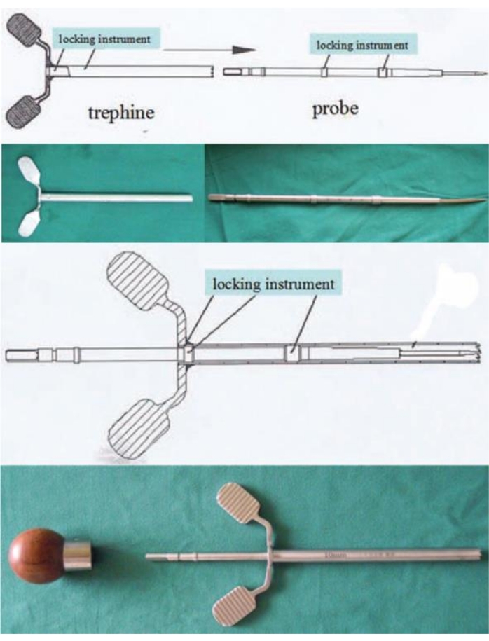
Figure 1: The modified trephine with the locking instrument. Use the probe to stab into the cancellous bone, and then rotate and push the trephine until it is locked. Put on the handle, and then pull out the probe and trephine together. This figure has been modified from Wang et al.11. Please click here to view a larger version of this figure.
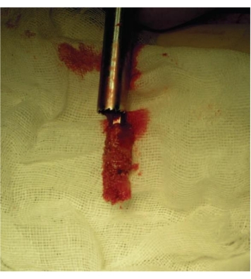
Figure 2: The bone removed by the trephine. Grab the handle of the probe and pull it out, and a columnar bone can be harvested. This figure has been modified from Wang et al.11. Please click here to view a larger version of this figure.
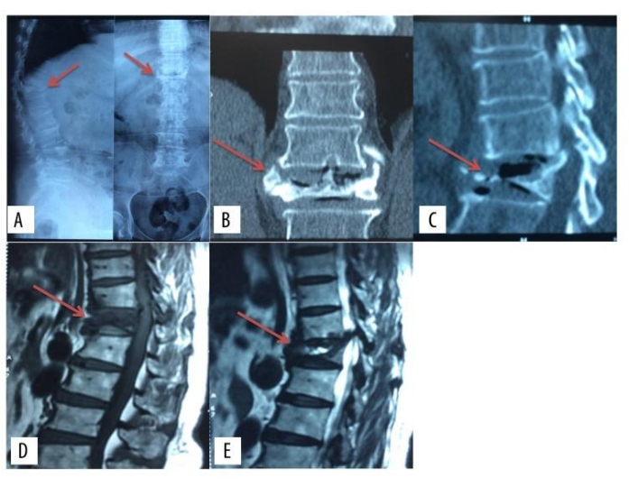
Figure 3: Preoperative images of the unilateral PVCR technique. Preoperative images of X-ray, computed tomography, and magnetic resonance imaging of a patient with Kummell Disease. This figure has been modified from Yang et al.9. Please click here to view a larger version of this figure.
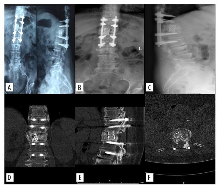
Figure 4: Postoperative images of the unilateral PVCR technique. Postoperative images of X-ray and computed tomography of a patient with Kummell Disease after the treatment of modified posterior vertebral column resection. This figure has been modified from Yang et al.9. Please click here to view a larger version of this figure.
The steps of placing the temporary fixation rod and the deformity correction mentioned in the protocol are the critical steps during the surgery. By preserving one side of the pedicle and loading a temporary fixation bar, stability is preserved during the osteotomy procedure. During the surgical progress of the deformity correction, the nerve roots must be protected to prevent serious postoperative neurological complications. If the surgeons are uncertain about the location of the nerve roots, the exposure of the nerve roots after removing the vertebral plate and the articular process is necessary. When removing the wall of the vertebral canal, blood loss can be huge due to the venous plexus. Although blood loss may be reduced using bipolar electrocoagulation to stop the bleeding, accompanied by gelatin sponges and brain cotton pieces layer-by-layer, the long surgery time is a big problem. The surgeons can prepare blood transfusion in advance and use an autologous blood transfusion system because of the long operation time and huge blood loss. If a dural matter tear happens during the surgery, the surgeons can use dural suture to fix the damage; a pedicled multifidus muscle flap is also useful according to previous research15. To prevent damage to the abdominal aorta or thoracic aorta, surgeons must be careful in removing the bone near the anterior longitudinal ligament. In addition, a C-arm can be used to detect the position of the pedicle probe to avoid over-insertion damage caused by the serrated top of the trephine. The cancellous bone collected by the trephine can be directly used in the titanium cage for the autologous bone graft.
The unilateral PVCR technique shortens the operation time compared to the classic PVCR as only one side of the vertebral pedicle access is required. Subsequently, the unilateral PVCR technique reduces the stimulation to the spinal cord and nerve roots. By using this approach, the contralateral vertebral pedicle and parts of the vertebra can be saved to increase the stability and bone fusion rate compared to total laminectomy. However, this technique is solely based on classic PVCR with the innovation of the tools. Therefore, similar to the original PVCR, the implant-related complications would still happen due to osteotomy16.
With the use of modified trephine, bone excision becomes easier and more controllable than the traditional use of a high-speed drill and osteotome. Surgeons with the ability of pedicle screw placement can master the refined technique quickly. The unilateral PVCR technique with the modified trephine provides a quicker way to resect and collect the bone, shortening the surgical time for surgeons to do more delicate operations. It is shown that the reconstruction of physiological curvature and the correction of spinal deformity can alleviate patients' symptoms, including low back pain17. Therefore, this unilateral PVCR technique can reconstruct spinal stability and rigid fixation to achieve satisfactory treatment effects with less trauma to the patients.
The authors have nothing to disclose.
None.
Adhesive | Biatain | 3420 | 12.5 cm x 12.5 cm |
Bipolar electrocoagulation tweezers | Juan'en Medical Devices Co.Ltd | BZN-Q-B-S | 1.2 mm x 190 mm |
Bone wax | ETHICON | W810T | 2.5g |
Curette | Qingniu | 20739.01 | 300 x Ø9 x 5° |
Double jointed forceps | SHINVA | 286920 | 240 mm x 8 mm |
High frequency active electrodes | ZhongBangTianCheng | GD-BZ | GD-BZ-J1 |
Laminectomy rongeur | Qingniu | 2054.03 | 220 x 3.0 x 130° |
Laminectomy rongeur | Qingniu | 2058.03 | 220 x 5.0 x 130° |
Pedicle screw | WEGO | 800386545 | 6.5 mm x 45 mm |
Pedicle screw | WEGO | 800386550 | 6.5 mm x 50 mm |
Pituitary rongeur | Qingniu | 2028.01 | 220 mm x 3.0 mm |
Pituitary rongeur | Qingniu | 2028.02 | 220 mm x 3.0 mm |
Rod | WEGO | 800386040 | 5.5 mm x 500 mm |
Surgical drainage catheter set | BAINUS MEDICAL | SY-Fr16-C | 100-400 mL |
Surgical film | 3L | SP4530 | 45 cm x 30 cm |
Titanium cage | WEGO | 9051228 | 19 mm x 80 mm |
Trephine | NATON MEDICAL GROUP | DJD04130 | 12 mm/10 mm |