This protocol presents the use of CRISPR SunTag-p65-HSF1 (SPH) in adipocytes (AdipoSPH) as an alternative strategy to adeno-associated virus (AAV) for investigating beige fat biology. In vivo injection of AAV-carrying sgRNA targeting the endogenous Prdm16 gene is sufficient to induce beige fat development and enhance the thermogenic gene program.
Clustered regularly interspaced short palindromic repeats (CRISPR) technology has prompted a revolution in biology, and recent tools have been applied far beyond the originally described gene editing. The CRISPR activation (CRISPRa) system combines the catalytically inactive Cas9 (dCas9) protein with distinct transcription modules to induce endogenous gene expression. SunTag-p65-HSF1 (SPH) is a recently developed CRISPRa technology that combines components of synergistic activation mediators (SAMs) with the SunTag activators. This system allows the overexpression of single or multiple genes by designing a customized single-guide RNA (sgRNA). In this study, a previously developed SPH mouse was used to generate a conditional mouse expressing SPH in adipocytes (adiponectin Cre lineage), named AdipoSPH. To induce a white-to-beige fat (browning) phenotype, an adeno-associated virus (AAV) carrying sgRNA targeting the endogenous Prdm16 gene (a well-established transcription factor related to brown and beige fat development) was injected into the inguinal white adipose tissue (iWAT). This mouse model induced the expression of endogenous Prdm16 and activated the thermogenic gene program. Moreover, in vitro SPH-induced Prdm16 overexpression enhanced the oxygen consumption of beige adipocytes, phenocopying the results of a previous Prdm16 transgenic mouse model. Thus, this protocol describes a versatile, cost-effective, and time-effective mouse model for investigating adipose tissue biology.
Beige (or brite) adipocytes are uncoupling protein 1 (UCP1)-expressing and mitochondrial-rich adipocytes that reside within white adipose tissue (WAT) depots. Beige fat emerges from a subset of adipocyte progenitors or mature white adipocytes in response to cold exposure and other stimuli1,2. Beige adipocytes can convert energy into heat in a UCP1-dependent or independent manner3. Regardless of its thermogenic function, beige fat can also improve metabolic health by other means, such as the secretion of adipokines and anti-inflammatory and anti-fibrotic activities. Studies in mice and humans have shown that the induction of beige fat improves whole-body glucose and lipid homeostasis3. However, although our knowledge of beige fat biology has evolved rapidly in recent years, most of its metabolic benefits and related mechanisms are still not fully understood.
Clustered regularly interspaced short palindromic repeats (CRISPR) were first described in eukaryotic cells as a tool capable of generating a double-strand break (DSB) at a specific site in the genome through the nuclease activity of the Cas9 protein4,5. Cas9 is guided by a synthetic single-guide RNA (sgRNA) to target a specific genomic region, leading to a DNA DSB. In addition to using the nuclease Cas9 for editing purposes, CRISPR-Cas9 technology has evolved to be used as a sequence-specific gene regulation tool6. The development of a catalytically inactive Cas9 protein (dCas9) and the association of transcriptional modules capable of enhancing gene expression has given rise to CRISPR activation (CRISPRa) tools. Several CRISPRa systems have emerged, such as VP647,8, synergistic activation mediator (SAM)9, SunTag10,11, VPR12,13, and SunTag-p65-HSF1 (SPH)14, which combines the components of SAM and SunTag activators. It has recently been demonstrated that the induced expression of neurogenic genes in N2a neuroblasts and primary astrocytes is higher using SPH compared to other CRISPRa systems14, demonstrating SPH as a promising CRISPRa tool.
Here, we took advantage of a previously developed SPH mouse14 to generate a conditional mouse model expressing SPH specifically in adipocytes using the adiponectin Cre lineage (AdipoSPH). Using an adeno-associated virus (AAV) carrying the gRNA targeting the endogenous Prdm16 gene, browning (white to beige conversion) of inguinal WAT (iWAT) was induced to increase the expression of the thermogenic gene program. Moreover, in vitro Prdm16 overexpression enhanced oxygen consumption. Therefore, this protocol provides a versatile SPH mouse model for exploring the mechanisms of beige fat development within adipose tissue.
Animal studies were performed in accordance with the University of Campinas Guide for the Care and Use of Laboratory Animals (protocol CEUA #5810-1/2021).
1. Molecular cloning
2. AAV packaging
NOTE: AAV packaging was performed according to previous publications15,16 with minor modifications.
3. Titration of the AAV by qPCR
4. In vivo injection of AAV into the inguinal white adipose tissue (iWAT)
5. In vitro differentiation of stromal vascular cells (SVFs) into beige adipocytes
6. In vitro AAV infection of SVFs
NOTE: SVFs derived from AdipoSPH mice iWAT were infected with AAV-carrying sgRNA-Prdm16 as previously described by Wang et al.18 with a few modifications.
AdipoSPH mice were developed by breeding SPH and Adipoq-Cre mouse strains. Both mouse strains were in a hybrid C57BL6J-DBA/2J background (according to the commercial supplier; see Table of Materials). The SPH mouse lineage was originally described by Zhou et al.14.
In vivo beige adipocyte development through AdipoSPH-mediated Prdm16 overexpression
To evaluate the capacity of the model described in this study to develop beige adipocytes in vivo, the AAV carrying the sgRNA targeting Prdm16 gene was injected into the iWAT of AdipoSPH mice. Prdm16 is a well-established transcription factor that determines beige adipocyte development and function20,21. It is important to mention that here we chose a previously tested and validated sgRNA targeting the endogenous Prdm16 gene14. AAV carrying an empty sgRNA was used as a control. At 10 days after AAV injection, iWAT was harvested for histological and gene expression analyses (Figure 1A). As expected, immunofluorescence images of iWAT from AdipoSPH mice demonstrated the expression of dCas9 in both the control and Prdm16 groups (Figure 1B). Additionally, mCherry expression confirmed the success of AAV infection of iWAT in both the control and Prdm16 groups (Figure 1B). SPH-induced Prdm16 expression clearly induced a widespread accumulation of multilocular beige adipocytes into iWAT (Figure 1B). Moreover, quantitative PCR (qPCR) revealed an increased expression of Prdm16 and the thermogenic gene program (Ucp1, Cox8b, Ppargc1a, and Cidea) in the Prdm16 group compared to the control (Figure 1C).
Promotion of beige adipocyte development and enhancement of oxygen consumption in vitro by AdipoSPH-mediated Prdm16 overexpression
Next, the suitability of this model for studying beige adipocyte biology in vitro was investigated. To this end, preadipocytes derived from the iWAT of AdipoSPH mice were transduced with AAV carrying the sgRNA sequence targeting Prdm16. At seven days after differentiation, beige adipocytes were used for the analysis of gene expression and oxygen consumption (Figure 2A). Empty sgRNA was used as a control. It is worth noting that the removal of STOP codon by CRE recombinase and activation of SPH machinery was under the control of the adiponectin promoter/enhancer, resulting in the activation of the endogenous Prdm16 gene in mature adipocytes. Gene expression analysis confirmed the increased expression of the endogenous Prdm16 gene and thermogenic genes in the Prdm16 overexpression group compared to the control (Figure 2B). To confirm that the SPH-induced beige adipocytes were functionally thermogenic, a high-resolution respirometry assay was performed on the primary adipocytes. The respirometry data analysis and interpretation was performed as recently described22. SPH-induced Prdm16 expression resulted in higher basal and maximal oxygen consumption than that in control cells (Figure 2C). Importantly, the data indicated enhanced uncoupled respiration (oligomycin-insensitive) in the Prdm16 group compared with that in control group (Figure 2C, Table 3). Taken together, these results revealed that SPH-induced expression of endogenous Prdm16 recapitulates the browning phenotype and enhances oxygen consumption rates observed in conventional Prdm16 Tg mice.
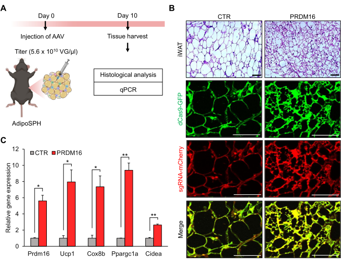
Figure 1: Beige fat development by injection of adeno-associated virus (AAV) targeting endogenous Prdm16 gene into inguinal white adipose tissue (iWAT) of AdipoSPH mice. (A) Schematic illustration of the in vivo beige adipocyte experimental design (created using Biorender.com). (B) Histological (hematoxylin and eosin [H&E] staining) images of iWAT. Scale bar = 50 µm. (C) Quantitative PCR (qPCR) of Prdm16 and thermogenic genes (Ucp1, Cox8b, Ppargc1α, and Cidea; n = 3). Data are presented as mean ± SEM. *p < 0.05; **p < 0.01 for Prdm16 versus control (CTR) by unpaired Student's t-test. Please click here to view a larger version of this figure.
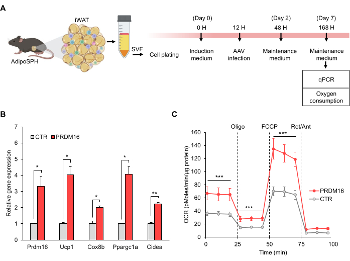
Figure 2: SPH-induced beige adipocyte differentiation by Prdm16 overexpression. (A) Schematic illustration of the in vitro beige adipocyte experimental design (created using Biorender.com). (B) Relative gene expression of Prdm16 and thermogenic genes (Ucp1, Cox8b, Ppargc1α, and Cidea; n = 3). (C) Oxygen consumption rate (OCR), n = 6. Data are presented as mean ± SEM. *p < 0.05; **p < 0.01; ***p < 0.001 for Prdm16 versus control (CTR) by (B) unpaired Student's t-test and (C) two-way repeated-measures ANOVA, followed by Tukey's test. Please click here to view a larger version of this figure.
Supplemental Figure 1: Sanger sequencing of Prdm16 sgRNA. (A) Schematic illustration demonstrating the alignment of single-stranded complementary Prdm16 oligonucleotides. (B) Sanger sequencing of Prdm16 sgRNA using universal primer. Please click here to download this File.
Gene | Species | Forward | Reverse | |||||
Gene expression | Prdm16 | mouse | CAGCACGGTGAAGCCATTC | GCGTGCATCCGCTTGTG | ||||
Ucp1 | mouse | TCTCAGCCGGCTTAATGACTG | GGCTTGCATTCTGACCTTCAC | |||||
Ppargc1a | mouse | AGCCGTGACCACTGACAACGAG | GCTGCATGGTTCTGAGTGCTAAG | |||||
Cox8b | mouse | GAACCATGAAGCCAACGACT | GCGAAGTTCACAGTGGTTCC | |||||
Cidea | mouse | ATCACAACTGGCCTGGTTACG | TACTACCCGGTGTCCATTTCT | |||||
36B4 | mouse | TCCAGGCTTTGGGCATCA | CTTTATCAGCTGCACATCACTCAGA | |||||
Molecular cloning | sgRNA Prdm16 sequence | mouse | CGAGCTGCGCTGAAAAGGGG | CCCCTTTTCAGCGCAGCTCG | ||||
universal primer | mouse | GAGGGCCTATTTCCC ATGATTCCTTCATAT |
Table 1: Primer sequences used in the study.
Medium | Composition | |||
Complete medium | Dulbecco's Modified Eagle Medium (DMEM) with L-Glutamine | |||
10% of fetal bovine serum (FBS) | ||||
2.5% of penicillin/streptomycin | ||||
Induction medium | Complete medium | |||
Indomethacin, final concentration 125 μM (0.125 M stock in ethanol). NOTE: Indomethacin must be heated to 90 °C for 10 seconds to be dissolved. | ||||
Insulin, final concentration 20 nM (1 mM stock, Add 1 μL of HCl to solubilize (5.73 mg/mL). Store stock at -20 °C. | ||||
Dexamethasone, final concentration 2 μg/mL (2 mg/mL stock in ethanol) | ||||
3-Isobutyl-1-methylxanthine (IBMX), final concentration 500 μM (0.25 M stock in 0.5 M KOH ) | ||||
3,3',5-Triiodo-L-thyronine (T3), final concentration 1 nM (10 μM stock, dissolve T3 in 1 M HCl and EtOH 1:4 to stock). | ||||
Rosiglitazone, final concentration 1 μg/mL (1 mg/mL stock in ethanol) | ||||
Maintenance medium | Complete medium | |||
Insulin, final concentration 20 nM (1 mM stock). Add 1 μL of HCl to solubilize (5.73 mg/mL). | ||||
Rosiglitazone, final concentration 1 μg/mL (1 mg/mL stock in ethanol) |
Table 2: Composition of media used in the study.
Parameters | CTR | PRDM16 | p-value | ||
No mitochondrial oxygen consumption (pMoles/min/µg protein) | 8.19 ± 1.40 | 10.80 ± 1.83 | 0.01 | ||
Basal respiration (pMoles/min/µg protein) | 28.82 ± 5.20 | 52.58 ± 13.73 | 0.001 | ||
Maximal respiration (pMoles/min/µg protein) | 63.81 ± 9.80 | 122.94 ± 22.31 | < 0.001 | ||
Spare respiratory capacity (pMoles/min/µg protein) | 34.98 ± 11.09 | 70.36 ± 26.06 | 0.006 | ||
Oligo insensitive (pMoles/min/µg protein) | 8.27 ± 2.29 | 15.85 ± 5.48 | 0.005 | ||
Oligo sensitive (pMoles/min/µg protein) | 20.54 ± 5.68 | 36.72 ± 14.79 | 0.016 | ||
Coupling efficiency | 71.28 ± 1.51 | 69.84 ± 3.05 | 0.163 | ||
Cell RCR | 7.70 ± 1.46 | 7.75 ± 2.43 | 0.485 |
Table 3: Respiratory parameters of SPH-induced beige adipocytes.
One of the most useful non-editing applications of CRISPR technology is the interrogation of gene function through the activation of endogenous genes using CRISPRa systems6. SPH is a powerful CRISPRa that was originally described to induce the conversion of astrocytes into active neurons by targeting several neurogenic genes14. In this study, AdipoSPH was demonstrated to be a suitable tool for investigating beige fat biology by activating the expression of endogenous Prdm16 in adipocytes. SPH-induced Prdm16 overexpression in adipocytes leads to activation of the thermogenic gene program and induces the browning phenotype, phenocopying a previous transgenic (Tg) Prdm16 mice model23. Prdm16 was chosen in this study as the target for inducing beige adipocytes based on the central role of this transcription factor in regulating brown and beige fat differentiation, as well as the regulation of the thermogenic gene program (e.g., enhancing Ucp1 expression) in differentiated adipocytes. Nevertheless, the current model allows for overexpression of any endogenous gene at the discretion of the researchers and the aim of the study.
Traditional transgenic models investigating adipose tissue biology rely on the development of genetically modified rodents overexpressing a transgene under the control of a promoter/enhancer restricted to adipocytes (for example, Fabp4 or adiponectin)24. Although current technology has accelerated the pace of the development of these models, the costs and time to develop them are still a common issue experienced by the scientific community. In contrast, AdipoSPH is a cheaper and more versatile model for gain-of-function approaches than traditional Tg mice. Moreover, AdipoSPH is well suited for studying long non-coding RNA and transcript isoforms, which are not particularly suitable for Tg mice models.
AdipoSPH also presents several advantages in investigating adipose tissue biology compared to the simple administration of AAV, a current alternative approach for gain-of-function experiments25. First, AAV delivered to adipose tissue promotes overexpression by introducing several copies of a transgene. In contrast, the SPH activation of endogenous genes represents a more natural mechanism of action. Second, transgenes delivered by AAV (using constitutive promoters) normally result in their overexpression in “any cell type” within the adipose tissue microenvironment. In contrast, AdipoSPH-induced expression of endogenous genes is restricted to adipocytes. Third, the packaging size (~4.5kb) is a typical limitation of AAV for some transgenes26. However, AAV-carrying sgRNA requires a simple and easy cloning strategy for customized 20 nucleotides. Finally, AAV administration typically reaches systemic circulation and infects other tissues and organs. Although some AAVs contain microRNAs to mitigate the expression of transgenes in particular tissues, such as the liver and heart25, this strategy cannot prevent AAV infection in other tissues and organs. In contrast, AdipoSPH-induced expression of endogenous genes is limited to adipocytes, even considering some leakage of AAV into systemic circulation. Thus, AdipoSPH is also a suitable model for the systemic administration of AAV-carrying sgRNA and the investigation of adipose tissue regulation of whole-body energy homeostasis.
Although the AdipoSPH model offers some advantages, it has limitations that are important to consider. AAV is currently the most common platform for in vivo delivery of sgRNA. However, some challenges in AAV manufacturing and immunological issues remain unresolved26,27. Moreover, different amounts and distributions of injected AAV throughout adipose tissue result in intra- and inter-tissue variability of gene expression. We consider that sgRNA vector cloning, AAV production, and the homogeneous administration of AAV into the iWAT are critical steps for the success of this protocol. Finally, different sgRNAs distinctively activate the expression of endogenous genes in the SPH system14. The main recommendation is to design and test three to five sgRNA sequences within 200 bp upstream of the transcription start site (TSS) of the gene of interest. Therefore, defining the best sgRNA sequence for a target gene typically requires laborious in vitro assays prior to in vivo experiments.
AdipoSPH is also an attractive model for investigating other aspects of adipose-tissue biology. For instance, adipocytes promote the secretion of numerous adipokines28,29; however, the whole-body effects of most of these molecules remain poorly understood. AdipoSPH is a suitable model to address this issue by overexpressing single or multiple endogenous genes in mature adipocytes and investigating their local or systemic effects. Thus, AdipoSPH is a unique tool for studying the biology and physiology of adipose tissue.
The authors have nothing to disclose.
The authors thank the support received from Centro Multidisciplinar para Investigação Biológica na Área da Ciência em Animais de Laboratório (Cemib), Unicamp, for the generation of AdipoSPH mice, the Inmmunometabolism and Cell Signaling Laboratory, and National Institute of Science and Technology on Photonics Applied to Cell Biology (INFABIC) for all experimental support. We thank the financial support from Sao Paulo Research Foundation (FAPESP): 2019/15025-5; 2020/09308-1; 2020/14725-0; 2021/11841-2.
3,3',5-Triiodo-L-thyronine | Sigma-Aldrich | T2877 | |
3-Isobutyl-1-methylxanthine | Sigma-Aldrich | I5879 | |
AAVpro 293T Cell Line | Takarabio | 632273 | |
Amicon Ultra Centrifugal Filter | Merckmillipore | UFC510008 | 100 KDa |
Dexamethasone | Sigma-Aldrich | D1756 | |
Dulbecco's Modification of Eagles Medium (DMEM) | Corning | 10-017-CV | |
Dulbecco's Modified Eagle Medium (DMEM) F-12, GlutaMAX™ supplement | Gibco | 10565-018 | high concentrations of glucose, amino acids, and vitamins |
Dulbecco's phosphate buffered saline (DPBS) | Sigma-Aldrich | D8662 | |
Excelta Self-Opening Micro Scissors | Fisher Scientific | 17-467-496 | |
Fetal bovine serum | Sigma-Aldrich | F2442 | |
Fisherbrand Cell Scrapers (100 pk) | Fisher Scientific | 08-100-241 | |
Fisherbrand High Precision Straight Tapered Ultra Fine Point Tweezers/Forceps | Fisher Scientific | 12-000-122 | |
Fisherbrand Sharp-Pointed Dissecting Scissors | Fisher Scientific | 08-940 | |
Glycerol | Sigma-Aldrich | G5516 | |
HEPES | Sigma-Aldrich | H3375-25G | |
Hexadimethrine bromide (Polybrene) | Sigma-Aldrich | H9268 | |
Indomethacin | Sigma-Aldrich | I7378 | |
Insulin | Sigma-Aldrich | I9278 | |
LigaFast Rapid DNA Ligation System | Promega | M8225 | |
Maxiprep purification kit | Qiagen | 12162 | |
Microliter syringe | Hamilton | 80308 | Model 701 |
NEB 10-beta/Stable | New England Biolabs | C3019H | E. coli competent cells |
pAAV2/8 | Addgene | 112864 | |
pAAV-U6-gRNA-CBh-mCherry | Addgene | 91947 | |
pAdDeltaF6 | Addgene | 112867 | |
PEG 8000 | Sigma-Aldrich | 89510 | |
Penicillin/streptomycin | Gibco | 15140-122 | |
Polyethylenimine | Sigma-Aldrich | 23966 | Linear, MW 25000 |
Povidone-iodine | Rioquímica | 510101303 | Antiseptic |
Rosiglitazone | Sigma-Aldrich | R2408 | |
SacI enzyme | New England Biolabs | R0156 | |
Surgical Design Premier Adson Forceps | Fisher Scientific | 22-079-741 | |
Syringe | Hamilton | 475-40417 | |
T4 DNA Ligase | Promega | M180B | |
T4 DNA ligase buffer | New England Biolabs | B0202S | |
T4 PNK enzyme kit | New England Biolabs | M0201S | |
Tramadol Hydrochloride | SEM | 43930 | |
Vidisic Gel | Bausch + Lomb | 99620 |