Blood transports oxygen (via the hemoglobin protein) and other important nutrients to tissues in our bodies. When the flow of blood through tissues is interrupted (ischemia), severe damage to the tissues can occur, the most immediate effects of which are due to a lack of oxygen (hypoxia). Ischemic stroke is the result of interrupted blood flow to a certain region of the brain. The brain damage resulting from an ischemic stroke can occur within minutes of a vessel blockage, and can often have debilitating and lasting effects1,2. A highly valuable strategy to evaluate the physiopathology after ischemic stroke and identify and test new treatments is the use of small-animal models in the lab. Treatments discovered in the lab aim to be translated to clinical use and improve patients' lives. However, the use of animals in biomedical research needs to be carefully evaluated according to Russell and Burch’s 3Rs principles: replacement, reduction, and refinement3. The objective of the reduction component is to reduce the number of animals without compromising data collection. With this in mind, being able to longitudinally evaluate the lesion evolution via noninvasive imaging allows a great advantage in reducing the number of animals required, as well as maximizing the information obtained from each animal4.
Photoacoustic tomography (PAT) is a hybrid imaging modality that combines optical absorption contrast with ultrasound imaging spatial resolution5. The imaging mechanism of PAT is as follows. An excitation laser pulse is illuminated on the target being imaged. Assuming the target absorbs light at the wavelength of the excitation laser, it will increase in temperature. This quick increase in temperature results in a thermoelastic expansion of the target. The expansion causes an ultrasound wave to propagate out from the target. By detecting the ultrasound wave at many positions, the time required for the wave to propagate from the target to the detectors can be used to create an image through a reconstruction algorithm. The ability of PAT to detect optical absorption in deep tissue regions differentiates PAT from ultrasound imaging, which detects boundaries of differing acoustic impedances of tissues5. In the visible and near-infrared spectra, the primary highly absorbing biomolecules that are abundant in organisms are hemoglobin, lipids, melanin, and water7. Of particular interest in the study of stroke is hemoglobin. Since oxyhemoglobin and deoxyhemoglobin have different optical absorption spectra, PAT can be used with multiple excitation laser wavelengths to determine the relative concentration of the two states of the protein. This allows the oxygen saturation of hemoglobin (sO2), or blood oxygenation, to be quantified in and outside of the infarct region8,9. This is an important measure in ischemic stroke, as it can indicate the level of oxygen in the damaged brain tissue following ischemia.
Acoustic angiography (AA) is a contrast-enhanced ultrasound imaging method that is particularly useful for imaging the morphology of vasculature in vivo10. The method relies on the use of a dual-element wobbler transducer (a low frequency element and a high frequency element) in conjunction with microbubbles injected into the circulatory system of the imaging subject. The low-frequency element of the transducer is used for transmitting at the resonant frequency of the microbubbles (e.g., 2 MHz), while the high-frequency element is used to receive the super harmonic signals of the microbubbles (e.g., 26 MHz). When excited at a resonant frequency, the microbubbles have a strong nonlinear response, resulting in the production of super harmonic signals that surrounding body tissues do not produce11. By receiving with a high-frequency element, this ensures that only the microbubble signals are detected. Since the microbubbles are confined to the blood vessels, the result is an angiographic image of blood vessel morphology. AA is a powerful method for imaging ischemic stroke, as the microbubbles that flow through the circulatory system are not be able to flow through blocked vessels. This allows AA to detect regions of the brain that are not perfused due to ischemic stroke, which indicates the infarct region.
Preclinical ischemic stroke research generally relies on the use of histology and behavioral testing to assess the location and severity of the stroke. Triphenyltetrazolium chloride (TTC) staining is a common histological analysis used to determine the stroke infarct volume. However, it can only be used at an endpoint, since it requires the animal to be euthanized12. Behavioral tests can be used to determine motor function impairment at multiple time points, but they cannot provide quantitative anatomical or physiological values13. Biomedical imaging provides a more quantitative approach to studying the effects of ischemic stroke noninvasively and longitudinally9,14,15. However, existing imaging technologies (such as small-animal magnetic resonance imaging [MRI]) can come at a high cost, be unable to provide concurrent structural and functional information, or have limited penetration depth (as most optical imaging techniques).
Here, we combine photoacoustic, ultrasound, and angiographic tomography (PAUSAT; see system diagram in Figure 1), which allows for complementary structural and functional information of blood perfusion and oxygenation following ischemic stroke16. These are two important aspects in assessing the severity of injury and monitoring the recovery or response to treatments. Using these integrated imaging methods can increase the amount of information obtained by each animal, reducing the number of animals required and providing more information in the study of potential treatments for ischemic stroke.
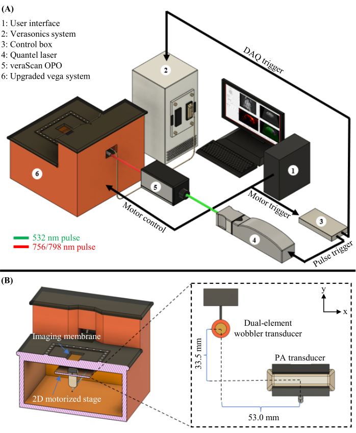
Figure 1: PAUSAT diagram. (A) Complete schematic of the PAUSAT system, including the laser and OPO used for PAT. (B) Inside view of the PAUSAT system, including two ultrasound transducers. The dual-element wobbler transducer is used for both B-mode ultrasound and AA, and the linear-array transducer is used for PAT. Both transducers are mounted on the same 2D motorized stage, allowing for scanning to generate volumetric data. This figure has been modified from16. Please click here to view a larger version of this figure.
