This article describes a protocol for diatom DNA extraction using a modified common DNA extraction kit.
Diatom testing is an essential auxiliary means in forensic practice to determine whether the corpse drowned in water and to infer the drowning location. Diatom testing is also an important research content in the field of the environment and plankton. The diatom molecular biology testing technology, which focuses on diatom DNA as the primary research object, is a new method of diatom testing. Diatom DNA extraction is the basis of diatom molecular testing. At present, the kits commonly used for diatom DNA extraction are expensive, which increases the cost of carrying out related research. Our laboratory improved the general whole blood genomic DNA rapid extraction kit and obtained a satisfactory diatom DNA extraction effect, thus providing an alternative economical and affordable DNA extraction solution based on glass beads for related research. The diatom DNA extracted using this protocol could satisfy many downstream applications, such as PCR and sequencing.
In forensic practice, determining whether a corpse found in the water drowning or was thrown into the water after death is essential for the proper resolution of the case1. It is also one of the difficult issues that need to be solved urgently in forensic practice2. Diatoms are abundant in the natural environment (especially in water)3,4. In the process of drowning, due to hypoxia and stress response, people will have intense breathing movements and inhale a large amount of drowning liquid. Therefore, the diatoms in the water enter the lung with the drowning liquid, and some diatoms can enter the blood circulation through the alveolar-capillary barrier and spread to internal organs with the blood flow5,6. The detection of diatoms in internal tissues and organs such as lung, liver, and bone marrow is a strong evidence of drowning before death7,8. Currently, forensic diatom testing is mainly based on morphological testing methods. After a series of pre-digestion of the tissue, the morphological qualitative and quantitative estimations of undigested diatoms are carried out under the microscope. During this period, dangerous and environmentally unfriendly reagents such as nitric acid need to be used. This process is time-consuming and requires researchers to have solid taxonomic expertise and extensive experience. These all bring certain challenges to the forensic staff9. Diatom DNA testing technology is a new technology for diatom testing developed in recent years10,11,12. This technology realizes the species identification of diatoms by analyzing the specific DNA sequence composition of diatoms13,14. PCR technology and sequencing technology are commonly used technical methods, but their basis is the successful extraction of DNA from diatoms. However, diatoms have a special structure different from other organisms, making their DNA extraction techniques also different.
The cell wall of diatom has a high degree of silicification, and its main component is silicon dioxide15,16,17. The siliceous cell wall is very hard, and it must be destroyed before extract the DNA. Ordinary DNA extraction kits are often difficult to use directly for the extraction of diatom DNA because they cannot destroy the siliceous shell of diatoms18. Therefore, destroying the siliceous shell of diatoms is one of the key technical problems to be solved in extracting diatom DNA.
At the same time, since the number of diatoms contained in forensic research samples, whether water samples or organs and tissues of drowned bodies, is often limited, it is necessary to enrich diatoms. The essence of enrichment is the separation of substances. While trying to gather diatoms together, minimize the content of other material components (interfering components). In forensic work, laboratories often use centrifugation or membrane filtration enrichment methods to separate diatom cells19. However, since vacuum pumping equipment is not widely used, the membrane enrichment method is not often used in ordinary primary forensic laboratories. So, the centrifugation method is still commonly diatom enrichment in forensic laboratories20.
DNA extraction from diatoms is currently used primarily in forensic practice, and there are significant limitations to its application. At present, there are few diatom DNA extraction kits used in forensic science on the market and they are generally expensive21. This article provides an improved diatom DNA extraction method, making diatom DNA extraction simple, convenient and cost effective. This increases the application of subsequent molecular biology testing of diatoms and can better solve problems related to drowning in forensic medicine through diatom testing. This method breaks the siliceous cell walls of diatoms by adding glass beads and setting an appropriate time for vortex. In this way, the proteinase K and the binding solution rapidly lyse the cells and inactivate various enzymes in the cells. The genomic DNA is absorbed in the matrix membrane in the adsorption column and finally eluted by the elution buffer. Such an improved whole blood gene extraction kit improves the diatom DNA extraction effect of the blood kit in forensic examination materials, reduces the cost of diatom DNA extraction in forensic practice, and can be better applied to grassroot forensic research.
This study was approved by the Ethics Committee of Hainan Medical University. The tissue samples used in this study are not considered to be studies involving human subjects. These specimens were obtained for the purpose of forensic pathological diagnosis, and the rest were used for the extraction of diatom DNA in this experiment. Researchers cannot readily identify individuals to obtain informed consent from relevant stakeholders.
NOTE: To ensure the general applicability of the research method reported in this experiment, this experiment basically followed the operating instructions of the kit used, and only some steps were modified. The water samples used in this experiment were randomly taken from ponds near the laboratory (Supplementary Figure 1A). In this experiment, the lung tissue of the drowned body was confirmed as the research tissue to demonstrate the extraction protocol (Supplementary Figure 1B). In forensic practice, it is also sometimes necessary to use other organs and tissues of drowned bodies (such as liver, spleen, kidney, bone marrow, etc.) to extract diatoms DNA, which requires minor corresponding improvements to this experimental method, which will be explained in the corresponding section of the experiment. The lung tissue samples used in this experiment came from corpses that were clearly drowned in forensic cases. Morphological tests have been conducted to prove that the lung tissue contains diatoms (Supplementary Figure 2).
1. Pretreatment of samples
2. DNA extraction
NOTE: All centrifugation steps are completed at room temperature. Using a desktop centrifuge with a centrifugal force of 14,500 x g; a water bath (or metal bath) preheated to 70 °C needs to be prepared before the experiment starts. All steps must strictly follow the principles of aseptic operation.
3. PCR test
NOTE: Since the content of diatoms in forensic samples is often low, the tissue sample extracts of the drowned bodies may also contain varying degrees of tissue and organs (such as the lungs in this experiment) with their own DNA. Therefore, direct detection of total DNA in DNA extracts does not reflect the situation of diatom DNA extraction. In this experiment, diatom-specific primers were selected, and PCR products were used to evaluate the diatom DNA extraction in the extract. The products can be observed and analyzed by agarose gel electrophoresis and can also be analyzed by real-time fluorescent quantitative PCR-melting curve, which has higher sensitivity.
Since the DNA solution extracted by the currently used DNA extraction method contains all DNA components from different sources in the sample, the DNA obtained by this protocol was no exception. So, the DNA solution was not just a solution of diatom genomic DNA. The primers that can specifically amplify diatom 18S rDNA fragments were selected by consulting the literature22,23,24. The primers were verified by NCBI-Blast Primer, and the results showed that the forward primer D512 and reverse primer D978 of 18S rDNA were algae-specific primers and covered many diatom species. The amplification results did not show any human genes. They were used a reliable DNA biomarker for diatom testing, so they were selected for verification of the results of this experiment. Using the extracted DNA as a template for diatom-specific PCR amplification, the amplification product by agarose gel electrophoresis showed that water samples and tissues had diatom DNA bands, these electrophoresis bands were between 250- 500 bp (closer to 500 bp), in line with the primer target amplification product length size (390 -410 bp). This showed that the extraction protocol was successful in extracting diatom DNA (Figure 1A,B).
Real-time fluorescence quantitative PCR technology is a kind of gene testing technology with stringent specificity and high sensitivity. By adding fluorescent groups in the PCR reaction system, the accumulation of fluorescent signals can make the whole PCR process be monitored in real-time, and the sensitivity is high25,26. Products specifically amplified by conventional PCR can sometimes be difficult to observe in agarose gel electrophoresis due to the low content and other reasons27 (Figure 1C). Through the specific amplification of real-time fluorescent quantitative PCR, a typical amplification curve with four characteristic stages was obtained (Figure 2). At the same time, through the real-time fluorescent quantitative PCR-melting curve technology, the DNA double strands of the amplified product were melted into single strands at high temperature, the dye was freed from the double strands, and the fluorescence value decreased. By detecting the change in the fluorescence value, the melting curve was obtained (Figure 3). The obtained melting curves had characteristic peaks, and the peak value was around 85.5 °C, which proved that there was a specific amplification of the target DNA, indicating that there was diatom DNA in the extracted DNA solution. The dye could also be directly added to the conventional PCR amplification product, and the melting curve could be obtained by real-time fluorescence quantitative PCR-melting curve technology to prove whether there was specific amplification. Therefore, when conventional PCR-agarose gel electrophoresis could not prove whether there was diatom DNA in the extracted DNA solution, the real-time fluorescent quantitative PCR technology with higher sensitivity could be selected for verification and evaluation. In this experiment, the real-time fluorescent quantitative PCR-melting curve technology was only used for qualitative analysis.
As shown in a previous report21, the traditional soil DNA extraction kit and the modified blood DNA extraction kit were basically the same for diatom DNA extraction from water samples and tissues. The cost difference of diatom DNA extraction was mainly the price difference of the two kits, and the cost of other consumables was basically the same. In this study, a simple improvement of the blood DNA extraction kit commonly used in molecular biology experiments could obtain better experimental results, which not only increased the scope for the researchers to select the test kit in research activities related to diatom DNA extraction but also reduced the cost of testing.
The improvement process of this protocol has been published21. By designing the combination of different mass ratios of glass beads and vortex oscillating time, the optimal DNA extraction conditions of the oscillation of glass beads were added into the conventional kit, therefore, the method could improve the extraction effect of diatom DNA in forensic samples, especially in tissue samples. The optimal combination of DNA extraction was obtained when the vortex oscillating frequency was 3000 rpm; the vortex oscillating time was 4 min, the mass ratio of large glass beads with diameter of 1.5-2.0 mm and small glass beads with diameter of 0.4-0.6 mm was 1:1. A comparison between the conventional method of the kit and the improved method was performed. It was shown that the used kit could meet the needs of amplifying diatom DNA, and the brightness of electrophoretic bands after amplification performed by the improved method in water samples was similar to the brightness of the conventional method, and the electrophoretic bands after amplification performed by the improved method in tissue samples were brighter (Figure 4).
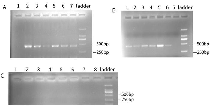
Figure 1: Electrophoresis results of DNA extracted using the protocol after PCR amplification. A total of 2 µL of DNA extract was used as a DNA template for PCR-specific amplification. The PCR amplification products were electrophoresed on 1x TAE buffer solution through 2% agarose gel, and run at a constant voltage of 100V for 25-30 min. A D2000 DNA ladder was used for all gels. (A) The electrophoresis results of diatom DNA in water samples after PCR amplification, lane 1 is blank control; lanes 2-7 are water samples from different locations in the same area. (B) The results of electrophoresis after PCR amplification of diatom DNA in the tissue. Lanes 1-6 are the tissues excised from different positions of the same lung tissue; lane 7 is the blank control. (C) In an unsuccessful experiment,after diatom DNA amplification, the electrophoresis results did not show electrophoresis bands at the corresponding positions, indicating that there were no amplification products or too few amplification products. At this time, the electrophoresis results could not indicate whether there was diatom DNA in the proposed DNA solution, which needed to be carried out by more sensitive methods (such as a PCR-melting curve). Please click here to view a larger version of this figure.
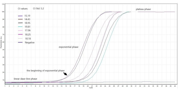
Figure 2: Diatom DNA qPCR amplification curve. A total of 2 µL of DNA solution extract that showed no bands by conventional PCR and agarose gel electrophoresis was used as a template to prepare a real-time fluorescence quantitative PCR system and set up a program. Finally, typical amplification curves with four characteristic stages of linear baseline phase, the beginning of exponential phase, exponential phase, and plateau phase were obtained. The Ct value of each curve was also obtained through analysis, indicating that DNA extraction was successful. Each number represents the Ct value of the corresponding curve. The threshold is 5.2. The unit of the y-axis is Rn. Please click here to view a larger version of this figure.
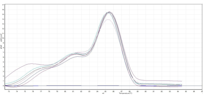
Figure 3: Diatom DNA melting curve. Using a real-time fluorescence quantitative PCR system, a program was set where the temperature rises from 60-95 °C. This generated a melting curve and the specifically amplified double-stranded DNA melted into single strands at high temperatures, the dye was free from the double strands, and the fluorescence value decreased until it was 0. The graph was obtained by taking the negative reciprocal of the original graph. The abscissa represented the temperature, the abscissa corresponding to the highest peak value was the Tm value, and the Tm value of the melting curve in the graph was 85.5 °C. The numbering of each curve corresponds to the numbering of each lane in Figure 1C. Please click here to view a larger version of this figure.
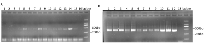
Figure 4: Electrophoresis results of the improved process. (A) Electrophoresis results for different combinations of glass bead ratios and vortex oscillation times. Lanes 1, 4, 7, 10, and 13 where the mixed mass ratio of large and small glass beads is 1:2, lanes 2, 5, 8, 11, and 14 where the mixed mass ratio of large and small glass beads is 1:1, lanes 3, 6, 9, 12, and 15 where the mixed mass ratio of large and small glass beads is 2:1, and the oscillation times were 2 min for lanes 1-3, 3 min for lanes 4-6, 4 min for lanes 7-9, 5 min for lanes 10-12, and 6 min for lanes 13-15. Lane 16 is the blank control. (B) Electrophoresis results after PCR amplification of the kit extraction products before and after improvement. Lanes 1-3 are conventional methods for extracting water samples, lanes 4-6 are improved methods for extracting water samples, lanes 7-9 are conventional methods for extracting tissues, lanes 10-12 are improved methods for extracting tissues, lane 13 is a blank control. The results have been modified from21. Please click here to view a larger version of this figure.
Supplementary Figure 1: Water samples and tissue samples. The water samples used were taken from a pond near the laboratory, and the tissue samples used were confirmed as lung tissue of the drowned body. (A) The experimental water sample collection site was a pond near the laboratory. (B) Enriched water samples and shredded lung tissue samples. Please click here to download this File.
Supplementary Figure 2: Micrograph of diatom. Images (A) and (B) were taken under a 400x optical microscope. The arrow points to diatoms. Images (C) and (D) were taken under an electron microscope. Images (A) and (B) show the diatoms in the water at the drowning site. The diatoms named Fragilaria and Navicula. Images (C) and (D) show diatoms in the lungs, which are called Nitzschia and Navicula. Please click here to download this File.
Target | Primer name | Sequence |
18S rDNA | forword primer D512 | ATTCCAGCTCCAATAGCG |
reverse primer D978 | GACTACGATGGTATCTAATC |
Table 1: List of primers used. This table describes the sequences, names, and target regions of specific primers used in this paper.
PCR reaction system | |||
2x Taq Mix Pro | 10 μL | ||
forword primer D512 | 0.5 μL (10 μmol/L) | ||
reverse primer D978 | 0.5 μL (10 μmol/L) | ||
template DNA | 2 μL | ||
Nuclease-free water | to 20 µL | ||
Note: The blank control template DNA is replaced by the same amount of Nuclease-free water. | |||
PCR program | |||
temperature | time | cycles | |
94 °C | 10 min | 1 | |
94 °C | 45 s | 40 | |
50 °C | 45 s | ||
72 °C | 1 min | ||
72 °C | 10 min | 1 |
Table 2: PCR components. This table describes the configuration of the reaction system for conventional PCR and the settings of the reaction temperature, time, and number of cycles for PCR. The total volume of the reaction system used was 20 µL.
Real-time quantitative PCR reaction system | |||
2x qPCR mix | 10 μL | ||
forword primer D512 | 0.45 μL (10 μmol/L) | ||
reverse primer D978 | 0.45 μL (10 μmol/L) | ||
template DNA | 2 μL | ||
Nuclease-free water | to 20 μL | ||
Note: The blank control template DNA is replaced by the same amount of Nuclease-free water. | |||
Real-time quantitative PCR program | |||
temperature | time | cycles | |
94 °C | 10 min | 1 | |
94 °C | 45 s | 40 | |
50 °C | 45 s | ||
72 °C | 1 min |
Table 3: Real-time quantitative PCR components. This table describes the configuration of the reaction system for real-time quantitative PCR and the settings of the reaction temperature, time, and number of cycles for PCR. The total volume of the reaction system used was 20 µL.
Diatom cells are protected by hard siliceous cell walls17, and this structure must be destroyed to extract diatom DNA. Ordinary kits do not easily destroy the siliceous shell of diatoms; thus it is difficult to successfully extract diatom DNA21. Our laboratory improved the most commonly used blood DNA extraction kit, adding glass beads of different diameters and different mass ratio in the process of diatom extraction. Vortex oscillation is carried out at the same time, which can fully break the diatom shell and improve the diatom DNA extraction effect. As shown in the previous report21, the diameter of glass beads, oscillation intensity and time would directly affect the quality of DNA extraction. The vortex oscillation time should not be too long (no more than 10 min). If the time is too long, the DNA will break due to excessive vibration. At the same time, the time should not be too short. If the time is too short (not less than 2 min), the diatom shell will not be completely removed, which will make the DNA extraction inefficient. While selecting glass beads choose beads with different circumferential diameters, the large glass beads ensure the collision strength, and the small glass beads ensure that there is no dead collision between the glass beads during the collision fragmentation process, which improves the efficiency of shell breaking.
To extract diatom DNA from samples, it is generally necessary to enrich diatoms. This process will have a great impact on the extraction of diatom DNA. The purpose of enrichment is to increase the concentration of diatom in samples so that the concentration of diatom DNA extracted can be in detectable range; for the next-step applications such as real-time fluorescent quantitative PCR and high-throughput sequencing.
Since diatoms are widely distributed in nature4, preventing laboratory contamination is a top priority28. Contamination during DNA extraction process will lead to deviations from the actual results and loss of use value. Therefore, always pay attention to the principle of aseptic operation when extracting diatom DNA and take all possible measures to reduce and avoid contamination of exogenous diatoms. When the tissue is extracted from the dead body, measures should be taken to prevent it from being contaminated by exogenous diatoms. After the tissue is extracted, it should be stored in a container or sample bag free of diatom contamination. The tissue should not be treated with formaldehyde. The equipment for extracting and processing tissues should be washed 2-3 times with distilled water repeatedly, and then sterilized by ultraviolet light or high temperature to ensure that there is no contamination of diatoms from other sources. The tissue before the test should be cut off from the superficial tissue with a diatom-free instrument, and then the instrument should be replaced to pick out the lower tissue for examination. At the same time, standardizing the operation process can also effectively avoid the problem of laboratory contamination.
Since diatoms are the food of many aquatic organisms, they will be lost during long-term storage29. Diatoms can also grow and reproduce under light conditions. Therefore, the DNA extraction step should be initiated as soon as possible after sampling and quick sample processing should be done to prevent loss of diatoms in the sample. If the DNA extraction cannot be carried out immediately, all samples should be frozen and kept in the dark to prevent the reduction of diatoms as much as possible.
Our research selected a common and cheap blood DNA extraction kit based on the proteinase K digestion method, which has been improved to extract diatom DNA from water samples and tissues with good results30. The tissue is sufficiently cut to be in full contact with proteinase K and the tissue gets fully digested. The possible principle of successful extraction is as follows: during the shaking step, the hard shell of diatom is repeatedly impacted and fragmented between fast-moving glass beads, exposing the cell membrane; the configuration of glass beads of different sizes can fill the gap between the collision of single size glass beads, so as to cover the collision contact surface as fully as possible; after losing the silicon shell, proteinase K helps to release DNA from the diatom. Compared with the currently used soil DNA extraction kit, the improved blood DNA extraction kit can meet the requirements of diatom DNA extraction, which is more economical and easier to obtain. It not only facilitates the use of forensic diatom inspection, but is suitable for application to other diatom research-related disciplines and can also be used for other plankton DNA extraction. When extracting diatom DNA from water samples, the amount of proteinase K used needs to be adjusted according to the number of plankton in the water. The amount of proteinase K can be increased or decreased based on preliminary experiments. When extracting diatom DNA from tissues, different organ tissue types, tissue contamination, and other factors will affect the amount of proteinase K required for digestion. In the experiment, the proteinase K in the kit should be stored at -20 °C after use, otherwise, the inactivation of proteinase K will affect the extraction process.
In conclusion, diatom DNA extraction should be performed as soon as possible after sampling and sample processing, otherwise stored at -20 °C for preservation. Sample handling, storage, and the entire extraction process should be done carefully to prevent contamination by exogenous diatoms. Otherwise, it cannot be assessed that the extracted diatom DNA was in the sample or exogenous contamination. When using proteinase K to digest tissue, its dosage may be a problem. Insufficient tissue digestion can increase the amount of proteinase K. Therefore, it is important to shear the tissue adequately. Sufficient digestion of the tissue also prevents the tissue debris from clogging the plasma membrane inside the adsorption column. After the membrane is blocked, it is not possible to use the pipette tip to pick out the blockage or to centrifuge repeatedly, which will damage the membrane. The final DNA solution contains many impurities, which will affect the detection of diatom DNA. Oscillation intensity and time are also key issues. The first vortex oscillation time is controlled at 4-5 min, and the oscillation frequency is controlled at 3000 rpm, otherwise, the DNA extraction will be affected. If the electrophoretic bands are not clear after PCR, it may be necessary to further increase the amount of DNA template. The DNA extracted in this study is a mixture of DNA, including diatom DNA and DNA from other species, so the concentration of diatom DNA cannot be determined. Therefore, it is not suitable for applications requiring high purity of DNA templates.
Studies have shown that different diatom species can be amplified by different primers31,32. In this study, only the diatom-specific primers D512 and D978 on the diatom 18S rDNA were used to test whether the diatom DNA was successfully extracted, which could not cover all diatom species22,33. In addition, since the length of the amplification product fragment of the pair of primers is 390-410 bp, the improved method selects all fragment sizes of diatom DNA to test whether there is an impact on the experimental parameters, which needs further study. Subsequent studies will also use primers of different regions and different amplification lengths to explore the effect of extraction.
Compared with the traditional diatom DNA extraction method, this method has the main advantages of low-cost and large-scale diatom DNA extraction. It can complement the advantages of diatom morphological methods and can meet the needs of drowning diagnosis in primary forensic laboratories. At the same time, it also expands the selection range of diatom DNA extraction kits and has a good application prospect. In future, this method will be not only limited to the extraction of diatom DNA but be used for the extraction of DNA from other algae.
The authors have nothing to disclose.
This work is supported by the National Natural Science Foundation of China (82060341,81560304) and by the Academician Innovation Platform Scientific Research Project of Hainan Province (YSPTZX202134).
Binding Buffer | BioTeke | B010006022 | rapidly lysing cells |
ChemoHS qPCR Mix | Monad | 00007547-120506 | qPCR Mix |
D2000 DNA ladder | Real-Times(Beijing) Biotechnology | RTM415 | Measure the position of electrophoretic bands |
D512 | Taihe Biotechnology | TW21109196 | forword primer |
D978 | Taihe Biotechnology | TW21109197 | reverse primer |
Elution buffer | BioTeke | B010006022 | A low-salt elution buffer washes off the DNA |
Glass bead | Yingxu Chemical Machinery(Shanghai) | 70181000 | Special glass beads for dispersing and grinding |
Import adsorption column | BioTeke | B2008006022 | Adsorption column with silica matrix membrane |
Inhibitor Removal Buffer | BioTeke | B010006022 | Removal of Inhibitors in DNA Extraction |
Isopropanol | BioTeke | B010006022 | Precipitate or isolate DNA |
MIX-30S Mini Mixer | Miulab | MUC881206 | oscillatory action |
Proteinase K | BioTeke | B010006022 | Inactivation of intracellular nucleases and other proteins |
Rotor-Gene Q 5plex HRM | Qiagen | R1116175 | real-time fluorescence quantification PCR |
Speed Micro-Centrifuge | Scilogex | 9013001121 | centrifuge |
Tanon 3500R Gel Imager | Tanon | 16T5553R-455 | gel imaging |
Taq Mix Pro | Monad | 00007808-140534 | PCR Mix |
Thermo Cycler | Zhuhai Hema | VRB020A | ordinary PCR |
Wash Buffer | BioTeke | B010006022 | Remove impurities such as cell metabolites |