This protocol describes a multicolor immunofluorescence technique for evaluating the rat model of allergic rhinitis.
Allergic rhinitis (AR) is a chronic, non-infectious inflammatory disease of the nasal mucosa, primarily mediated by specific immunoglobulin E (IgE), affecting approximately 10%-20% of the world’s population. While immunofluorescence (IF) staining has long been a standard technique for detecting disease-specific protein expression, conventional IF techniques are limited in their ability to detect the expression levels of three or more proteins in the same sample. Consequently, multicolor IF techniques have been developed in recent years, which allow the simultaneous labeling of multiple targets in cells or tissues.
This protocol provides a comprehensive overview of the process for establishing a rat model of AR, obtaining nasal mucosal samples, and the technical procedures for multicolor immunofluorescence. All rats in the AR group exhibited typical symptoms such as sneezing, a runny nose, and an itchy nose, with behavioral observations scoring ≥5 points. Hematoxylin and eosin (H&E) staining revealed increased inflammatory cell counts and disrupted nasal mucosal integrity in the AR group. Multicolor immunofluorescence (mIF) demonstrated increased expression of RORγt and TICAM-1, while Foxp3 expression decreased in the nasal mucosa tissue of AR rats.
Allergic rhinitis (AR) is a chronic, non-infectious inflammatory disease of the nasal mucosa primarily mediated by specific immunoglobulin E (IgE)1,2. It is characterized by symptoms such as sneezing, a runny nose, nasal congestion, and nasal itching. With industrialization and urbanization, the prevalence of AR is gradually increasing, affecting approximately 10%-20% of the world's population1. The immunofluorescence (IF) technique is a fluorescent staining method that utilizes an antibody-antigen binding reaction. It can be employed to detect and quantify the distribution and expression levels of specific proteins in biological tissues or cells. In AR research, IF can simultaneously detect multiple targets, including AR-related cytokines, inflammatory cells, receptors, and more, facilitating the exploration of AR pathogenesis and the effects of drugs3,4,5,6.
The multicolor immunofluorescence (mIF) staining process closely resembles traditional IF, with the addition of an antibody elution step during each round of staining. This modification enables the simultaneous detection of multiple biomarkers on the same tissue section through sequential single-labeling and multiple rounds of re-staining. mIF is based on tyramide signal amplification (TSA), allowing for repeated cycles of TSA fluorescent staining and the use of microwave heating to remove antibodies while retaining fluorescent signals7,8. In comparison to conventional IF, mIF offers several advantages: (1) it can detect weakly expressed antigens that are challenging to identify with conventional IF9,10; (2) it provides high-quality staining with an improved signal-to-noise ratio; (3) it allows for the quantification of tissue-specific structures and regions of interest11; (4) multiplexing multiple pathways efficiently utilizes tissues and conserves limited pathological resources12; (5) multi-parameter analysis through mIF offers deeper insights into tissues, uncovering hidden biological information13.
Overall, mIF allows the observation of different antigen expressions and distributions within the same sample, facilitating the study of target proteins. In the future, researchers seeking to understand the expression and distribution of multiple target proteins will find this technique a valuable choice. This study demonstrates the application of mIF for staining nasal mucosa samples from rats with AR and evaluate the establishment of a rat model of AR.
The experimental protocol and procedures have received approval from the Administrative and Animal Research Committee of Chengdu University of Traditional Chinese Medicine (Record number: 2022DL-010). Eight-week-old male Sprague Dawley (SD) rats, weighing 180-200 g, were commercially obtained (see Table of Materials) and housed under a natural light/dark cycle with controlled temperature (23 ± 2 °C) and relative humidity (55% ± 10%). Twelve rats were randomly divided into two groups: the control group and the AR group. All rats were acclimated to these conditions and provided with free access to food and water for one week before the trial.
1. Establishment of a rat model of AR
2. Behavioral scoring of rats
3. Acquisition of nasal mucosa samples
4. Pre-processing of nasal mucosa samples
5. H&E staining of nasal mucosal tissue
6. Multicolor immunostaining of nasal mucosal tissue
Six SD rats were successfully induced into the AR model through OVA intraperitoneal injection and nasal challenge. AR was induced in all rats in the AR group, accounting for 100% of the group. All rats in the AR group exhibited typical symptoms such as sneezing, a runny nose, and an itchy nose. All behavioral observations scored ≥5 points (Table 2).
H&E staining results on the 21st day of modeling revealed that in the control rats, the nasal mucosa's epithelial cells and cilia were well-arranged, with no signs of inflammatory cell infiltration. Conversely, in the AR group, the nasal septum's mucosa was damaged and detached, with notable neutrophil infiltration (Figure 3).
When comparing the nasal mucosa tissue of AR rats with the control group, it was observed that the expression of RORγt (a Th17 cell-specific transcription factor) and TICAM-1 (Toll-like receptor linker molecule-1) was increased, while Foxp3 (a Treg cell-specific transcription factor) was decreased (Figure 4).
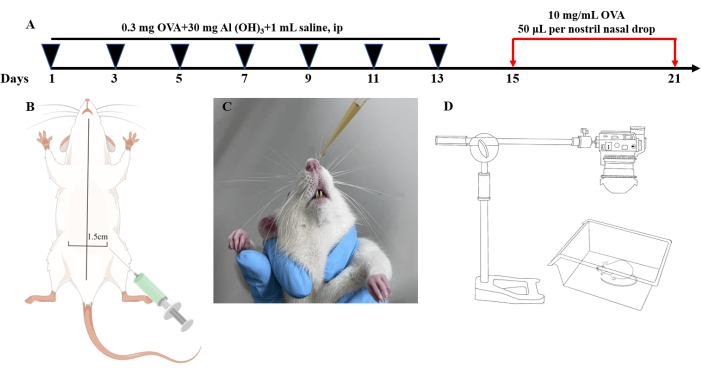
Figure 1: The experimental workflow. (A) Schematic diagram of intraperitoneal injection. (B) Schematic diagram of nasal drip. (C) Flowchart of modeling of allergic rhinitis rats. (D) Schematic diagram of behavioral observation. Please click here to view a larger version of this figure.
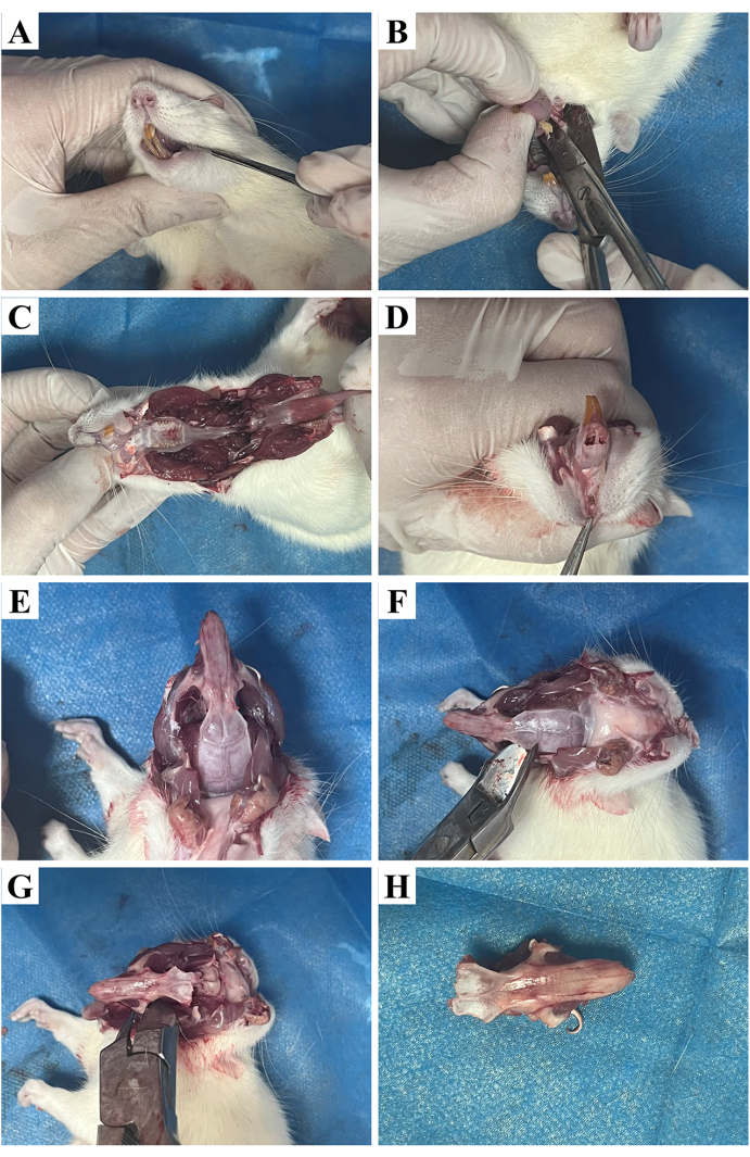
Figure 2: Nasal removal process. (A) Cutting of the muscle tissue at the corners of the mouth. (B) Cutting the connection between the cheekbone and the mandible. (C) Separation of the mandible. (D) Removal of the skin of the maxilla. (E) Exposing the nasal cavity. (F) Cutting the connection between the nasal cavity and the maxilla. (G) Cutting the connection between the nasal cavity and the orbital bone. (H) The removed nasal cavity. Please click here to view a larger version of this figure.
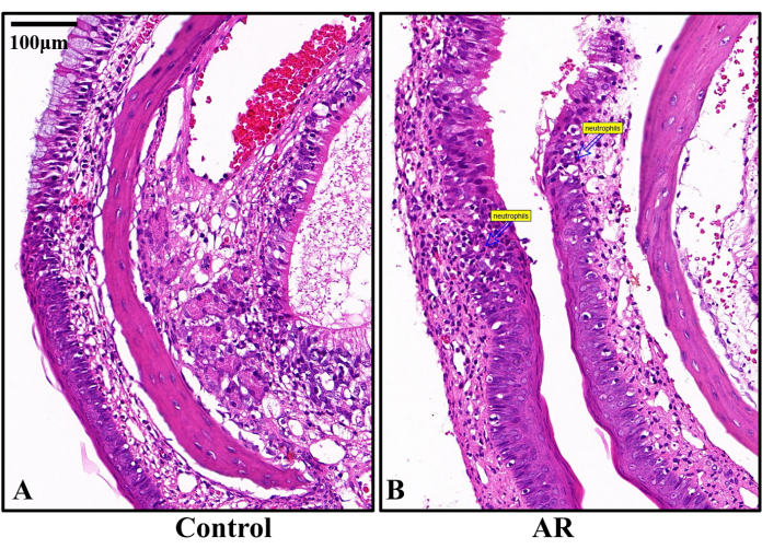
Figure 3: Representative histopathological H&E staining images on day 14 after AR modeling (n = 6). (A) The epithelial cells and cilia in the nasal mucosa of the control rats were well arranged, and no inflammatory cell infiltration was observed. (B) The mucosa of the nasal septum of the AR group was damaged and detached with neutrophil infiltration. Scale bars = 100 µm. Please click here to view a larger version of this figure.
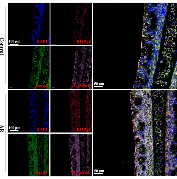
Figure 4: MIF staining analysis of RORγt, Foxp3, and TICAM-1 (n = 6). Compared with the control group, the expression of RORγt and TICAM-1 in the nasal mucosa tissues of rats in the AR group was elevated, while the expression of Foxp3 was decreased. Scale bars = 100 µm (left panels); 50 µm (right panels). Please click here to view a larger version of this figure.
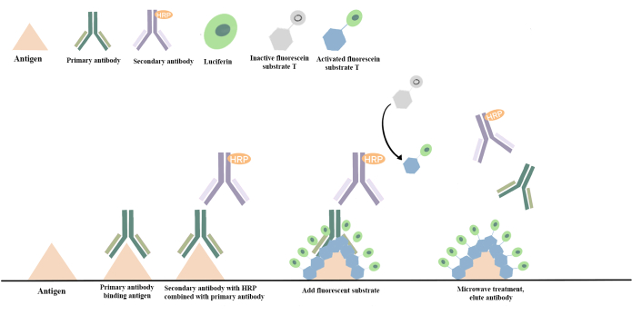
Figure 5: Principle of mIF technique. The mIF technique is based on the TSA technique, which involves covalently binding the fluorescent signal to the antigen. In this process, horseradish peroxidase-labeled on the secondary antibody catalyzes the transition of the fluorescein substrate from an inactive to an activated state. This activated state can covalently bind to the tyrosine on the antigen, resulting in a stable covalent attachment of fluorescein to the sample. Subsequently, non-covalently bound antibodies are removed through thermal repair. The procedure is then repeated with additional primary antibodies, secondary antibodies, and fluorescein to enable the detection of a completely different antigen. This image was redrawn and color-matched with reference to the mIF schematic diagram17. Please click here to view a larger version of this figure.
Scores | Sneezing frequency | Rhinorrhea | Nasal rubbing |
1 | <3 | watery discharge within the nasal cavity | slight and occasional nasal rubbings |
2 | 4~10 | watery discharge spilling out of the anterior naris | repeated nasal rubbings |
3 | ≥11 | the face covered with abundant watery discharge | rubbings from nose to face |
Table 1: Quantistive scale table of rat behavioral test.
Individiuals | Day 1 | Day 21 |
Control 1 | 0 | 0 |
Control 2 | 0 | 0 |
Control 3 | 0 | 0 |
Control 4 | 0 | 0 |
Control 5 | 0 | 0 |
Control 6 | 0 | 0 |
AR 1 | 0 | 7 |
AR 2 | 0 | 6 |
AR 3 | 0 | 5 |
AR 4 | 0 | 5 |
AR 5 | 0 | 5 |
AR 6 | 0 | 6 |
Table 2: Results of behavioral scoring.
Allergic Rhinitis (AR) is a non-infectious inflammatory disease of the nasal mucosa resulting from a combination of environmental and genetic factors. It has become a global health concern, impacting work efficiency, diminishing the quality of life, impairing sleep, cognitive function, and causing irritability and fatigue. AR affects 10%-20% of the world's population¹ and carries substantial economic costs, causing annual losses of up to 30-50 billion euros in EU countries18. Moreover, several studies have established a strong association between AR and asthma18,19,20, with individuals having AR being seven times more likely to develop asthma than those without AR21. Current research suggests that an imbalance between type 1 helper T cells (Th1) and type 2 helper T cells (Th2) with a bias towards Th2 cells is a significant contributor to AR. Th2 cells, stimulated by interleukin-4, produce Th2-like cytokines such as IL-5 and IL-13, triggering inflammation in B lymphocytes, mast cells, eosinophils, and dendritic cells22. Furthermore, recent AR research indicates a strong connection between an imbalance in helper Th17/regulatory T cell (Treg) populations23. Th17 and Treg cells, both derived from CD4+ T cells, play critical roles in the immune system. RORγt is essential for Th17 cell differentiation24, while Treg cells, with immunosuppressive functions, are key to maintaining immune tolerance. Changes in Foxp3 expression influence Treg cell function25,26. Thus, alterations in RORγt and Foxp3 levels reflect Th17/Treg imbalances, potentially inducing an inflammatory response primarily driven by Th17 cells27. Toll-like receptors (TLRs) are vital proteins in the body's innate immune system, mediating intracellular signaling pathways to activate specific gene expression28. Research by He Shan et al.29 found that variations in the TLR4 gene, specifically CT heterozygous and TT pure mutations at the rs10759930 locus, were associated with AR development. Given TICAM-1's role as the bridging molecule between TLR3 and TLR4, it was hypothesized that AR might be linked to TICAM-1. Indeed, the results showed elevated TICAM-1 expression in the AR group, consistent with Xu et al.'s study30.
The mIF (Multiplex immunofluorescence) technique employed in this study is a relatively new detection method developed over the past 20 years. It offers several advantages over conventional immunofluorescence (IF) techniques, including increased specificity, sensitivity, and throughput, along with the capability for visual analysis. This technique is based on the TSA (Tyramide Signal Amplification) method, which covalently bonds fluorescent signals to antigens and remains unaffected by microwave heating. This allows for the multicolor labeling of tissues through repeated cycles of TSA fluorescent staining, followed by microwave heating to remove antibodies while retaining the fluorescent signal17,31,32 (Figure 5). Advanced algorithms are then applied for the automatic identification and segmentation of target tissue regions displaying specific structures in multilabeled images. This enables quantitative statistical analysis of regions of interest, allowing for the quantitative assessment of cellular immune phenotypes and the functional localization of immune cells. It also provides information on tissue context and spatial distribution33,34.
However, technically, mIF still relies on traditional IF techniques and faces challenges such as antibody cross-reactivity and optical crosstalk. The use of multiple antibodies can lead to cross-reactivity and false-positive results, while overlapping fluorescence spectra may result in the blending of fluorescence signals from multiple targets, making naked-eye differentiation challenging and increasing reliance on specialized image analysis algorithms35. Furthermore, as fluorescent dyes are used to label antibodies, mIF can be affected by fluorescence quenching and bleaching, leading to diminished signal intensity. Notably, while conventional IF techniques evaluate the entire tissue, mIF techniques focus on specific regions of interest within the tissue section for analysis. This may introduce deviations from reality due to tissue heterogeneity, potentially resulting in inaccurate histological quantification or the omission of rare events.
The authors have nothing to disclose.
This work was supported by the Sichuan Provincial Department of Science and Technology (2021YJ0175).
Al(OH)3 | Sollerbauer Biotechnology Co., Ltd | A7130 | |
75% ethanol | Anhui Yiren An Co., Ltd | 20210107 | |
Ammonia | Chengdu Kolon Chemical Co., Ltd | 2021070101 | |
Anhydrous ethanol | Chengdu Kolon Chemical Co., Ltd | 2022070501 | |
Anti-fluorescence quenching sealer | SouthernBiotech | 0100-01 | |
Automatic dyeing machine | Thermo scientific | Varistain Gemini ES | |
Carrier slides | Nantong Mei Wei De Experimental Equipment Co., Ltd | 220518001 | |
Citrate-phosphate buffer | Servicebio biotechnology co., Ltd | G1201 | |
Citric acid antigen repair solution (PH 6.0) | Xavier Biotechnology Co., Ltd | G1201 | |
Coverslip | Nantong Mei Wei De Experimental Equipment Co. | 220518001 | |
Coverslip | Nantong Mewtech Life Science Co., Ltd | CS01-2450 | |
CY3-Tyramide | Sawell Biotechnology Co., Ltd | G1223-50UL | |
DAPI | Sawell Biotechnology Co., Ltd | G1012 | |
Decoloring shaker | SCILOGEX | S1010E | |
EDTA decalcification solution | Wuhan Xavier Biotechnology Co., Ltd | CR2203047 | |
Electric heating blast dryer | Shanghai Yiheng Scientific Instruments Co., Ltd | DHG-9240A | |
Embedding box marking machine | Thermo scientific | PrintMate AS | |
Embedding machine | Wuhan Junjie Electronics Co., Ltd | JB-P5 | |
Fast tissue dewatering machine | Thermo scientific | STP420 ES | |
Film sealer | Thermo scientific | Autostainer 360 | |
FITC-Tyramide | Sawell Biotechnology Co., Ltd | G1222-50UL | |
Fluorescence microscope | Sunny Optical Technology Co.Ltd | CX40 | |
Foxp3 | Affinity Biosciences Co., Ltd | bs-10211R | |
Freezing table | Wuhan Junjie Electronics Co., Ltd | JB-L5 | |
Goat Anti-Rabbit IgG H&L (HRP) | Liankebio Co., Ltd | GAR0072 | |
Goat serum | Biosharp | BL210A | |
H&E staining kit | Leagene | DH0020 | |
Hemostatic forceps | Shanghai Medical Devices Co., Ltd | J31010 | |
Hydrochloric acid | Sichuan Xilong Science Co., Ltd | 210608 | |
Immunohistochemical pen | Biosharp | BC004 | |
Microwave oven | Midea | M1-L213B | |
Neutral gum | Sinopharm Group Chemical Reagent Co., Ltd | 10004160 | |
Ovalbumin | Sollerbauer Biotechnology Co., Ltd | A804010 | |
Oven | Shanghai Yiheng Scientific Instruments Co., Ltd | DHG-9240A | |
Palm centrifuge | SCILOGEX | D1008E | |
Paraformaldehyde | Beyotime Biotechnology Co., Ltd | P0099-100ml | |
Pathology section scanner | 3DHISTECH Kft | Pannoramic SCAN | |
PBS buffer | Biosharp | G4202 | |
Pipette | Dragon | KE0003087/KA0056573 | |
Rorγt | Affinity Biosciences Co., Ltd | DF3196 | |
Scalpel | Quanzhou Excellence Medical Co., Ltd | 20170022 | |
Self-fluorescent quenching agent Sudan Black B | Bioengineering Co., Ltd | A602008-0025 | |
Slicer | Thermo scientific | HM325 | |
Slicing machine | Thermo scientific | HM325 | |
Slide | Nantong Mewtech Life Science Co., Ltd | PC2-301 | |
Sprague Dawley rats | Sichuan Academy of Traditional Chinese Medicine | SYX 2023-0100 | |
TICAM-1 | Affinity Biosciences Co., Ltd | DF6289 | |
Tissue scissors | Shanghai Medical Devices Co., Ltd | J22120 | |
Tissue spreading baking sheet machine | Wuhan Junjie Electronics Co., Ltd | JK-6 | |
TYR-690 fluorescent dyes | Shanghai Rutron Biotechnology Co., Ltd | RC0086-34RM | |
Vortex mixer | SCILOGEX | SLK-O3000-S | |
Water bath-slide drier | Wuhan Junjie Electronics Co., Ltd | JK-6 | |
Wax trimmer | Wuhan Junjie Electronics Co., Ltd | JXL-818 | |
Xylene | Chengdu Kolon Chemical Co., Ltd | 2022051901 |