This protocol is based on LPS and ATP-induced death of PMA-differentiated THP-1 macrophages. We use flow cytometry to analyze Annexin V and 7-AAD double staining to detect cell death, using the whole cell and employing scanning electron microscopy to observe the cell membrane morphology.
Cell death is a fundamental process in all living organisms. The protocol establishes a lipopolysaccharide (LPS) and adenosine triphosphate (ATP)-induced phorbol-12-myristate-13-acetate (PMA)-differentiated lipid deposition in human monocyte (THP-1) macrophage model to observe cell death. LPS combined with ATP is a classic inflammatory induction method, often used to study pyroptosis, but apoptosis and necroptosis also respond to stimulation by LPS/ATP. Under normal circumstances, phosphatidylserine is only localized in the inner leaflet of the plasma membrane. However, in the early stages of pyroptosis, apoptosis, and necroptosis, the cell membrane remains intact and exposed to phosphatidylserine, and in the later stages, the cell membrane loses its integrity. Here, flow cytometry was used to analyze Annexin V and 7-Aminoactinomycin D (AAD) double staining to detect the cell death from the whole cells. The results show that substantial cells died after stimulation with LPS/ATP. Using scanning electron microscopy, we observe the possible forms of cell death in individual cells. The results indicate that cells may undergo pyroptosis, apoptosis, or necroptosis after stimulation with LPS/ATP. This protocol focuses on observing the death of macrophages after stimulation with LPS/ATP. The results showed that cell death after LPS and ATP stimulation is not limited to pyroptosis and that apoptosis and necrotic apoptosis can also occur, helping researchers better understand cell death after LPS and ATP stimulation and choose a better experimental method.
Cell death is a fundamental physiological process in all living organisms. In recent years, substantial studies have shown that cell death is involved in the immunity and balance within the organism. Studying cell death helps us better understand the onset and development of diseases. Several forms of programmed cell death have been described, and some key targets in these processes have been identified. Pyroptosis, apoptosis, and necroptosis are the three genetically defined programmed cell death pathways involved in internal balance and disease1.
Pyroptosis is characterized by the formation of membrane pores and the release of cell contents. Activated caspases or granzymes cleave gasdermins to separate their N-terminal domains, which then oligomerize, bind to the membrane, and form pores2,3,4. The gasdermin pore provides an atypical secretion channel across cellular membranes, resulting in downstream cell responses, including content release and ion influx2,3,4. Ultimately, the cells eventually experience plasma membrane rupture and pyroptotic lysis facilitated by ninjurin-15. In apoptosis, activated Bax and Bak form oligomers on the mitochondrial outer membrane and release cytochrome C, which is regulated by a balance between proapoptotic and antiapoptotic proteins of the BCL-2 family, initiator caspases (caspase-8, -9 and -10) and effector caspases (caspase-3, -6 and -7)1,6,7. The morphological changes of apoptosis include membrane blebbing, cell shrinkage, nuclear fragmentation, chromatin condensation, and apoptotic body formation6,8. The receptor-interacting serine/threonine-protein kinase 1 (RIPK3) and mixed-lineage kinase domain-like (MLKL) are two downstream core components of the necroptotic mechanism1. RIPK3 recruits and phosphorylates MLKL, p-MLKL oligomerizes, associates with the cell membrane, initiates membrane perforations, causes ion influx, increases intracellular osmolarity, and eventually cell rupture6,9. Gasdermins and MLKL bind to the plasma membrane and mediate pyroptosis and necroptosis, respectively, while BAX/BAK mediates apoptosis by binding to the outer membrane of mitochondria6.
Although each pathway has specific mechanisms and outcomes, they lead to similar changes on the cell membrane. Under normal circumstances, phosphatidylserine (PS) is only localized in the inner leaflet of the plasma membrane. However, in the early stages of pyroptosis, apoptosis, and necroptosis, PS will be exposed, outside of the plasma membrane. Caspase-3/caspase-7 activates TMEM16 and XKR families, which leads to an asymmetrical cell membrane and externalizes PS during apoptosis10. Gasdermin D-mediated and MLKL-mediated Ca2+ influx leads to loss of the symmetry of the phospholipid bilayer on the cell membrane and the exposure of PS. The exposure occurs before the loss of cell membrane integrity11,12. Based on the similar changes in the membrane of these three types of programmed cell death, we use flow cytometry to analyze Annexin V/7-Aminoactinomycin D (7-AAD) double staining to detect cell death. Annexin V, a calcium-dependent phospholipid-binding protein, has a high affinity for PS, which can serve as a sensitive probe to detect exposed PS on the surface of the plasma membrane13. 7-AAD is a nucleic acid stain that cannot pass through the entire cell membrane. It is similar to propidium iodide (PI), a commonly used nucleic acid dye. They have similar fluorescence characteristics, but 7-AAD has a narrower emission spectrum and less interference with other detection channels. Owing to these similarities, it is not sufficient to distinguish between pyroptosis, apoptosis, and necroptosis. We used flow cytometry to detect the cell death from whole cells. A second method is used to capture the cell membrane using scanning electron microscopy (SEM) to observe the possible forms of cell death in individual cells.
We established a lipopolysaccharide (LPS) and adenosine triphosphate (ATP)-induced phorbol-12-myristate-13-acetate (PMA)-differentiated lipid deposition in human monocyte (THP-1) macrophage model to observe cell death. This protocol focuses on observing cell death rather than investigating mechanisms.
1. Cell line and cell culture
2. Differentiation of THP-1 cells
3. Treatment of THP-1cells
4. Flow cytometry analysis (Method 1)
5. SEM imaging (Method 2)
The cell samples were treated as described in the protocol and flow cytometry detection was done. Normal cells cannot be stained with Annexin V and 7-AAD (Annexin V-/7-AAD-). In the early stages of pyroptosis, apoptosis, and necroptosis, PS was exposed and bound to Annexin V, but the cell membrane was still intact and excluded 7-AAD from the extracellular space (Annexin V+/7-AAD-). In the later stages, the cell membrane loses its integrity, cells are simultaneously stained by Annexin V and 7-AAD, showing double positive results (Annexin V+/7-AAD+). The necrotic cells were only stained with 7-AAD (Annexin V-/7-AAD+). Figure 1 shows that after LPS/ATP stimulation, the cells in the Q2 region increased significantly, indicating cell death.
We use SEM to capture the plasma membrane surface, directly observing the characteristics of membrane from a single cell. Figure 2A shows the control cells, Figure 2B-D show the cells after LPS/ATP stimulation. We observed cells with characteristics of pyroptosis, including membrane blebbing and rupture membrane (Figure 2B), cells with apoptotic characteristics, including membrane blebbing and cell shrinkage (Figure 2C), and cells with necroptosis characteristics, including cell swelling and rupture membrane (Figure 2D).
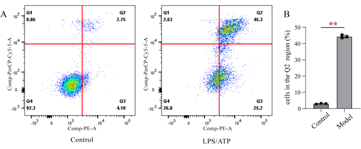
Figure 1: Detection of Annexin V/7-AAD double staining using flow cytometry. (A) Annexin V negative and 7-AAD negative staining (Q4), Annexin V positive and 7-AAD negative double staining (Q3), Annexin V positive and 7-AAD positive staining (Q2), and Annexin V negative and 7-AAD positive staining (Q1). (B) Quantitative data for (A). Data are presented as mean ± SD (n=3), unpaired t test, **p < 0.01. Please click here to view a larger version of this figure.
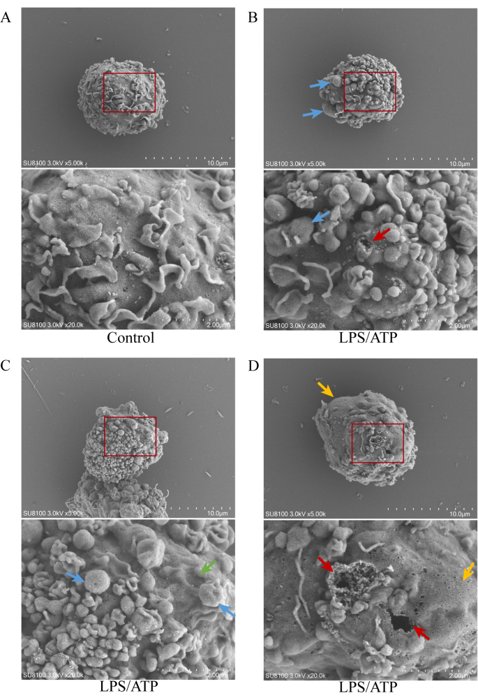
Figure 2: Scanning electron microscopy images of PMA-differentiated THP-1 macrophages. (A) Control group, 5,000x/20,000x magnification. (B, C, D) LPS/ATP group show membrane blebbing, rupture of membrane (↑), cell swelling (↑), and cell shrinkage (↑) at 5,000x/20,000x magnification. Please click here to view a larger version of this figure.
In this manuscript, two methods were used to detect LPS and ATP-induced death of PMA-differentiated THP-1 macrophages. Annexin V/7-AAD double staining was used, and the results were analyzed by flow cytometry from overall staining. As with other flow cytometric analysis, a group of unstained cells and two groups of single-stained cells were set up to exclude false positive and false negative results. The results show that after LPS/ATP stimulation, several cells lost membrane integrity, indicating that cell death may have occurred. This detection method allows us to evaluate the cell death from whole cells. It is worth noting that the Annexin V/7-AAD double staining is not sufficient to distinguish between pyroptosis, apoptosis, and necroptosis. In the early stages of these three forms, PS will be exposed outside the plasma membrane, and Annexin V staining is positive. The integrity of membrane is lost during the late stage of these three forms, and the staining results show double-positive staining. Therefore, we used SEM to further observe the possible forms of cell death that might occur.
In apoptosis, BAX/BAK pore formation on the mitochondrial outer membrane, subsequent cell shrinkage and cell membrane blebbing occurs. Necroptosis employs MLKL to open pores in the cell membrane, while pyroptosis signal activates gasdermin to form pores in the cell membrane. It is worth noting that both pyroptosis and necroptosis are accompanied by cell membrane rupture in the final stage. However, gasedermin pores are much larger than MLKL pores, so gasdermin pores do not have ion selectivity. Necroptotic cells exhibit significant swelling in morphology15. They ultimately end in membrane rupture. Regardless of the signaling pathway upstream of cell membrane permeability activation, changes in the cell membrane accompany these three forms of cell death. We used SEM to further observe the cell membrane, which provide an intuitive way to examine cell death. We observed the characteristics of pyroptosis, including membrane blebbing and the rupture membrane, and necroptosis, including cell swelling and the rupture membrane, consistent with characteristics studied by previous researchers15. We observed membrane blebbing and cell shrinkage, consistent with the characteristics of apoptosis studied by previous researchers16.
In previous studies, the combination of LPS and ATP was often used to induce pyroptosis. Interestingly, we observed features of apoptosis and necroptosis. Although all three forms may occur in LPS/ATP-induced cell death, previous studies have shown that pyroptosis remains the dominant mode of death. LPS is one of the most characterized pathogen-associated molecular patterns. ATP released from dying cells, has a high concentration within the cell. Extracellular ATP is one of the danger-associated molecular patterns17. LPS/ATP is a classic two-step stimulation method to induce pyroptosis. Research has shown that the addition of ATP to LPS-stimulated cells can rapidly activate caspase-118 while silencing the caspase-1 gene can significantly reverse LPS/ATP-induced cell death19. In previous studies, we have also found LPS/ATP-induced activation of apoptotic and necroptotic target proteins20,21. Although LPS/ATP is a classic pyroptosis inducer, it may also induce apoptosis and necroptosis.
In fact, these three forms often do not exist independently, and crosstalk between signal stages. For example, gasdermin proteins target the mitochondria to promote cytochrome c release to enhance the mitochondrial apoptotic pathway22. Activation of caspase-3 can cleave gasdermin E and then trigger pyroptosis23. A study suggests that MLKL/PIPK3 signaling is involved in the activation of NLRP3 inflammasomes24. This complex cross-talk led to the concept of PANoptosis, defined as an inflammatory programmed cell death pathway regulated by the PANoptosome complex with key features of pyroptosis, apoptosis, and/or necroptosis that cannot be accounted for by any of these programmed cell death pathways alone25. As the concept of PANoptosis was proposed, scientists described the occurrence of programmed cell death more comprehensively, this also highlights the complexity of programmed cell death. Meanwhile, this also indicates the limitations of this research method. Although pyroptosis is considered dominant in LPS/ATP-induced cell death, one cannot determine the proportion of these death forms or whether they all exist, even involving other forms of programmed death. This can be an area to investigate in the future.
This protocol is based on LPS and ATP-induced death of macrophages. We have described a method of testing from the whole to the individual, which is also applicable to other models and cells. The results indicate that cells may have undergone pyroptosis, apoptosis, or necroptosis, which is not limited to pyroptosis, helping researchers better understand cell death after LPS and ATP stimulation and choose a better experimental method. In previous studies, Annexin V/7-AAD or Annexin V/PI double staining was commonly used to indicate the occurrence of apoptosis26, 27, some researchers have also used it to study apoptosis28. In fact, we cannot distinguish between the three types of cell death through these double staining. This protocol reminds researchers, especially beginners, that Annexin V/7-AAD or Annexin V/PI double staining cannot qualitative the type of cell death. It can only show cell damage and death as a whole or indicate that the cell may have undergone some form of death. In more targeted research, it is necessary to introduce a control group that inhibits other deaths to show that conclusively program death is at hand. Similarly, further mechanistic research will investigate the critical targets of different programmed deaths.
In addition, during the preparation of the experiment, we found that there are currently many detailed experimental procedures for SEM of adherent cells, with little description of suspended cells. Here, we provide a more detailed description of the sample preparation method for suspended cells. Step 5.5 is a necessary step to assist in immobilizing suspended cells onto cell slides. Subsequent operations must be performed gently to prevent cell detachment. In addition to the chromium alum solution, poly-L-lysine can also immobilize cells.
The authors have nothing to disclose.
We express our great appreciation to Jiayi Sun and Lu Yang at Innovative Institute of Chinese Medicine and Pharmacy, Chengdu University of Traditional Chinese Medicine, for the assistance with flow cytometry, Cuiping Chen at State Key Laboratory of Southwestern Chinese Medicine Resources, for the help with scanning electron microscopy. This work was supported by the National Natural Science Foundation of China [82104491], the Natural Science Foundation of Sichuan [2023NSFSC0674], and the Post-doctoral Science Foundation of China [2021M693789].
0.25% pancreatic enzyme solution (excluding EDTA) | BOSTER Biological Technology co.ltd | PYG0068 | |
5 mL Polystyrene Round-Bottom Tube | CORNING | 352235 | |
5 mL centrifuge tube | Labgic Technology Co., Ltd. | BS-50-M | |
6-well plate | Sorfa Life Science Research Co.,Ltd | 220100 | |
Annexin V-PE/7-AAD apoptosis analysis kit | Absin (Shanghai) Biological Technology co.ltd | abs50007 | Annexin V-PE, 7-AAD, 5×Binding buffer, Apoptosis Positive Control Solution |
celculture CO2 incubator | Esco (Shanghai) Enterprise Development Co., Ltd. | N/A | |
cell culture dish, 100 mm | Sorfa Life Science Research Co.,Ltd | 230301 | |
Cellometer K2 Fluorescent Cell Counter | Nexcelom Bioscience LLC | Cellometer K2 | |
Cellometer SD100 Counting Chambers | Nexcelom Bioscience LLC | CHT4-SD100-002 | |
centrifuge machine | Hunan Xiangyi Laboratory Instrument Development Co., Ltd | L530 | |
chromium alum | Guangdong Wengjiang Chemical Reagent Co., Ltd. | PA04354 | |
cover glasses, 9 mm | Labgic Technology Co., Ltd. | BS-09-RC | |
critical point dryer | Quorum Technologies | K850 | |
dimethyl sulfoxide | BOSTER Biological Technology co.ltd | PYG0040 | |
electron microscope fixative | Servicebio Technology co.ltd | G1102 | 2.5% glutaric dialdehyde, 100 mM phosphorous salts |
electronic balance | SHIMADZU | ATX124 | |
ethanol absolute | Chengdu Kelong Chemical Co., Ltd | 2021033102 | |
flow cytometer | Becton,Dickinson and Company | FACSCanto | |
flow cytometry analysis software | Becton,Dickinson and Company | BD FACSDivaTM Software | |
gelatin | Guangdong Wengjiang Chemical Reagent Co., Ltd. | PA00256 | |
High resolution cold field emission scanning electron microscope | TITACHI | Regulus 8100 | |
human monocytic cell line THP-1 | Procell Life Science&Technology Co.,Ltd. | CL0233 | |
inverted microscope | Leica Microsystems Co., Ltd | DMi1 | |
IR Vortex Mixer | VELP Scientifica Srl | ZX4 | |
lipopolysaccharide | Beijing Solarbio Science & Technology Co.,Ltd. | L8880 | LPS is derived from Escherichia coli 055:B5 |
Na2ATP | Beijing Solarbio Science & Technology Co.,Ltd. | A8270 | |
phorbol-12-myristate-13-acetate | Beijing Solarbio Science & Technology Co.,Ltd. | P6741 | |
phosphate-buffered saline | Servicebio Technology co.ltd | G4202 | |
Pipette | Eppendorf AG | N/A | |
pipette tips, 10 μL | Servicebio Technology co.ltd | T-10PL | |
pipette tips, 1 mL | Servicebio Technology co.ltd | T-1250L | |
pipette tips, 200 μL | Servicebio Technology co.ltd | T-200L | |
RPMI-1640 complete culture media | Procell Life Science&Technology Co.,Ltd. | CM0233 | RPMI-1640 + 10% FBS + 0.05mM β-mercaptoethanol + 1% P/S |
RPMI-1640 culture media | Shanghai BasalMedia Technologies Co., LTD. | K211104 | |
sheath fluid | BECKMAN COULTER | 8546733 | |
sputter coater | Cressington Scientific Instruments Ltd | 108 | |
thermostatic water bath | GUOHUA Electric Appliance CO.,Ltd | HH-1 |