The present protocol describes pH measurements in human tissue-derived gastric organoids using microelectrodes for spatiotemporal characterization of intraluminal physiology.
The optimization and detailed characterization of gastrointestinal organoid models require advanced methods for analyzing their luminal environments. This paper presents a highly reproducible method for the precise measurement of pH within the lumina of 3D human gastric organoids via micromanipulator-controlled microelectrodes. The pH microelectrodes are commercially available and consist of beveled glass tips of 25 µm in diameter. For measurements, the pH microelectrode is advanced into the lumen of an organoid (>200 µm) that is suspended in Matrigel, while a reference electrode rests submerged in the surrounding medium in the culture plate.
Using such microelectrodes to profile organoids derived from the human gastric body, we demonstrate that luminal pH is relatively consistent within each culture well at ~7.7 ± 0.037 and that continuous measurements can be obtained for a minimum of 15 min. In some larger organoids, the measurements revealed a pH gradient between the epithelial surface and the lumen, suggesting that pH measurements in organoids can be achieved with high spatial resolution. In a previous study, microelectrodes were successfully used to measure luminal oxygen concentrations in organoids, demonstrating the versatility of this method for organoid analyses. In summary, this protocol describes an important tool for the functional characterization of the complex luminal space within 3D organoids.
Organoids-miniature multicellular structures derived from stem cells-have revolutionized our ability to study human physiology and are starting to replace animal models, even in regulatory settings1. Since the initial description of intestinal organoids by Sato et al. in 2009, organoid technology has become immensely popular2. A large number of studies have characterized the cellular composition and function of organoid models in great detail3,4,5,6. However, the luminal space of these 3D multicellular structures remains largely undefined7,8. The lumen is the central cavity of organoids derived from mucosal tissues that is surrounded by the apical portions of polarized epithelial cells. Since cellular secretion and absorption predominantly occur at the apical epithelial surface, the luminal microenvironment of organoids is controlled by these important physiological processes. Currently used organoid models demonstrate variations in cell signaling patterns, overall stemness, metabolite concentration gradients, and environmental conditions9. Understanding organoid luminal physiology is therefore necessary for the accurate modeling of organ function and pathology. Unfortunately, the relative inaccessibility of the lumen significantly hinders functional analyses of luminal physiology in 3D organoids10.
The ability to examine pH profiles is especially important in the stomach, which is notorious for having the steepest proton gradient in the body, ranging from approximately 1-3 in the lumen, to near neutral at the epithelium11,12,13.There remains a significant gap in our understanding of the microscale maintenance of the gastric pH gradient, and the relevance of organoid models in recapitulating this dynamic milieu across the gastric mucus layer. Traditional approaches for the analysis of organoid pH have involved the use of pH-sensitive dyes, which can be fluorescent or colorimetric indicators. McCracken et al. used a luminal injection of SNARF-5F-a ratiometric pH indicator-into organoids to analyze a drop in luminal pH in response to histamine treatment. Such dyes can be incorporated into the culture media, allowing for real-time, non-invasive monitoring of pH. Not only do pH-sensitive dyes require complex calibration steps that contribute to poor reliability and accuracy with measurements, but such dyes also tend to operate within specific detection ranges that may not be representative of the full pH range within the microenvironment of interest14,15. It could be considered reasonable, however, to use indicator dyes for confirmatory experiments. Optical nanosensors that use fluorescent optode-based, pH-sensing approaches have also been developed; however, such sensing techniques require microscopic imaging and are also susceptible to photobleaching, phototoxicity, as well as imaging bias16,17. Additionally, Brooks et al. have 3D-printed multiwell plates containing microelectrodes on top of which organoids may be plated18. This approach, however, does not allow for measurements directly inside the organoid lumen.
Electrode-based pH measurements can achieve improved accuracy compared to other methods, as well as provide real-time pH monitoring. In addition, pH electrodes mounted on micromanipulators allow for superior spatial resolution of pH measurements as the precise location of the electrode tip can be finely controlled. This enables the highest possible flexibility and reproducibility in the analyses of organoid models. The electrodes used here are miniaturized pH microelectrodes that operate based on the diffusion of protons through selective pH glass that surrounds a thin platinum wire. The microelectrode is connected to an external Ag-AgCl reference electrode and then connected to a high-impedance millivolt meter. The electrical potential between the two electrode tips when submerged in the same solution will reflect the pH of the solution19. Such microprofiling systems have been used in the metabolic analysis of biofilms20,21, planktonic algae22, human sputum samples23, and even in mesenchymal stem cell spheroids24. Both our lab and Murphy et al. have previously used micromanipulator-controlled O2 microelectrodes to evaluate the oxygen concentrations in the luminal spaces of organoids. Murphy et al. paired this method with mathematical modeling to reveal an oxygen gradient within their spheroids. Our group was able to find reduced luminal oxygen levels in tissue-derived gastric organoids compared to the surrounding extracellular matrix25.
Here, we provide a detailed method for the manual microelectrode profiling of the luminal pH in spherical gastrointestinal tract organoids that will enable enhanced physiological understanding of their complex luminal microenvironment. We anticipate that this technique will add a new dimension to the exploration of organoid physiology through real-time, high-resolution measurements of pH levels at a microscale. Furthermore, the following protocol could be easily adapted for the analysis of O2, N2O, H2, NO, H2S, redox, and temperature in various types of organoid models. Physiological profiling serves as a valuable tool for optimizing organoid culture conditions to better mimic in vivo environments, thereby enhancing the relevance and utility of organoid models in biomedical research.
This protocol requires 3D organoids of at least 200 µm in diameter that have a distinct lumen and that are embedded in an artificial extracellular matrix (ECM, e.g., Matrigel). Human gastric tissues for organoid derivation were obtained with approval from the Institutional Review Board of Montana State University and informed consent from patients undergoing upper endoscopy at Bozeman Health (protocol # 2023-48-FCR, to D.B.) or as exempt whole stomach or sleeve gastrectomy specimens from the National Disease Research Interchange (protocol #DB062615-EX). Information about the organoid lines and passage numbers used for this study is provided in Table 1, and the media composition is listed in Table 2. Refer to previously published protocols for the generation and maintenance of gastrointestinal organoid lines6,26,27.
1. Preparation of human gastric organoids for pH profiling
2. Unpacking and calibration of microelectrodes
NOTE: To enable microscale measurements, a separate reference electrode is used in addition to the pH sensor microelectrode rather than using an integrated (and hence bulkier) design. Both pH microelectrode and reference electrode must be stored wet. Do not allow exposure to air for more than 10 min at a time. Determine the appropriate tip size for the application. Here, we used a potentiometric pH microelectrode with a beveled tip diameter of 25 µm.
3. pH profiling of human gastric organoids
NOTE: The following protocol is described for a right-handed user. CAUTION: Disable all power-saving options on your PC as ongoing measurements will be disrupted if the PC enters sleep mode.
4. Motorized profiling (optional)
NOTE: This option requires a micromanipulator that is mounted on a mechanical motor stage, which is ultimately controlled by computer software via a motor controller31.
5. Cleaning of electrodes
6. Storage of electrodes
NOTE: Both electrodes are to be stored at room temperature in a low-activity location, safe from accidental damage.
7. Methyl red injection (optional)
NOTE: Methyl red is a colorimetric indicator dye that can be used to validate the microelectrode measurements.
Secretion of acid is a crucial function of the human stomach. However, to what extent acid secretion can be modeled in organoids is still a matter of debate6,32,33,34. We therefore developed the protocol detailed above to accurately measure acid production in gastric organoids. Notably, we used unstimulated adult stem cell-derived organoids cultured under standard expansion conditions that had been passaged several times, which led to parietal cell loss35. Therefore, the presence of acid-secreting parietal cells and active acid release was not expected in our model system.
For our experiments, we used organoids with diameters between 200 µm and 1,000 µm seeded on glass-bottom dishes. First, we tested two different tip sizes for the microelectrodes-a pH-25 with a tip diameter of 25 µm and a pH-50 with a tip diameter of 50 µm. As shown in Figure 2A, there was no significant difference between measurements obtained with the smaller compared to the larger tips. Interestingly, the baseline pH in the organoids tended to be slightly alkaline at ~pH 8. Using the 25 µm pH-25 tip, we next assessed the variation in luminal pH within different ten organoids maintained on the same culture plate, with three measurements obtained from each organoid (Figure 2B). Within one plate, the luminal pH of individual organoids showed only slight, non-significant variations (8.16 ± 0.12; p ≥0.0445-0.99) (Figure 2B). We also compared intraluminal pH measurements within five different organoid lines with passages ranging from 3 to 16 (Figure 2C). Again, we found consistent pH measurements within each culture but significant variability between organoid lines. However, luminal pH generally remained between pH 7.3 and pH 8.2, within one order of magnitude, and there was no apparent trend for average pH when comparing early to late passage numbers (Figure 2C). We next asked whether the pH of the organoid lumen was directly related to the pH of the organoid expansion media and extracellular matrix (ECM). Comparison between the media, the ECM surrounding the organoids, and the organoid lumen revealed significant differences in pH, with the luminal pH of the organoids lower than that of the ECM, and the ECM pH lower than that of the surrounding organoid expansion media (Figure 2D), suggesting that the luminal pH of the organoids was physiologically relevant rather than directly determined by the culture environment. Across six independent experiments, we measured an average luminal pH that was near neutral at 7.13 ± 0.09.
A motorized micromanipulator was used to obtain pH measurements of the organoid lumen with greater spatial resolution. This approach is relevant if pH or other gradients within the mucus-filled organoid lumen29 need to be recorded. Figure 2E shows two representative series of pH measurements in large organoids (>1,000 µm diameter), demonstrating the entry point into each organoid and ending at a depth of ~800 µm. As the two organoids profiled did not have the same diameter, the measurements are not immediately comparable. Regardless, we show evidence of a slight pH gradient of Δ0.6 between the epithelial surface and the deeper organoid lumen (Figure 2E). To determine the feasibility of performing pH measurements in the organoids over time, which would enable measurements of treatment responses in real time, we recorded the intraluminal pH inside a representative organoid for approximately 20 min and found that the reading remained highly consistent after an initial adaptation period (Figure 2F). To validate the luminal pH measurements with an independent method, we used a pH-sensitive colorimetric dye (methyl red) that we injected into the organoid lumen using a micromanipulator-mounted nanoliter autoinjector (Figure 2G). The yellow coloring of the dye confirmed that the organoid lumen had a pH >6.2, consistent with the microelectrode measurements that showed a near-neutral pH. Overall, these representative results illustrate the feasibility and reproducibility of microelectrode-based pH measurements in organoid cultures.
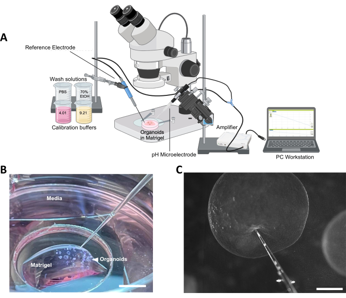
Figure 1: Overview of the method. (A) Schematic diagram of profiling setup. The microelectrode in Figure 1A was taken from the Unisense website.36 (B) Representative image of organoid culture ready for profiling, surrounding ECM, and surrounding media (scale bar = 5 mm). (C) Example of correct microelectrode positioning in preparation for organoid probing (scale bar = 500 µm). Abbreviation: ECM = extracellular matrix. Please click here to view a larger version of this figure.
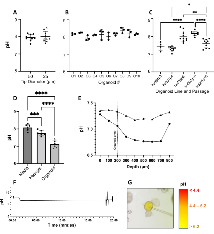
Figure 2: Validation of microsensor profiling in human gastric organoids. (A) Comparison of mid-lumen pH measurements using 25 µm (pH-25) and 50 µm (pH-50) microelectrode tips. Data from 10 individual organoids, each from a single representative experiment. (B) Three replicate pH measurements were obtained for 10 individual organoids. One-way ANOVA with multiple comparisons (p = 0.1229). (C) pH measurements obtained with a pH-25 electrode are consistent within each of five organoid lines analyzed at different passage numbers; 4-10 organoids measured per line based on size and availability. One-way ANOVA with multiple comparisons (p < 0.0001). (D) pH measurements within gastric organoids and the surrounding Matrigel and media (n = 6 independent experiments). Measurements obtained by penetrating gastric epithelial organoids with a pH-25. Each data point is the mean of 10 individual pH measurements within a single organoid. One-way ANOVA (p < 0.0001). (E) Epithelial-to-lumen pH profiles of two representative organoids using a motorized micromanipulator. (F) Stability of pH over time (~19.8 min) in one representative organoid. (G) Microinjection of methyl red pH indicator dye and HCl into the gastric organoid lumen. Please click here to view a larger version of this figure.
Donor | |||||
Figure | Line | Passage | Sex | Age | Ethnicity |
2A | 35 | 6 | F | 40 | B |
2B | 7 | 15 | F | 26 | B |
7 | 4 | F | 26 | B | |
1 | 16 | F | 45 | C | |
34 | 3 | F | 44 | Unknown | |
35 | 6 | F | 40 | B | |
2C | 7 | 15 | F | 26 | B |
2D | 36 | 1 | Unknown | ||
7 | 5 | F | 26 | B | |
37 | 1 | Unknown | |||
31 | 3 | F | 45 | H | |
7 | 9 | F | 26 | B | |
7 | 4 | F | 26 | B | |
2E | 10 | 15 | F | 43 | Unknown |
2F | 34 | 3 | F | 33 | C |
2G | 7 | 5 | F | 26 | B |
Table 1.
Human Gastric Organoid Expansion Medium (L-WRN) | |
L-WRN conditioned medium | 50% |
Advanced DMEM/F12 | 37% |
Fetal bovine serum | 10% |
Penicillin/streptomycin | 1% |
L-glutamine | 1% |
Gentamycin | 0.10% |
Amphotericin B | 0.10% |
HEPES buffer | 0.40% |
Y-27632 | 0.10% |
SB-431542 | 0.10% |
Table 2.
Supplemental Figure S1: Ion transporter expression in human gastric organoids. Relative copy numbers normalized to 18S rRNA. Please click here to download this File.
Supplemental Figure S2: Human gastric organoid with deformed architecture following puncture with the pH microsensor probe. Scale bar = 1 mm. Please click here to download this File.
Supplemental Video S1: Unsuccessful profiling attempt in a gastric organoid culture. The 25 µm pH microelectrode is manually advanced toward the organoid of interest. After a failed attempt due to an angle misjudgment, a different angle is attempted and it is revealed that the organoid lacks the structural integrity to be profiled (spherical structure is not sturdy and deforms upon penetration with the electrode). The user then redirects to attempt a different organoid. Please click here to download this File.
Limited access to the luminal space of organoids has severely restricted our understanding of the physiological dynamics of this microenvironment. A reliable tool for functional analyses of luminal physiology will expand our ability to leverage organoids as in vitro models for physiology, pharmacology, and disease research. Organoids are highly tunable, physiologically relevant models with the added potential to replicate genetic variability within the human population. Existing methods for pH measurement inside the organoid lumen have used pH-sensitive dyes or nanoparticles, methods that often must be coupled with fluorescence microscopy or spectroscopy16,17,37. The described microsensor-based profiling method provides a new route for measuring the intraluminal pH of gastrointestinal organoids with new spatial precision. Our method demonstrates consistent and reliable measurement of gastrointestinal organoid luminal pH, as well as evidence of a pH gradient. In unpublished experiments, we confirmed the expression of SLC family genes SLC4A2 and 26A7, which are known to be responsible for bicarbonate secretion as well as SLC9A3, 9A4, and 9A8 that are responsible for proton exchange (Supplemental Figure S1)38,39,40,41. Our studies suggest a predominance of bicarbonate secretion and a lack of parietal cells in our organoids, which may explain the alkalinity.
It is important to ensure that the microelectrodes used are well-suited for organoid microprofiling – this includes selecting the correct tip diameter. We tried both 25 µm and 50 µm tips and obtained similar results (Figure 2A) but decided to move forward with the smaller tip size per the manufacturer's advice since it was expected to be less invasive and provide a higher spatial resolution, as measurements are averaged across the beveled tip area. As a downside, however, smaller tips are more fragile. Organoid size must be considered in determining the desired spatial resolution.
A critical step in the protocol was ensuring the ability to distinguish between the organoid intraluminal pH from the pH in the ECM, and subsequently, in the surrounding culture medium. One would expect a reasonable amount of metabolite flux into and out of the organoids42. We expect an organoid's pH to be influenced by its environment and found that the organoids were consistently less alkaline than their surroundings in six independent experiments (Figure 2D). In addition, stability is just as important in profiling as it is during sensor calibration.
Tissue-derived organoids are expected to exhibit a certain degree of donor-to-donor variability, so it is essential to profile any organoid line at baseline before applying any experimental interventions such as drug treatment43. We hypothesize that fluctuations in pH within any one organoid culture may be due to local ion gradients at the microscale and the heterogeneous distribution of luminal mucus as determined by immunofluorescence and immunohistochemical staining (unpublished data). The observed variations in pH between individual organoid lines and passages may be due to individual differences in cell composition and secretory activity of the organoids. We also found that some organoid lines were inherently more difficult to penetrate than others. Similarly, some may lose their structural integrity and collapse upon electrode entry (Supplemental Figure S2 and Supplemental Video S1). This does not seem to be associated with organoid passage but has been observed most commonly in organoids over ~1.5 mm in diameter. In a previous study, we showed that the mucus inside the organoids was heterogeneously distributed29. Because of this heterogeneity, it becomes difficult to determine whether (or the extent to which) mucus distribution has impacted a measurement.
Microsensors are minimally invasive tools that can profile organoid intraluminal physiology with high speed and high spatial resolution. Such sensors also do not disturb chemical gradients and are minimally sensitive to turbulence in the microenvironment of interest. The small design is made possible by separating the sensor electrode from the reference electrode-standard laboratory pH probes combine these two electrodes, leading to a larger size. Optical pH and O2 nanosensors for intracellular measurements have been developed by the Cash group at the Colorado School of Mines and have been successful in measuring metabolites in both mouse and biofilm models. Because of the heterogeneity in the organoid lumen, however, such sensors may lead to uninterpretable results17. McCracken et al. analyzed luminal pH in embryonic stem cell-derived gastric organoids by injecting SNARF-5F and visualizing the pH-sensitive dye with real-time confocal microscopy37. Notably, measurements reported by McCracken were highly similar to the pH values obtained in our current study (Figure 2B,C). Our technique could perhaps be applied in a monolayer architecture, with more of a vertical setup as in biofilm profiling, though this may pose a risk of damaging the electrodes44. Future studies also could involve sequential measurements of pH over multiple days to assess changes associated with cell development. Since medium-sized organoids generally recover and heal after probing, one organoid could theoretically be probed multiple times as long as the sterility of the cultures can be maintained. As we refine our understanding of pH dynamics in organoid models, spatial resolution must be a priority for the precise mapping of the complex and historically inaccessible 3D microenvironment within.
In summary, this protocol provides a detailed method for accurate measurements of pH with high spatiotemporal resolution using pH microelectrodes mounted on a micromanipulator and a stereomicroscope. This method was validated with human adult stem cell-derived organoids and is suitable for epithelial organoids of at least 200 µm in diameter that have a distinct lumen. We anticipate that by selecting alternative microelectrodes, this method can be easily adapted to other types of organoids and the measurement of alternative compounds, such as NO or O2.
The authors have nothing to disclose.
The authors would like to acknowledge Dr. Ellen Lauchnor, Dr. Phil Stewart, and Bengisu Kilic for their previous work and assistance with the O2 microsensors; Andy Sebrell for training in organoid culture and micromanipulation; Lexi Burcham for assistance in organoid culture, media preparation, data recording, and organization; and Dr. Susy Kohout for general advice in electrophysiology. We would like to thank Dr. Heidi Smith for her assistance with imaging and acknowledge the Center for Biofilm Engineering Bioimaging Facility at Montana State University, which is supported by funding from the National Science Foundation MRI Program (2018562), the M.J. Murdock Charitable Trust (202016116), the US Department of Defense (77369LSRIP & W911NF1910288), and by the Montana Nanotechnology Facility (an NNCI member supported by NSF Grant ECCS-2025391).
Special thanks to the entire Unisense team who made this work possible, especially Dr. Andrew Cerskus, Dr. Laura Woods, Dr. Lars Larsen, Dr. Tage Dalsgaard, Dr. Line Daugaard, Dr. Karen Maegaard, and Mette Gammelgaard. Funding for our study was provided by the National Institutes of Health grants R01 GM13140801 (D.B., R.B.) and UL1 TR002319 (K.N.L), and a Research Expansion Award from the Montana State University Office for Research and Economic Development (D.B.). Figure 1A was created with BioRender.
3 M KCl | Unisense | ||
5 mL Wobble-not Serological Pipet, Individually Wrapped, Paper/Plastic, Bag, Sterile | CellTreat | 229091B | |
10 mL Wobble-not Serological Pipet, Individually Wrapped, Paper/Plastic, Bag, Sterile | CellTreat | 229092B | |
15 mL Centrifuge Tube – Foam Rack, Sterile | CellTreat | 229412 | |
24 Well Tissue Culture Plate, Sterile | CellTreat | 229124 | |
25 mL Wobble-not Serological Pipet, Individually Wrapped, Paper/Plastic, Bag, Sterile | CellTreat | 229093B | |
35 mm Dish | No. 1.5 Coverslip | 20 mm Glass Diameter | Uncoated | MatTek | P35G-1.5-20-C | |
50 mL Centrifuge Tube – Foam Rack, Sterile | CellTreat | 229422 | |
70% Ethanol | BP82031GAL | BP82031GAL | |
70 μm Cell Strainer, Individually Wrapped, Sterile | CellTreat | 229483 | |
1,000 µL Extended Length Low Retention Pipette Tips, Racked, Sterile | CellTreat | 229037 | |
Amphotericin B (Fungizone) Solution | HyClone Laboratories, Inc | SV30078.01 | |
Biosafety Cabinet | Nuaire | NU-425-600 | Class II Type A/B3 |
Bovine Serum Albumin | Fisher Bioreagents | BP1605-100 | |
Cell recovery solution | Corning | 354253 | Cell dissociation solution |
DMEM/F-12 (Advanced DMEM) | Gibco | 12-491-015 | |
Dulbecco's Modification of Eagles Medium (DMEM) | Fisher Scientific | 15017CV | |
Fetal Bovine Serum | HyClone Laboratories, Inc | SH30088 | |
G418 Sulfate | Corning | 30-234-CR | |
Gentamycin sulfate | IBI Scientific | IB02030 | |
HEPES, Free Acid | Cytiva | SH30237.01 | |
HP Pavillion 2-in-1 14" Laptop Intel Core i3 | HP | M03840-001 | |
Hydrochloric acid | Fisher Scientific | A144C-212 | |
Incubator | Fisher Scientific | 11676604 | |
iPhone 12 camera | Apple | ||
L-glutamine | Cytiva | SH3003401 | |
Large Kimberly-Clark Professional Kimtech Science Kimwipes Delicate Task Wipers, 1-Ply | Fisher Scientific | 34133 | |
M 205 FA Stereomicroscope | Leica | ||
Matrigel Membrane Matrix 354234 | Corning | CB-40234 | |
MC-1 UniMotor Controller | Unisense | ||
Methyl red | |||
MM33 Micromanipulator | Marzhauser Wetzlar | 61-42-113-0000 | Right handed |
MS-15 Motorized Stage | Unisense | ||
Nanoject-II | Drummond | 3-000-204 | nanoliter autoinjector |
Penicillin/Streptomycin (10,000 U/mL) | Gibco | 15-140-148 | |
pH Microelectrodes | Unisense | 50-109158, 25-203452, 25-205272, 25-111626, 25-109160 | SensorTrace software is not compatible with Apple computers |
Reference Electrode | Unisense | REF-RM-001652 | SensorTrace software is not compatible with Apple computers |
SB 431542 | Tocris Bioscience | 16-141-0 | |
Smartphone Camera Adapter | Gosky | ||
Specifications Laboratory Stand LS | Unisense | LS-009238 | |
Trypsin-EDTA 0.025%, phenol red | Gibco | 25-200-056 | |
UniAmp | Unisense | 11632 | |
United Biosystems Inc MINI CELL SCRAPERS 200/PK | Fisher | MCS-200 | |
Y-27632 dihydrochloride | Tocris Bioscience | 12-541-0 | |
µSensor Calibration Kit | Unisense/ Mettler Toledo | 51-305-070, 51-302-069 | pH 4.01 and 9.21, 20 mL packets |
.