JoVE Educazione Scientific
Physical Examinations II
This content is Free Access.
JoVE Educazione Scientific Physical Examinations II
Lymph Node Exam
  • 00:00Panoramica
  • 01:47Lymph Nodes of the Head and Neck
  • 04:23Axillae and Upper Extremities
  • 06:19Lower Extremities
  • 07:34Summary

Exame do Linfonodo

English

Condividere

Panoramica

Fonte: Richard Glickman-Simon, MD, Professor Assistente, Departamento de Saúde Pública e Medicina Comunitária, Tufts University School of Medicine, MA

O sistema linfático tem duas funções principais: devolver o fluido extracelular de volta à circulação venosa e expor substâncias antigênicas ao sistema imunológico. À medida que o fluido coletado passa por canais linfáticos em seu caminho de volta à circulação sistêmica, encontra múltiplos nós que consistem em aglomerados altamente concentrados de linfócitos. A maioria dos canais linfáticos e nódulos residem profundamente dentro do corpo e, portanto, não são acessíveis ao exame físico(Figura 1). Apenas nós próximos à superfície podem ser inspecionados ou palpatados. Os linfonodos são normalmente invisíveis, e nós menores também não são palpáveis. No entanto, nós maiores (>1 cm) no pescoço, axilas e áreas inguinais são frequentemente detectáveis como massas macias, lisas, móveis, não macias, em forma de feijão embutidas em tecido subcutâneo.

A linfadonopatia geralmente indica uma infecção ou, menos comumente, um câncer na área de drenagem linfática. Os nódulos podem ser ampliados, fixos, firmes e/ou macios, dependendo da patologia presente. Por exemplo, um linfonodo macio e macio palpável perto do ângulo da mandíbula pode indicar uma amígdala infectada, enquanto um nódulo linfático firme, ampliado e não macio palpável na axila de uma paciente do sexo feminino pode ser um sinal de câncer de mama.

Linfonodos regionais que drenam a área de uma infecção localizada normalmente permanecem invisíveis, mas podem se tornar ampliados e macios na palpação. Uma ferida infectada ou celulite também pode resultar em linfadenite ou linfadenite, uma condição na qual a infecção se espalha ao longo da cadeia de canais linfáticos e nódulos. Isso pode ser acompanhado pelo aparecimento de listras vermelhas e sintomas sistêmicos, como febre, calafrios e mal-estar. Em casos raros, a intensidade da reação inflamatória pode fazer com que os nós aderam ao tecido mole circundante, fixando-os no lugar.

Muitos cânceres metastáticos se espalham primeiro para linfonodos regionais. Ao contrário das infecções, as células malignas que invadem linfonodos podem fazer com que elas se sintam irregulares e firmes (até mesmo duras) mas permanecem não-macias. Se o câncer invadir as cápsulas externas, os nódulos podem ficar fixos no tecido mole circundante ou emaranhados. Linfoma, um câncer primário intrínseco ao sistema linfático, pode estar presente em qualquer lugar do corpo como linfonodos únicos ou múltiplos aumentados, que podem se tornar grandes o suficiente para ver na inspeção, e geralmente são difíceis e não macios na palpação. Além do linfoma, a linfanopatia difusa pode ser uma indicação de doenças infecciosas ou inflamatórias generalizadas, como HIV, mononucleose ou sarcoidose.

Figure 1
Figura 1. O Sistema Linfático.

Procedura

Como os linfonodos são distribuídos por todo o corpo, sua avaliação geralmente ocorre como parte dos exames regionais da cabeça e pescoço, mama e axilas, extremidades superiores, genitália externa e/ou extremidades inferiores. É melhor usar as almofadas do índice e dos dedos médios para notar o tamanho, forma, número, plibilidade, textura, mobilidade e ternura de nódulos bilateralmente. 1. Linfonodos da Cabeça e pescoço Figura 2. Linfonodos da Cabeça e pescoço. Com o pescoço do paciente flexionado ligeiramente para a frente, inspecione para um alargamento de nódulos visivelmente visível. Para cada um dos passos a seguir, planeje palpar os nós da cabeça e pescoço(Figura 2) com ambas as mãos, uma de cada lado. Em muitos casos, os nós não são palpáveis. Palpar os nódulos préauriculares, posteriores e mastoides na frente da orelha, atrás da orelha, e superficial ao processo mastoide, respectivamente. Palpa os nódulos occipitais posteriormente na base do crânio. Palpa os nós tonsiláconos localizados no ângulo da mandíbula, os nós submandibulares no meio do caminho entre o ângulo e a ponta, e os nós submentais a poucos centímetros da ponta. Note que os nódulos submandibulares precisam ser distinguidos da glândula submandibular subjacente, que é maior e lobulada. Palpa os nódulos cervicais anteriores e superficiais na frente e sobrevaste o músculo estemastoide, respectivamente. Nódulos cervicais profundos, sob o músculo esternostoide, raramente são palpáveis. Palpa os nódulos cervicais posteriores entre a borda anterior do trapézio e a borda posterior do esternostoide. Palpa os nódulos supraclaviculares no fundo do ângulo formado pelo músculo estemastoide e clavícula. Alguns cânceres de pulmão e abdominais metástases para esses nódulos, então eles podem ser descobertos durante o exame. Palpa os nódulos infraclaviculares na parte inferior da clavícula. 2. Axillae e Extremidade Superior Figura 3. Linfonodos Axilares. Três grupos de nós axilares – braquial, subscapular e peitoral – drenam seu linfático nos nódulos axilares centrais que estavam profundamente dentro da axila contra a parede do peito no meio do caminho entre as dobras axilares anteriores e posteriores(Figura 3). Esses nódulos, por sua vez, drenam para os nódulos infraclaviculares (apical) e supraclaviculares. Dos quatro grupos axilares, apenas os nós centrais são geralmente palpáveis. Como a maioria dos cânceres de mama drenam aqui, os linfáticos axilares precisam ser examinados cuidadosamente, especialmente nas mulheres. A maioria das partes das extremidades superiores drenam mais ou menos diretamente para os linfonodos axilares. Uma exceção é a drenagem dos aspectos ulnar da mão e do antebraço, que primeiro encontra os nódulos epitrocleares acima do cotovelo. Para examinar os nódulos axilares esquerdos, fique na frente e à esquerda do paciente sentado, apoiando o braço esquerdo relaxado do paciente no pulso ou cotovelo. Informe o paciente que o exame pode parecer um pouco desconfortável. Alcance sua mão direita no alto da axila esquerda, logo atrás do músculo peitoral, com os dedos apontando para a clavícula média. Pressione os dedos contra a parede do peito e deslize-os para baixo para sentir os nódulos centrais. Se ainda não terminar, palpa os nós infraclaviculares (apical) e supraclaviculares. Enquanto ainda sustenta o braço esquerdo do paciente, palpa os nós epitrocráis, que estão localizados mediadamente cerca de 3 cm acima do cotovelo. Repita os passos 2.1 – 2.5 para a axila direita do paciente e extremidade superior usando a mão esquerda. 3. Extremidades Inferiores Figura 4. Linfonodos inguinais superficiais. Linfonodos inguinais superficiais (Figura4)estão localizados no alto da coxa anterior e drenam várias regiões das pernas, abdômen e períneo. Esses nós são muitas vezes grandes o suficiente para palpar, mesmo quando normal. Que o paciente coloque supino com quadris totalmente estendidos ou ligeiramente flexionados. Palpate um grupo horizontal de nódulos ao longo e apenas inferior ao ligamento inguinal. Esses nódulos drenam a nádega superficial e o abdômen inferior, genitália externa (excluindo os testículos), vagina inferior, canal anal e área perianal. Palpate um grupo vertical de nódulos medial para o grupo horizontal apenas inferior ao pulso da artéria femoral. Esses nódulos ficam ao longo da veia superior safena e drenam as mesmas regiões das extremidades inferiores.

Applications and Summary

Most lymph nodes lie too deep to be accessible via physical examination. The superficial nodes are most efficiently assessed during regional examinations of the head and neck, breasts and axillae, upper extremities, lower extremities, and/or external genitalia. Because lymph nodes are constantly interacting with extracellular fluid draining from nearby tissues, their examination can provide information about the presence and status of infections or malignancies in the area. Nodes draining the site of a soft tissue infection are apt to become enlarged and tender but generally remain soft, smooth, and mobile. Hard, non-tender, matted, or fixed nodes are more typical of a spreading malignancy. Diffuse lymphadenopathy may indicate systemic diseases such as lymphomas, HIV, mononucleosis, or sarcoidosis. Finding a single abnormal node should prompt an examination of all nodes.

Trascrizione

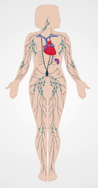
The lymph node examination forms an essential part of the evaluation for infectious diseases and cancer. The lymphatic system is comprised of organs including the spleen, lymphatic channels and lymph nodes. The channels are responsible for returning the lymph formed from extracellular fluid back to the venous circulation.

On its way, the lymph encounters multiple lymph nodes. These nodes consist of highly concentrated clusters of lymphocytes, which play a critical role in maintaining immunity. Most lymph channels and nodes reside deep within the body and are too small to be assessed by physical examination. However, superficial, larger nodes, close to or more than one centimeter in diameter-primarily located in the head and neck region, the axillae and the inguinal areas-can be palpated and assessed.

Lymph nodes in these areas normally present as soft, smooth, movable, non-tender, bean-shaped structures imbedded in the subcutaneous tissue. However, sometimes nodes may become enlarged, fixed, firm, and/or tender depending on the pathology present. This condition is referred to as lymphadenopathy and it usually indicates an infection or, less commonly, a cancer in the area of lymph drainage. This video will review the anatomical location of the key lymph nodes as well as demonstrate the procedural steps of this examination.

Let’s begin by briefly reviewing the lymph nodes in the head and neck area. The list of palpable nodes in this region is extensive including the preauricular and posterior auricular nodes located in front and behind the ear, respectively, the mastoid node positioned superficial to the mastoid process and the occipital nodes found at the base of the skull. Around the mandible are the tonsillar nodes, the submandibular nodes, and the submental nodes. Another group of nodes surround the sternomastoid muscle. These include the superficial and deep cervical nodes. The last groups of nodes are the clavicular nodes, including the supra- and the infra-clavicular nodes. The infraclavicular nodes are also known as the apical nodes.

Upon entering the examination room, introduce yourself and briefly explain the maneuvers you’re going to conduct. Before beginning with the examination, sanitize your hands by using topical disinfectant solution. Start by asking the patient to flex their neck slightly forward and inspect for noticeably enlarged nodes. Following inspection, palpate the preauricular node located in front of the ear. Throughout the exam, palpate using the pads of your index and middle fingers to note the size, shape, number, pliability, texture, mobility, and tenderness of nodes bilaterally.

Next, move to the posterior auricular node located behind the ear followed by the mastoid node located superficial to the mastoid process, and the occipital nodes found posteriorly at the base of the skull. Then move onto the tonsillar nodes located at the angle of mandible, the submandibular nodes that lie midway between the angle and tip of the mandible, and the submental nodes located a few centimeters from the tip. Next, palpate the superficial cervical nodes situated beneath and anterior to the sternomastoid muscles. The deep cervical nodes are rarely palpable. This is followed by palpation of the posterior cervical nodes located between the anterior edge of the trapezius and posterior edge of the sternomastoid muscles. Finally, palpate the supraclavicular nodes found deep within the angle formed by the sternomastoid muscle and clavicle, and the infraclavicular, or the apical nodes, located on the underside of the clavicle.

Following palpation of the head and neck nodes, move to the axillae and upper extremities. The three groups of axillary nodes-lateral, subscapular, and pectoral-drain their lymph into the central axillary nodes that lay deep within the axillae. The central nodes, in turn, drain lymph into the apical and supraclavicular nodes. Of the four axillary groups, only the central nodes are usually palpable. Since most breast cancers drain here, the axillary and clavicular lymphatics need to be examined more carefully in women. Most parts of the upper extremities drain more or less directly into the axillary lymph nodes. One exception is drainage from the ulnar aspects of the hand and forearm, which first encounters the epitrochlear nodes above the elbow.

To examine the left axillary nodes, position yourself in front and to the left of the seated patient. Gently grasp the patient’s left wrist or elbow and slightly abduct the arm. Inform the patient that the following maneuver may feel slightly uncomfortable. Move your right hand high up into the left axilla, just behind the pectoralis muscle. With your fingers pointing toward the mid-clavicle, press them against the patient’s chest wall, and slide them downward to feel the central nodes. Subsequently, you can palpate the apical and supraclavicular nodes if missed during the head and neck examination. While supporting the patient’s left arm in the same position, palpate the patient’s epitrochlear nodes, which are located medially about three centimeters above the elbow. Repeat the entire examination on the patient’s right using your left hand.

With the patient’s axilla and upper extremity examination complete, proceed to the lower extremities. This region includes the superficial inguinal lymph nodes, which are located high in the anterior thigh and drain various regions of the legs, abdomen, and perineum. These nodes are often large enough to palpate, even when normal, and can be subdivided into two groups: the horizontalgroup located just below the inguinal ligament and the vertical group located just below the femoral artery pulse.

In order to palpate these nodes, ask the patient to lay supine with their hips fully extended or slightly flexed. Once the patient is comfortable begin palpating the horizontalgroup of nodes just below the inguinal ligament. Move your hand along the entire length of the ligament, taking note of the nodes’ size, shape and firmness. Finally, palpate the vertical group of nodes, which is medial to the horizontal group and just below the femoral artery pulse. This concludes the lymph node examination. Thank the patient for their cooperation.

You have just watched JoVE’s video documenting the lymph node examinations of patients’ head and neck areas, axillae, upper extremities and lower extremities. You should now understand the systematic sequence of steps that every physician should follow in order to conduct an effective lymph node exam. As always, thanks for watching!

Tags

Cite This
JoVE Science Education Database. JoVE Science Education. Lymph Node Exam. JoVE, Cambridge, MA, (2023).