- 00:00Panoramica
- 01:07Principles of X-ray Fluorescence
- 02:37Preparing the Silicon Nitride Windows
- 03:09Plating and Fixing Cells on the Windows
- 04:10Generating X-ray Fluorescence Images
- 05:39Representative Results
- 06:09Applications
- 07:26Summary
Fluorescenza a raggi X (XRF)
English
Condividere
Panoramica
Fonte: Laboratorio della Dott.ssa Lydia Finney — Argonne National Laboratory
La fluorescenza a raggi X è una radiazione indotta ed emessa che può essere utilizzata per generare informazioni spettroscopiche. La microscopia a fluorescenza a raggi X è una tecnica di imaging non distruttiva che utilizza l’emissione di fluorescenza indotta dei metalli per identificare e quantificare la loro distribuzione spaziale.
Principi
Procedura
Risultati
The X-ray fluorescence map of an adherent cell is shown in Figure 1. Each panel shows the distribution of a particular element (e.g., copper, iron, zinc, etc) over the cell. The panel labeled 's_a' shows the absorption of X-rays.
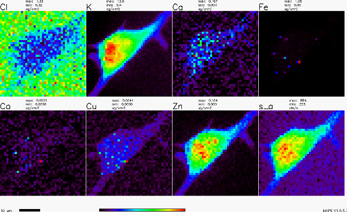
Figure 1. X-ray fluorescence map of an adherent cell. Please click here to view a larger version of this figure.
Applications and Summary
X-ray fluorescence imaging can be a useful tool in many fields including geosciences, forensic science, materials science, biology, and even in studying our cultural heritage. In materials science, it can help find defects in chips and catalysts made with metals. In cultural heritage work, it has been used to identify poisonous metals in the hair of famous dead people (e.g., Beethoven), and to identify the source of paints used in art. In biology, it is used to study the natural metals that perform important biochemistry. In geosciences, it is often used to study events chronicled in the rock record. Two particular characteristics that make X-ray fluorescence imaging useful in so many fields are 1) its non-destructive, so many items that are rare, or of high value can be imaged, and 2) while the sample preparation described here for cells is complex — because the cells must be dried-for many materials such as rocks, art, or other items, there is very little sample preparation required, other than it should be flat and free of dust. Although a synchrotron is required which is best accessed through collaboration with scientists at these facilities, the technique can be very accessible.
Trascrizione
X-ray fluorescence, or XRF, spectroscopy is a non-destructive analytical technique that is used to perform elemental analysis of samples at room temperature.
XRF can be applied to a wide range of samples, including biological, forensic, environmental, and even works of art. The samples can also take a variety of forms, such as powders, crystals, and liquids. In XRF, a sample is bombarded with a beam of X-rays causing it to emit secondary X-rays at a lower energy, which are called fluorescent radiation.
Though it is referred to as a fluorescence technique, XRF differs from traditional fluorescence microscopy in that it does not use lower energy visible light, or light-active molecules.
This video will introduce the basics of XRF, and demonstrate how to collect elemental maps of a biological sample.
When a photon of sufficient energy collides with an atom, the energy is absorbed, exciting one of the outer shell electrons. As the electron relaxes, it emits a secondary photon, typically of lower energy. This process is known as fluorescence. Unlike lower-energy photons, like those used in fluorescence microscopy, X-ray photons are energetic enough to completely expel tightly held electrons from an inner shell. An electron from a higher-energy shell will then fall into the vacancy. A photon proportional to the energy difference between the two shells is released. Each element emits a unique set of photons, or spectrum, that can be used to identify the element and determine the quantity present. This phenomenon is known as X-ray fluorescence.
Once an elemental spectrum has been collected, the signals of elements of interest can be isolated. Measurements can be taken at multiple locations across a sample, generating an image, one pixel at a time. This process is known as raster scanning. Images of all elements of interest can be subsequently generated. These elemental maps provide valuable information about the sample. With an understanding of XRF, you are now ready to prepare a biological cell sample to generate elemental maps.
To begin, first prepare and sterilize a silicon nitride window, which will hold the sample in place for the scan. Use care, as they are very fragile. Orient the window so that it is flat side up. Then place the window in a culture dish, and adhere it to the dish using small pieces of adhesive tape.
Finally, sterilize the silicon nitride window with UV radiation for 1 hr.
Now that the window has been sterilized, the sample can be fixed to it. First, hold the culture dish at an angle and add media to the side of the dish. Slowly relieve the tilt to coat the window. Add cells to the dish in the same manner, and incubate.
Periodically observe the cells under a light microscope until they are ready to use.
In a laminar flow hood, gently aspirate the media from the dish.
Then, rinse the cells with phosphate buffered saline to remove excess media.
Aspirate the PBS, and fix the cells with paraformaldehyde. After 20 min, remove the mixture and dispose as hazardous waste.
Remove the window from the dish, and quickly blot the edges and back indentation of the window with a Kimwipe. Set the window on a clean surface to dry.
Once the sample is dry, verify the presence of cells on the window with a light microscope.
Using clear nail polish, secure the window to an aluminum holder.
Insert the sample holder into the instrument mount, then place the mount on the X-ray microscopes’ positioning stage.
Position the sample window at the focal point of the X-ray microscope optics, with a 45-degree angle to the incident beam.
Exit the instrument area and conduct the remaining steps remotely to minimize X-ray exposure.
Open the shutter, and use optics to focus the monochromatic X-ray beam down to a sub-micrometer spot size.
The position of the spot can be imaged with a pre-calibrated camera. Using the positioning stages, determine the appropriate width and height needed to raster over the sample.
Collect a test spectrum of the element of interest with a dwell time of 1 – 2 seconds.
From the test spectrum, choose an appropriate scan time in order to provide a sufficient signal-to-noise ratio for the elements of interest.
Then, determine the resolution needed for the sample. The resolution should be smaller than the features of interest, but larger than the spot size. Finally, program the scan into the scanning software, and collect the image.
In this experiment, an elemental map of a cell was performed for several different elements. Many metals, such as copper, iron, and zinc are important nutrients in the cell, and were clearly identified within the cell.
By determining where each metal is found in the cell, valuable information can be elucidated about its normal cellular processes. In addition, metal-based diseases can be understood.
X-ray fluorescence is used in a wide range of scientific fields. The non-destructive nature of XRF enables its use in the study of historical artifacts. Art historians utilize the technique to determine the pigments originally used in works of art. This can elucidate information about the work, such as the provenance, colors that have faded over time, and the authenticity. Forensic scientists also use XRF in crime scene investigation. When a gun is fired, the surrounding area is coated with gun shot residue. Gun shot residue contains gun powder, ignition primer, and metal from the casing and bullet. The information collected with XRF can identify the culprit and weapon used.
Another field of study that lends itself to X-ray fluorescence is paleontology. Here, elemental information is collected from a trilobite fossil, a marine arthropod that lived over 250 million years ago.
By characterizing the elemental composition of fossils, new information can be gained about long extinct life. Better-preserved samples can even provide the composition of soft tissues that have deteriorated long ago.
You’ve just watched JoVE’s introduction to X-ray fluorescence. You should now understand the theory of X-ray spectroscopy and how to collect elemental information from a wide range of sources.
Thanks for watching!