Science Education
>

Measurement of Tissue Oxygenation Using Near-Infrared Spectroscopy in Patients Undergoing Hemodialysis

PREPARAZIONE ISTRUTTORI
CONCETTI
Student Protocol
JoVE Journal
Medicina
È necessario avere un abbonamento a JoVE per visualizzare questo.  Accedi o inizia la tua prova gratuita.
JoVE Journal Medicina
Measurement of Tissue Oxygenation Using Near-Infrared Spectroscopy in Patients Undergoing Hemodialysis

All participants provided written informed consent. The study was approved by the Institutional Review Board of the Saitama Medical Center, Jichi Medical University, Japan (RIN 15–104).

1. Device for the monitoring of rSO2

  1. Prepare a NIRS device for measuring tissue oxygenation. This device has four channels and can perform measurement in up to four organs at the same time.
  2. Prepare a measurement sensor for NIRS monitoring, to evaluate rSO2 values in each organ via transmitting near-infrared light at two wavelengths of attachment.

2. Attaching the measurement sensor

  1. Allow each patient to rest in a supine position for at least 5 minutes before HD.
  2. Attach measurement sensors to the forehead, the right hypochondrium and lower legs to evaluate rSO2 in the brain, liver and lower leg muscles, respectively.
  3. Monitoring of cerebral oxygenation
    1. Attach measurement sensors to the forehead of the dominant hemisphere.
  4. Monitoring of hepatic oxygenation
    1. Prepare echography to measure the depth to the patients’ liver from the body surface. Confirm that this measurement is within 20–30 mm from the body surface. Next, attach the measurement sensors to the right hypochondrium.
      NOTE: In this device, rSO2 values should be obtained in deep tissue 20–30 mm from the body surface. In some instances, the liver may be located in more than 30 mm from the body surface due to the presence of thick subcutaneous fat.
  5. Monitoring of muscle oxygenation
    1. Attach measurement sensors to the right or bilateral lower legs.
  6. Sensor connection and powering the device
    1. Connect each sensor to the leads from the device. Next, turn on the device, and start measuring oxygenation.

3. Puncturing the dialysis shunt and starting monitoring

  1. Puncturing the dialysis shunt
    1. Puncture the patients’ dialysis shunt to start HD therapy. At this time, measure BP using a digital blood pressure monitor equipped with the dialysis machine and collect blood samples using syringes.
  2. Start monitoring
    1. After starting HD therapy, start monitoring the tissue oxygenation of the three organs: the brain, liver and lower leg muscle.
  3. Monitoring of rSO2 during HD
    1. Observe changes in rSO2 values of each organ and measure BP regularly in addition to the usual monitoring performed during HD therapy including heart rate, venous pressure and blood volume. Confirm the attachment area and connection between the sensors and leads.

Measurement of Tissue Oxygenation Using Near-Infrared Spectroscopy in Patients Undergoing Hemodialysis

Learning Objectives

Cerebral rSO2 values before HD were lower than those in healthy subjects and cerebral rSO2 in HD patients with diabetes mellitus (DM) were lower than those in HD patients without DM (Figure 1)16. Furthermore, although tissue oxygenation continues without a decrease of BP during HD, we incidentally observed changes in cerebral and hepatic rSO2 due to intradialytic hypotension (Figure 2). Due to the continuous monitoring, the changes in tissue oxygenation were observed more quickly than by intermittently monitored BP. Data were expressed as means ± standard error. The analysis of variance for non-paired values was used to compare three groups.

Figure 1
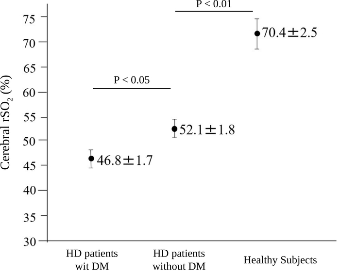
Figure 1: Comparison of cerebral rSO2 before HD among HD patients with diabetes mellitus (n = 27), HD patients without diabetes mellitus (n = 27) and healthy subjects (n = 28). The patients included 38 men and 16 women with mean age of 67.7 ± 1.2 years and HD duration of 6.5 ± 1.9 years. The causes of chronic kidney disease were DM (27 patients), chronic glomerulonephritis (14 patients), nephrosclerosis (4 patients), polycystic kidney disease (4 patients), and other (5 patients). The error bars indicate the standard error. The data were based on and the figure has been modified from a previous report16. DM; diabetes mellitus, HD; hemodialysis, rSO2; regional oxygen saturation. Please click here to view a larger version of this figure.

Figure 2
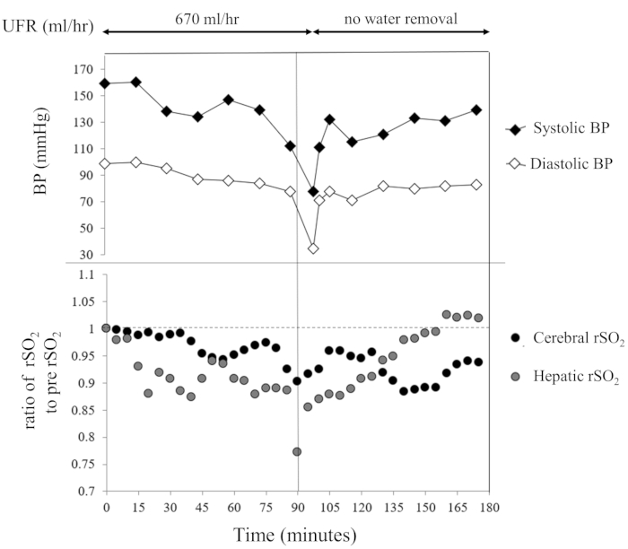
Figure 2: Changes in cerebral and hepatic rSO2 in a patient with acute intradialytic hypotension. BP; blood pressure, hr; hour, rSO2; regional oxygen saturation, UFR; ultrafiltration rate. Please click here to view a larger version of this figure.

List of Materials

DBB-100NX Nikkiso DBB-100NX Dialysis machine
INVOS 5100c Covidien Japan INVOSTM 5100c tissue oxygenation device
SOMASENSER Covidien Japan CV-SAFB-SM/INTL NIRS sensor

Lab Prep

Near-infrared spectroscopy (NIRS) has recently been applied as a tool to measure regional oxygen saturation (rSO2), a marker of tissue oxygenation, in clinical settings including cardiovascular and brain surgery, neonatal monitoring and prehospital medicine. The NIRS monitoring devices are real-time and noninvasive, and have mainly been used for evaluating cerebral oxygenation in critically ill patients during an operation or intensive care. Thus far, the use of NIRS monitoring in patients with chronic kidney disease (CKD) including hemodialysis (HD) has been limited; therefore, we investigated rSO2 values in some organs during HD. We monitored rSO2 values using a NIRS device transmitting near-infrared light at 2 wavelengths of attachment. The HD patients were placed in a supine position, with rSO2 measurement sensors attached to the foreheads, the right hypochondrium and the lower legs to evaluate rSO2 in the brain, liver and lower leg muscles, respectively. NIRS monitoring could be a new approach to clarify changes in organ oxygenation during HD or factors affecting tissue oxygenation in CKD patients. This article describes a protocol to measure tissue oxygenation represented by rSO2 as applied in HD patients.

Near-infrared spectroscopy (NIRS) has recently been applied as a tool to measure regional oxygen saturation (rSO2), a marker of tissue oxygenation, in clinical settings including cardiovascular and brain surgery, neonatal monitoring and prehospital medicine. The NIRS monitoring devices are real-time and noninvasive, and have mainly been used for evaluating cerebral oxygenation in critically ill patients during an operation or intensive care. Thus far, the use of NIRS monitoring in patients with chronic kidney disease (CKD) including hemodialysis (HD) has been limited; therefore, we investigated rSO2 values in some organs during HD. We monitored rSO2 values using a NIRS device transmitting near-infrared light at 2 wavelengths of attachment. The HD patients were placed in a supine position, with rSO2 measurement sensors attached to the foreheads, the right hypochondrium and the lower legs to evaluate rSO2 in the brain, liver and lower leg muscles, respectively. NIRS monitoring could be a new approach to clarify changes in organ oxygenation during HD or factors affecting tissue oxygenation in CKD patients. This article describes a protocol to measure tissue oxygenation represented by rSO2 as applied in HD patients.

Procedura

Near-infrared spectroscopy (NIRS) has recently been applied as a tool to measure regional oxygen saturation (rSO2), a marker of tissue oxygenation, in clinical settings including cardiovascular and brain surgery, neonatal monitoring and prehospital medicine. The NIRS monitoring devices are real-time and noninvasive, and have mainly been used for evaluating cerebral oxygenation in critically ill patients during an operation or intensive care. Thus far, the use of NIRS monitoring in patients with chronic kidney disease (CKD) including hemodialysis (HD) has been limited; therefore, we investigated rSO2 values in some organs during HD. We monitored rSO2 values using a NIRS device transmitting near-infrared light at 2 wavelengths of attachment. The HD patients were placed in a supine position, with rSO2 measurement sensors attached to the foreheads, the right hypochondrium and the lower legs to evaluate rSO2 in the brain, liver and lower leg muscles, respectively. NIRS monitoring could be a new approach to clarify changes in organ oxygenation during HD or factors affecting tissue oxygenation in CKD patients. This article describes a protocol to measure tissue oxygenation represented by rSO2 as applied in HD patients.

Tags