Sample collection was approved by the Latvian University Life and Medical Science Research Ethics Committee (decision N0-71-35/54)
NOTE: The materials used in this study are included in the Table of Materials file.
1. Three-dimensional (3D) printed mold fabrication
2. Preparation of the PDMS molds
3. Preparation of OSTE-COC top channel
4. Preparation of OSTE-COC bottom channel and device assembly
5. Device evaluation
6. Device setup
7. Device testing with standardized latex beads
8. Device testing with urine samples
9. Device testing with conditioned media
10. Isolation of EVs using ultracentrifugation
11. Isolation of EVs using size exclusion chromatography (SEC)
12. EV characterization
13. NTA
14. dsELISA for EV markers
We fabricated a microfluidic device using a 3D printed double negative mold (Figure 1) via soft-lithography (Figure 2A) for high throughput EV separation based on the bifurcated A4F principle (Figure 2B,C). The setup requires a pump and a flow-through station, as can be seen in Figure 3, for the isolation of EVs in an automated manner. Firstly, to evaluate the proof of concept of the devices, a mixture of polystyrene beads with diameters of 100 nm and 1000 nm was prepared to represent vesicles and fine cell debris, respectively10. Experiments were conducted with varying flow rates for the bead mix, both with and without bifurcating flow, to investigate the effect of linear velocity on separation efficiency. Across all experiments, the recovery of small beads remained consistent and above 90%10,showing the potential of the device to recover EVs.
Then, we assessed and compared the potential of the COC-OSTE device in isolating EVs from large volumes (>1 mL) from complex biofluids with minimal pre-processing. As such, urine from 10 healthy donors (Figure 4) and cell media from two different prostate cell lines (Figure 5) were used as a template to simultaneously isolate EVs following three different procedures: ultracentrifugation (UC), size exclusion chromatography (SEC), and the A4F microfluidics-based OSTE-COC device. After isolation, the total number of particles and their size distribution were assessed using nanoparticle tracking analysis (NTA). On average, the OSTE-COC device showed better total particle recovery from biofluids compared to UC, but the statistical significance was only achieved when combining the particle numbers from both ports (Figure 4A). In order to compare the device performance with other systems, R & L outlet together should be taken into consideration. As shown in Figure 4, L & R outlet together performance on recovering EVs outperforms UC and SEC. Separately, the L-port was designed to capture the small EV fraction, while the R-port was designed to collect the bigger EV fraction with other molecules of similar size. Interestingly, the recovery using the R-PORT of the OSTE-COC device was slightly higher than SEC and UC alone (Figure 4A). CD63 expression showed a similar pattern (Figure 4C). This finding indicates that the OSTE-COC device was more effective in total EV recovery. Equivalent size distribution was found between the different methodologies, except for UC, which shows a bigger particle size distribution (Figure 4B).
Comparable results were observed in cell media cultures. In both scenarios, the total particle recovery from both device ports exhibited superior performance compared to SEC or UC methodologies (as depicted in Figure 5A,B). Notably, EVs derived from PC3 cells demonstrated a distinct size distribution, with greater homogeneity in the L-PORT distribution when contrasted with other experimental groups (Figure 5C,D). Furthermore, the analysis of CD63 expression confirmed the higher EV recovery rates achieved using the COC-OSTE device (as illustrated in Figure 5E,F). A summary comparing the isolation characteristics of the different methodologies examined in this study can be found in Table 1.
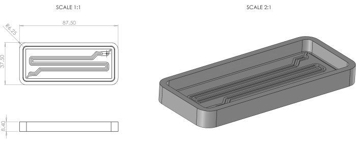
Figure 1: 3DP serpentine-shaped double negative mold dimensions and isometric view. Please click here to view a larger version of this figure.
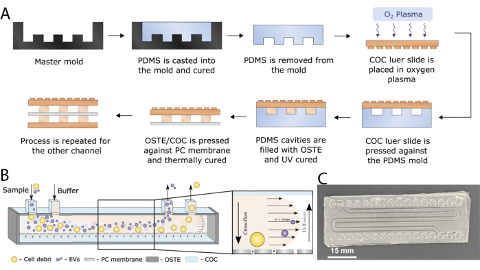
Figure 2: COC-OSTE microfluidic device. (A) Scheme of the different main steps of fabricating the OSTE-COC device. (B) Device working principle. (C) Image of the finished device. Scale bar: 15 mm. This figure has been modified with permission from Priedols et al.10 and Bajo-Santos et al.20. Please click here to view a larger version of this figure.
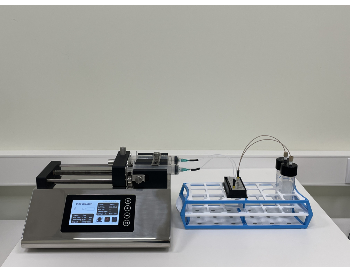
Figure 3: Experimental configuration of the device. The syringe pump is on the left, the OSTE-COC device is in the middle, and the recovery station is on the right. This figure has been modified with permission from Priedols et al.10 and Bajo-Santos et al.20. Please click here to view a larger version of this figure.
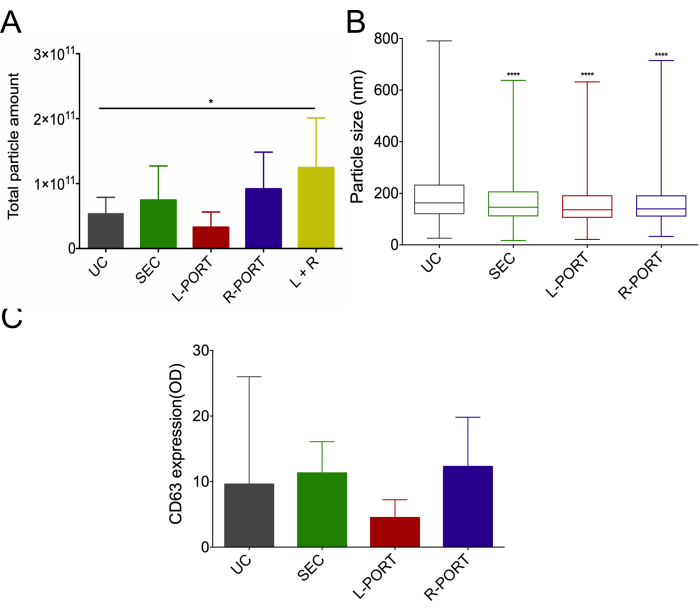
Figure 4: Urinary EV size distribution and particle recovery from 10 donors using ultracentrifugation (UC), size-exclusion chromatography (SEC), and the COC-OSTE device. (A) Particle amount recovered from each isolation method by nanoparticle tracking analysis (NTA). Data represented as mean +/- standard deviation. Statistical significance denoted by * (p<0.05). (B) Boxplots display the average particle size distribution among all urine samples assessed by NTA, with whiskers indicating the minimum and maximum values. P-values were derived from comparisons to UC, with **** indicating high statistical significance (p<0.0001). (C) The median and range of the average CD63 amount, assessed using double-sandwich enzyme-linked immunosorbent assay (dsELISA), for each isolation method, were calculated across all samples. L-port: Left port; R-port: Right port. This figure has been modified with permission from Bajo-Santos et al.20. Please click here to view a larger version of this figure.
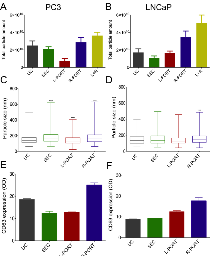
Figure 5: Characterization of EVs isolated from PC3 and LNCaP cells using different methods. (A) Particle amount recovered from PC3 media using nanoparticle tracking analysis (NTA) represented by mean+/- standard deviation. (B) Particle amount recovered from LNCaP cell media using NTA represented as mean +/- standard deviation. (C) Median particle size distribution of EVs from PC3 cultures, along with the range, was determined by NTA. (D) Median size distribution of EVs isolated from LNCaP cultures with range. (E) Median and range of CD63 expression for each isolation method for PC3 cell line-derived EV, using double-sandwich enzyme-linked immunosorbent assay (dsELISA). (F) Median and range of LNCaP-derived EV CD63 expression by dsELISA for each isolation method. UC: Ultracentrifugation; SEC: Size-exclusion chromatography; L-port: Left port; R-port: Right port. This figure has been modified with permission from Bajo-Santos et al.20. Please click here to view a larger version of this figure.
UC | SEC | COC-OSTE | |
Processing time/sample | ++/+++ | +++ | + |
Throughput | ++ | + | +++ |
Cost/sample | + | +++ | ++ |
Purity | + | +++ | ++ |
Automatization | ++ | + | +++ |
EV Yield | ++ | ++ | +++ |
Size selection | + | ++ | ++ |
Table 1. Comparison of isolation characteristics of EVs with the three methods UC SEC, and the COC-OSTE device. UC: Ultracentrifugation; SEC: Size-exclusion chromatography. COC-OSTE: cyclic olefin copolymer-off-stoichiometry thiol-ene. +: low; ++: medium; +++: high. * Device dependent.
0.1 µm carboxylate FluoSpheres | Invitrogen | #F8803 | Stock concentration: 3.6 x 10¹³ beads/mL (LOT dependent) |
0.5 mL microcentrifuge tubes | Starstedt | 72.704 | |
1 mL Luer cone syringe single use without needle | RAYS | TUB1ML | |
1.0 µm polystyrene FluoSpheres | Invitrogen | #F13083 | Stock concentration: 1 x 10¹⁰ beads/mL (LOT dependent) |
10 mL Serological pipettes | Sarstedt | 86.1254.001 | |
15 mL (100k) Amicon Ultra centrifugal filters | Merck Millipore | UFC910024 | |
2.0 mL Protein LoBind tubes | Eppendorf | 30108132 | |
20 mL syringes | BD PlastikPak | 10569215 | |
250 µm ID polyether ether ketone tubing | Darwin Microfluidics | CIL-1581 | |
3 kDa MWCO centrifugal filter units | Merck Millipore, | UFC200324 | |
5 mL Medical Syringe without Needle | Anhui Hongyu Wuzhou Medical | 159646 | |
50 mL conical tubes | Sarstedt | 62.547.254 | |
70 Ti fixed angle ultracentrifuge rotor | Beckman Coulter | 337922 | |
800 µm ID polytetrafluoroethylene tubing | Darwin Microfluidics | LVF-KTU-15 | |
96 well microplate, f-bottom, med. binding | Greiner Bio-One | 655001 | ELISA plate |
B-27 Supplement (50x), serum free | Thermo Fisher Scientific | 17504044 | |
Bovine serum albumin | SigmaAldrich | A7906-100G | |
COC Topas microscopy slide platform | Microfluidic Chipshop | 10000002 | |
COC Topas microscopy slide platform 2 x 16 Mini Luer | Microfluidic Chipshop | 10000387 | |
Elveflow OB1 pressure controller | Elvesys Group | ||
Luer connectors | Darwin Microfluidics | CS-10000095 | |
Mask aligner Suss MA/BA6 | SUSS MicroTec Group | ||
Mixer Thinky ARE-250 | Thinky Corporation | ||
NanoSight NS300 | Malvern Panalytical | NS300 | nanoparticle analyzer |
Optical microscope Nikon Eclipse LV150N | Nikon Metrology NV | ||
OSTE 322 Crystal Clear | Mercene Labs | ||
PBS TABLETS.Ca/Mg free. Fisher Bioreagents. 100 g | Fisher Scientific | BP2944-100 | |
PC membrane (50 nm pore diameter, 11.8% density) | it4ip S.A., Louvain-La Neuve, Belgium | ||
Petri dishes, sterile | Sarstedt | 82.1472.001 | |
Plasma Asher GIGAbatch 360 M | PVA TePla America, LLC | ||
qEVoriginal/35 nm column | Izon | SP5 | SEC column |
QSIL 216 Silicone Elastomer Kit | PP&S | ||
Resin Tough Black | Zortrax | ||
SW40 Ti swing ultracentrifuge rotor | Beckman Coulter | 331301 | |
Syringe pump | DK Infusetek | ISPLab002 | |
T175 suspension flask | Sarstedt | 83.3912.502 | |
TIM4-Fc protein | Adipogen LifeSciences | AG-40B-0180B-3010 | |
TMB (3,3',5,5'-tetramethylbenzidine) | SigmaAldrich | T0440-100ML | Horseradish peroxidase substrate |
Tween20 | SigmaAldrich | P1379-100ML | |
Ultracentrifuge Optima L100XP | Beckman Coulter | ||
Ultrasonic cleaning unit P 60 H | Elma Schmidbauer GmbH | ||
Universal Microplate Spectrophotometer | Bio-Tek instruments | 71777-1 | |
Urine collection cup, 150mL, sterile | APTACA | 2120_SG | |
Whatman Anotop 25 Syringe Filter | SigmaAldrich | 68092002 | |
Zetasizer Nano ZS | Malvern Panalytical | dynamic light scattering (DLS) system | |
Zortrax Inkspire | Zortrax |
Extracellular vesicles (EVs) hold immense potential for various biomedical applications, including diagnostics, drug delivery, and regenerative medicine. Nevertheless, the current methodologies for isolating EVs present significant challenges, such as complexity, time consumption, and the need for bulky equipment, which hinders their clinical translation. To address these limitations, we aimed to develop an innovative microfluidic system based on cyclic olefin copolymer-off-stoichiometry thiol-ene (COC-OSTE) for the efficient isolation of EVs from large-volume samples in a continuous manner. By utilizing size and buoyancy-based separation, the technology used in this study achieved a significantly narrower size distribution compared to existing approaches from urine and cell media samples, enabling the targeting of specific EV size fractions in future applications. Our innovative COC-OSTE microfluidic device design, utilizing bifurcated asymmetric flow field-flow fractionation technology, offers a straightforward and continuous EV isolation approach for large-volume samples. Furthermore, the potential for mass manufacturing of this microfluidic device offers scalability and consistency, making it feasible to integrate EV isolation into routine clinical diagnostics and industrial processes, where high consistency and throughput are essential requirements.