The described cellular assay is designed for the identification of CXC chemokine receptor 4 (CXCR4)-interacting agents that inhibit or stimulate, either competitively or allosterically, the intracellular Ca2+ release initiated by CXCR4 activation.
G protein-coupled receptors (GPCRs) are of great importance to the pharmaceutical industry as they are involved in many human diseases and include well-validated targets for therapeutic intervention. Discovery of lead compounds, including small synthetic molecules, that specifically inhibit the receptor's function, is an important initial step in drug development and relies on sensitive, specific, and robust cell-based assays. Here, we describe a kinetic cellular assay with a fluorescent readout primarily designed to identify receptor-specific antagonists that inhibit the intracellular Ca2+ release evoked upon the activation of the CXC chemokine receptor 4 (CXCR4) by its endogenous ligand, the CXC chemokine ligand 12 (CXCL12). A key advantage of this method is that it also enables screening of compounds endowed with intrinsic agonistic properties (i.e., compounds eliciting an increase in intracellular Ca2+ concentration in the absence of CXCL12) or compounds modulating the receptor's function via interaction with allosteric binding sites (i.e., positive and negative allosteric modulators (PAMs and NAMs, respectively)). On the down side, autofluorescent compounds might interfere with the assay's readout, thereby hampering reliable data interpretation. Most likely this assay can be implemented, with minimal adaptations, as a generic drug discovery assay for many other GPCRs of which the activation leads to a release of intracellular Ca2+.
GPCRs are an important superfamily of cell surface proteins that activate signal transduction cascades upon extracellular ligand binding. They can be activated by a large variety of stimuli including peptides, protein hormones, biogenic amines, and lipids, which results in the initiation of diverse intracellular signaling pathways and eventually biological responses1,2. Furthermore, GPCRs are involved in many, if not all, developmental and physiological processes and many human diseases are associated with dysfunctional GPCR signaling or receptor overexpression. GPCRs are therefore amongst the most validated pharmacological targets in medicine1,3.
Typically, a GPCR drug discovery workflow starts with cellular screening assays enabling the identification of compounds such as small molecules, monoclonal antibodies, and peptides that can modulate the activity of a particular GPCR. In GPCR drug discovery many different types of assays exist to search for such compounds, most of which are compatible with mid- to high-throughput screening campaigns. The most used assays include receptor binding experiments, fluorescence or luminescence based assays detecting fluctuations in the level of so-called secondary messengers (e.g., Ca2+, cyclic adenosine monophosphate (AMP)), phenotypic screening assays, and β-arrestin recruitment assays4. The choice for a particular type of assay may depend on multiple factors, but is also determined by prior knowledge concerning the signaling properties of a given GPCR. Agonist binding to a GPCR induces a conformational change catalyzing the exchange of guanidine diphosphate (GDP) for guanidine triphosphate (GTP) on the α-subunit of heterotrimeric G proteins. Subsequently the Gα-GTP subunit dissociates from the Gβγ subunit and both subunits will initiate further signaling pathways. Hydrolysis of the GTP-molecule and subsequent re-association of the Gα-GDP and Gβγ subunits will restore the G protein into its inactive conformation5,6. Based on sequence similarity of the Gα subunit different types of G proteins are defined (Gs, Gi, Gq, G12/13)7. Signaling via the Gα subunit gives rise to several typical responses such as the increase (via Gs) or decrease (via Gi) of cyclic AMP production and intracellular Ca2+ mobilization (via Gq)5,7. Gβγ subunits are also able to induce intracellular effector pathways. For instance, upon activation of Gi-coupled GPCRs, Gβγ can directly stimulate phospholipase C (PLC-β) to produce inositol triphosphate (IP3) that triggers the release of Ca2+ from intracellular stores7. Following receptor activation, GPCRs are phosphorylated by GPCR kinases (GRKs) which promotes interaction with β-arrestins. This process terminates G protein signaling and leads to receptor desensitization and eventually internalization. β-arrestins are also able to form multi-molecular complexes that can trigger other signaling pathways independent of G protein signaling8.
Within the subfamily of chemokine receptors, the Gi-coupled CXCR4 is a GPCR that has raised much interest as a promising target for drug discovery. Given its established role as a major co-receptor for human immunodeficiency virus 1 (HIV-1) viral entry and infection, compounds targeting CXCR4 were initially developed as anti-HIV drug candidates9. More recently, a growing body of evidence has pointed to an important role for CXCR4 in tumorigenesis and cancer metastasis making it a well-validated therapeutic target in oncology as well10. CXCR4 is highly expressed in more than twenty types of human cancer and controls tumor cell survival, proliferation, and migration as well as tumor-related angiogenesis10. CXCR4 antagonists of different chemical classes have previously been described11,12, but only the small molecule AMD3100 is currently approved for use in the clinic as a stem cell mobilization agent used during treatment of lymphoma and myeloma patients13,14. Clinical trials are ongoing to evaluate the safety and efficacy of several other CXCR4 antagonists in different human diseases, but with a strong focus on oncology12. Given the many potential applications for CXCR4 antagonists, the search for novel compounds with improved pharmacokinetic properties, improved bioavailability, or potentially less side effects is warranted.
Herein, a kinetic fluorescence-based cellular assay primarily used to screen for compounds capable of inhibiting CXCR4 is described. The fluorescent measurement of this method is based on the transient increase of the intracellular Ca2+ concentration evoked upon CXCR4 activation by its endogenous agonist, the chemokine ligand CXCL12 (formerly known as stromal cell derived factor 1α (SDF1-α)), and the potential inhibition of this CXCL12-induced Ca2+ response by particular compounds. In this assay, U87 human glioblastoma cells stably expressing the human CXCR4 receptor are used. At the same time, these cells lack endogenous expression of CXCR7, a related chemokine receptor that also binds CXCL1215,16,17. CXCR7 has previously also been shown to be capable of forming heterodimers with CXCR4, thereby modulating the signaling properties of this latter receptor18. Fluctuations in the level of intracellular Ca2+ mediated by CXCR4 are monitored by loading the CXCR4+ cells with fluo-2 acetoxymethyl (AM) ester, a cell-permeable high affinity fluorescent Ca2+-binding dye. Fluo-2 AM is a single wavelength fluorescent molecule that can be excited at 490 nm while its emission fluorescence is measured at 520 nm. This emission fluorescence increases upon Ca2+ binding, with a large dynamic range between the Ca2+-bound and unbound state. The increase in fluorescent signal is transient, occurring within a time interval of a few minutes, and will decay afterwards. The peak height of the fluorescent emission further correlates with the level of receptor activation. The assay itself is performed using a fluorescence microplate reader equipped with an Intensified CCD (ICCD) camera that possesses an integrated pipetting system that allows standardization of the pipetting steps in the assay (see Table of Materials). In addition, the simultaneous measurement of the fluorescent signal in all wells of a microplate is another key advantage of the fluorescence reader that is used. During the first part of the assay the compounds under investigation (e.g., a panel of small molecules at a fixed concentration or in a dilution series) are added to the fluo-2 AM loaded CXCR4+ U87 cells followed by a ~ 10 min incubation period during which the potential agonistic effect of the compounds is continuously measured in real time. Then, the endogenous agonist (i.e., CXCL12) is added to the cells to evoke a CXCR4-mediated transient increase in the level of intracellular Ca2+. During this part of the assay the potential antagonistic activity of the tested compounds can be evaluated. A schematic overview of the assay's general workflow is presented in Figure 1.
Although this Ca2+ mobilization assay has primarily been used to identify and determine the inhibitory potency of competitive CXCR4 antagonists (i.e., compounds that prevent the endogenous agonist to bind and stimulate the receptor), it also can identify receptor agonists and, in addition, compounds that exert their function by binding at allosteric sites (i.e., sites that topographically differ from the orthosteric binding site occupied by the endogenous agonist). Examples of the latter category of compounds include allosteric agonists and PAMs and NAMs19,20. Whereas receptor-specific antagonists as well as NAMs would inhibit the CXCL12-induced Ca2+ response, PAMs would enhance this response (see also Discussion section). Although the assay described herein specifically targets CXCR4, it is anticipated that this method can be applied to other GPCRs with minimal optimization effort, at least if they signal via the release of intracellular Ca2+.
NOTE: All steps described under sections 1 and 2 are carried out under sterile conditions in a laminar flow cabinet.
1. Maintenance of U87.CD4.hCXCR4 Cells
2. Seeding of the Cells for the Ca2+ Mobilization Assay
3. Loading of the Cells with a Fluorescent Ca2+ -sensitive Dye
4. Preparation of 96-well Polypropylene Plates Containing the Chemokine Ligand CXCL12 or the Compounds under Investigation
5. Protocol Settings on the Fluorescence Microplate Reader
NOTE: The fluorescence microplate reader used in this protocol is referred to in the Table of Materials.
6. Running the Fluorescence Assay
7. Data Analysis and Quality
The effect of CXCL12 stimulation on the intracellular Ca2+ mobilization in U87.CD4.CXCR4+ and U87.CD4 cells was evaluated with the Ca2+ mobilization assay. Instead of 20 µL of test compound that would normally be added during the first pipetting step of the protocol (Figure 1), assay buffer was added to the fluo-2 AM loaded U87.CD4.CXCR4+ cells in the measurement plate. During the second dispensing step, different concentrations of CXCL12 (0.2-102.4 nM, final concentration) were dispensed in the measurement plate. A dose-dependent increase in fluorescence, correlating with a dose-dependent increase of the release of Ca2+, is demonstrated (Figure 2A). Here, the negative control sample represents those wells in which at both additions only assay buffer was added to the measurement plate (i.e., 0 nM CXCL12), resulting in the absence of a response (Figure 2A). Increasing amounts of CXCL12 induce increased levels of fluorescence that decay over time (Figure 2A). From these data, a dose response curve was generated based on the "Max-Min response over baseline" between measurement 84 (i.e., the first measurement after CXCL12 addition) and measurement 243 (i.e., the final measurement in the protocol). Using nonlinear regression, the EC50 value (i.e., the concentration needed to evoke the half maximal response) was determined and corresponds to 1.14 nM (Figure 2B). No fluorescent Ca2+-related response was evoked by CXCL12, even at high concentration (102.4 nM), when U87.CD4 cells lacking functional CXCR4 expression were used. This demonstrates the receptor-specificity of the measurement evoked by CXCL12 (Figure 2C).
A key application for this cell-based assay is the identification of small molecules specifically targeting CXCR4 and capable of inhibiting the CXCL12-induced Ca2+ mobilization. If active compounds ("hits") are identified, they can be further characterized by testing serial dilutions of the compound and analyzing the inhibitory potency in more detail. As an example, the effect of the bicyclam AMD3100, a well-established small molecule CXCR4-specific receptor antagonist with potent anti-HIV-1 activity21,22, is illustrated in Figure 3. A concentration series of AMD3100 (n = 4, 3 µM down to 1.37 nM final concentration in a 1/3 dilution series) was dispensed on U87.CD4.CXCR4+ cells during the first step of the protocol. The compound was then allowed to incubate with the cells for ~ 10 min during which the fluorescence signal was recorded continuously. Then 6.4 nM of CXCL12 (50 ng/mL, final concentration) was added to all wells of the cell plate simultaneously to induce the CXCR4-mediated Ca2+ mobilization. Dose-dependent inhibition of this response by the different dilutions of AMD3100 is shown in Figure 3A. In this case, only the part of the graph following addition of CXCL12 is shown. As a negative control (blue line) only buffer was applied in the assay (no pre-incubation with AMD3100, no CXCL12 added to the wells), resulting in the expected lack of response. The positive control (red line) corresponds to the samples in which 6.4 nM CXCL12 was added to wells without prior incubation of AMD3100 (Figure 3A). Based on these defined negative and positive controls the Z' value in this assay typically is between 0.5 and 1. In order to determine the inhibitory potency of AMD3100 a dose-response curve was generated by non-linear regression resulting into a calculated IC50 value of 770.8 nM (Figure 3B). To further illustrate the behavior of a non-active compound, maraviroc was included in this experiment. Maraviroc is a small molecule specifically inhibiting the CC chemokine receptor 5 (CCR5) thereby blocking (CCR5)-tropic HIV-1 infection23 . Even at high concentration (10 µM final concentration) no inhibitory effect of this compound was observed on the CXCR4-mediated Ca2+ response (Figure 3A).
Finally, to demonstrate that this Ca2+ mobilization assay can also be applied to study other GPCRs, a serial dilution of the CC chemokine ligand 5 (CCL5), the endogenous ligand for CCR5, was added to cells expressing CCR5 (U87.CD4.CCR5+) using exactly the same experimental conditions and hardware settings. A dose-dependent fluorescent Ca2+ response is demonstrated after addition of a serial dilution of CCL5 (Figure 3C). In addition, and in contrast to its effect on CXCR4, pre-incubation with 100 nM (final concentration) of maraviroc strongly inhibited this response (Figure 3D).
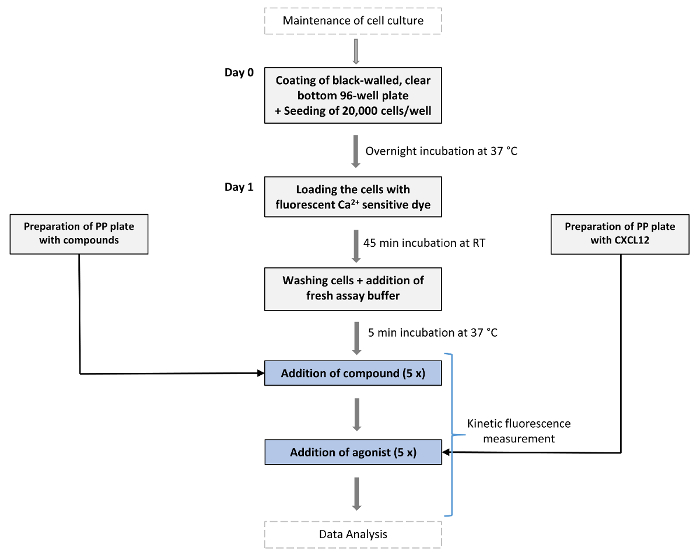
Figure 1: Schematic overview of the assay's workflow. On day 0 cells expressing the GPCR of interest (in this case CXCR4) are seeded in black-walled 96-well plates with clear bottom and are grown overnight at 37 °C and 5% CO2. At day 1 the fluorescence-based Ca2+ assay is performed. Cells are first loaded with a fluorescent Ca2+-sensitive dye (fluo-2 AM) and incubated for 45 min at RT in the dark. A "chemokine plate" and "compound plate" are prepared (PP = Polypropylene). After incubation with loading dye, seeded cells are washed once with assay buffer (150 µL/well) after which 80 µL/well of assay buffer is added. All plates are then incubated for 5 min in the device at 37 °C before starting the assay. Then, compounds of interest (e.g., small molecules) are dispensed at the desired concentration into the wells of the measurement plate and allowed to incubate for ~ 10 min while fluorescence is continuously measured. Next, a fixed concentration of the endogenous agonist of the GPCR (here CXCL12) is added to evoke Ca2+ release and fluorescence is further recorded over time. Please click here to view a larger version of this figure.
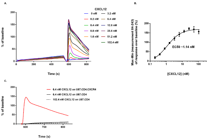
Figure 2: Agonist activity of CXCL12 on CXCR4+ cells and specificity of the detected response. (A) The dose-dependent effect of CXCL12 on the Ca2+ mobilization in U87.CD4.CXCR4+ cells. (B) Based on the Max-Min response over baseline between measurement 84 and 243 a dose-response curve was generated and the EC50 value calculated (n = 4, mean ± SD). (C) No response was induced by CXCL12 when it was dispensed on U87.CD4 cells lacking CXCR4. Please click here to view a larger version of this figure.
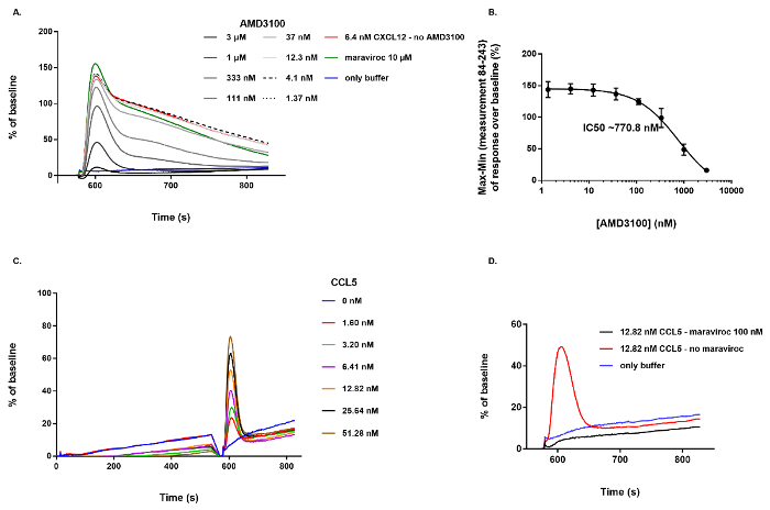
Figure 3: Illustration of the effect of a CXCR4 antagonist (AMD3100) and a non-active compound (maraviroc) on CXCR4+ cells and activation of CCR5 by its endogenous agonist, CCL5. (A) Dose-dependent inhibitory effect of AMD3100 on the Ca2+ mobilization evoked by adding 6.4 nM CXCL12 to U87.CD4.CXCR4+ cells. Maraviroc (at 10 µM final concentration) showed no inhibitory effect on the CXCL12-induced Ca2+ response. (B) Based on the Max-Min response over baseline between measurement 84 and 243, an inhibitory dose-response curve was generated and the IC50 value was calculated to be 770.8 nM (n = 4, mean ± SD). (C) Dose-dependent activation of CCR5 by its endogenous agonist, CCL5. (D) Inhibition of the CCR5-mediated Ca2+ response by pre-incubation of the cells with 100 nM of maraviroc. Please click here to view a larger version of this figure.
The Ca2+-mobilization assay described herein has previously been shown to be a valuable tool to identify and characterize receptor antagonists targeting CXCR417. It is, however, anticipated that this method can be more generally applied to a large group of other GPCRs that trigger a cytosolic Ca2+ release upon their activation, as illustrated for the related chemokine receptor CCR5. Whereas in the case of CCR5 exactly the same experimental conditions could be applied, several steps in the protocol (e.g., the cell number at plating, loading of the cells with a fluorescent dye) will need to be re-optimized before transferring the assay to other GPCRs in order to obtain a favorable signal-to-noise ratio. An important issue here is also the selection of a suitable in vitro cellular host allowing high-level expression of the GPCR of interest and functional coupling to the Ca2+ pathway. In case of CXCR4, human glioblastoma U87 cells were chosen because of: (1) the lack of endogenous expression of CXCR7 (a related chemokine receptor that occupies the same agonist as CXCR4), (2) their ability to couple CXCR4 activation to the Ca2+-signaling pathway, (3) the favorable dynamic range of the fluorescent response obtained after loading of the cells with the Ca2+-sensitive dye, and (4) their excellent adherence to the assay plates minimizing cell disturbance throughout the procedure. All of these parameters might need to be experimentally determined for each novel GPCR of interest and each cellular expression system.
When setting up a similar assay for another GPCR, several alternative steps or reagents in the protocol can be considered. For instance, other Ca2+-sensitive fluorophores (e.g., fluo-4, fluo-3) might be used as an alternative for the fluorophore (fluo-2) used in this protocol. In case of loosely adhering cells, washing of the cells might be omitted by introducing no-wash dyes to minimize cell dislodging24. In addition, excluding the washing step from the protocol would further increase the assay's throughput. Although this assay might also be performed with cells showing endogenous expression of CXCR4, for instance including several types of human cancer cell lines10,25, it should be noted that with stably transfected cell lines high GPCR expression levels that remain stable over time can be obtained. This will result in more pronounced assay measurements, a larger window for reliable data interpretation, and consistent assay performance over longer periods. When using cells endogenously expressing CXCR4 it will be much more difficult to achieve this result.
A major drawback of the assay is that it relies on a fluorescence measurement. Hence, autofluorescent compounds can interfere with the assay's measurement, which excludes them from analysis due to unreliable data interpretation. Also, this Ca2+ mobilization assay is ideally performed using a cellular screening system equipped with an integrated pipetting system that allows standardized mixing and dispension of compounds and receptor agonist into the measurement plate in all wells simultaneously. The assay could, however, be adapted for use with other, less expensive fluorescence microplate readers with kinetic measurement capabilities. The addition of compounds and agonist would then need to be performed manually using multichannel pipettes, which will increase hands-on-time and lowers the assay's throughput. Furthermore, obtaining a similar number of fluorescence measurements within a given time interval of a kinetic assay would be difficult to achieve as many microplate readers measure signals well-by-well whereas high-end screening systems measure all wells simultaneously.
Receptor antagonists preventing binding to the receptor and subsequent activation by the endogenous agonist (CXCL12) currently form the main category of compounds specifically targeting CXCR411,12. AMD3100, the small molecule that was used to illustrate the performance of the Ca2+ mobilization assay, is one of the most prominent examples of CXCR4 antagonists26. Besides receptor antagonists, molecules that regulate GPCR signaling differently raise general interest as well. These molecules include inverse agonists, as well as GPCR PAMs and NAMs19,20,27. An interesting feature of the assay described in this manuscript is that it not only can identify receptor antagonists, but also agonists and allosteric modulators. However, some minor adaptations to the assay and limitations need to be taken into account.
PAMs and NAMs bind to receptor sites topographically distinct from the orthosteric binding site occupied by the receptor's endogenous ligand. Whereas PAMs enhance the potency and/or efficacy of the receptor's endogenous ligand(s), NAMs inhibit the potency and/or efficacy of the endogenous ligand(s)19,20. PAMs have no intrinsic agonist activity. They are only active in the presence of the endogenous agonist, unlike so-called allosteric agonists. Because allosteric GPCR modulators would evoke less side effects than classical receptor antagonists when applied in clinical settings28,29, they are valuable pharmacological tools. In our assay, allosteric agonists would evoke a transient increase of the fluorescence signal immediately upon addition to the CXCR4+ cells. Receptor specificity of this signal should then be determined by testing the same compound with the same assay, but on cells lacking CXCR4. When specifically screening for PAMs, a lower concentration of endogenous agonist should be used in the assay to induce a fluorescent Ca2+ response. Although this lower amount of agonist will generate a smaller fluorescent signal, it leaves a larger window to detect additional receptor stimulation. Typically, an agonist concentration corresponding to the EC10-EC30 value of receptor stimulation is chosen30,31. Addition of a PAM during the first part of the assay would not result in an increased fluorescent measurement. However, the fluorescent Ca2+ signal after subsequent stimulation of the receptor with its endogenous agonist would increase. Similarly, a NAM does not alter the fluorescent measurement before agonist addition, but does inhibit the receptor's response after addition of the agonist. Hence, the activity profile of a receptor antagonist and a NAM will look similar. They both will result in the inhibition of the fluorescent response after agonist addition. Therefore, further experimental studies are required to discriminate between an antagonist and a NAM. Here, receptor binding studies whereby unlabeled compounds compete with a fixed amount of labeled CXCL12 for binding at CXCR417,32 can be a valuable strategy to discriminate between an antagonist, which would prevent the labeled agonist from binding to the receptor, and a NAM that would not as it would not share the same receptor binding site.
Ligand independent (or basal or constitutive) activity of GPCRs has been observed for numerous GPCRs33 although the level of basal activity is generally rather low when GPCRs are expressed recombinantly27. Basal GPCR activity can be enhanced by naturally occurring mutations33 and a point mutation leading to increased basal CXCR4 activity has been described earlier34. By coupling this constitutively active CXCR4 mutant to the pheromone response pathway in yeast and by [35S]GTPγS binding experiments, it was shown that T140, a peptide-based CXCR4 antagonist with potent anti-HIV activity17,35 significantly decreased this basal activity and thus was shown to behave as an inverse agonist34. Of note, in a Fura-2 fluorescence-based Ca2+ mobilization assay the same authors observed a small transient decrease in Ca2+-related fluorescence following addition of T140 to cells expressing mutant CXCR4, suggesting a decrease of basal receptor activity34. However, when analyzing an additional panel of cyclic pentapeptide-based CXCR4 inverse agonists designed from T140, detection of reduced basal activity with a Ca2+ mobilization assay was less straightforward36. When the effect of T140 on cells expressing wild type CXCR4 was analyzed using the Ca2+ mobilization assay as described in this manuscript, no change in basal fluorescence signal was observed17. Furthermore, fluorescence fluctuations are fast and transient and increases in basal Ca2+ are, for instance, not observed in cells expressing constitutively active Gαq-coupled receptors4. Taken together, the effect of inverse agonists would be very difficult, if not impossible to identify and evaluate reliably with our assay.
The authors have nothing to disclose.
The authors would like to thank Eric Fonteyn and Geert Schoofs for excellent technical assistance. This work has been supported by the KU Leuven (grant no. PF/10/018), the Fonds voor Wetenschappelijk Onderzoek (FWO, grant no. G.485.08), and the Fondation Dormeur Vaduz.
Fluo-2 AM | Abcam | ab142775 | fluorescent Ca2+ sensitive dye |
Pluronic F-127 | Sigma | P2443-250G | pluronic acid |
Gelatin | Sigma | G9391 | |
AMD3100 | Sigma | A5602-5 mg | specific CXCR4 antagonist |
Maraviroc | kind gift of AnorMed | antiretroviral drug, CCR5 antagonist | |
Chemokine ligand CXCL12 | PeproTech | 300-28A | |
Chemokine ligand CCL5 | PeproTech | 300-06 | |
Fetal Bovine Serum (FBS) | Gibco (Life Technologies) | 10270-106 | |
Bovine Serum Albumin (BSA) | Sigma | A1933-25G | |
Dulbecco's Modified Eagle's Medium (DMEM) | Gibco (Life Technologies) | 41965-039 | |
HBSS (10 x), calcium, magnesium, no phenol red | Gibco (Life Technologies) | 14065-049 | |
HEPES (1 M) | Gibco (Life Technologies) | 15630-056 | |
Trypsin-EDTA (0,25 %), phenol red | Gibco (Life Technologies) | 25200-056 | |
Dulbecco's Phosphate Buffered Saline (DPBS) | Gibco (Life Technologies) | 14190-094 | |
Falcon tubes, 50 mL | Greiner Bio-One | 227 261 | |
Tissue culture flask (T75) | Corning | 353024 | |
Black plate, 96-well, clear bottom, with lid | Costar/Fisher Scientific | 10530753 | assay plate (96-well), for cell seeding |
Polypropylene (PP) plates | Thermo Scientific (VWR) | 732-2661 | plates used to prepare the compound plates and chemokine plates, round bottom |
FLIPR Tetra high throughput cellular screening system | Molecular Devices | Fluorescent plate reader with integrated pipettor head and ICCD camera | |
FLIPR Tetra LED Module 470 – 495 nm | Molecular Devices | 0200-6128 | Light emitting diodes for excitation of the fluorescent Ca2+ sensitive dye |
FLIPR Tetra Emission Filter 515 – 575 nm | Molecular Devices | 0200-6203 | emission filter compatible with the fluorescent dye |
FLIPR Tetra 96 Head | Molecular Devices | 0310-4536 | 96-well pipettor head, integrated within the fluorescent plate reader |
ScreenWorks | Molecular Devices | software package used for data analysis and visualization on the FLIPR Tetra | |
Vi-CELL | Beckman Coulter | cell viability analyzer | |
Corning CellBIND 96 Well Flat Clear Bottom Black Polystyrene Microplates, with Lid, Sterile | Corning | 3340 | Pre-coated 96-well assay plates that may represent an alternative for manual coating of the assay plate. |