Wet-spinning-based Molding Process of Gelatin for Tissue Regeneration
Summary
We developed and describe a protocol based on the wet spinning concept, for the construction of gelatin-based biomaterials used for the application of tissue engineering.
Abstract
This article presents an inexpensive method to fabricate gelatin, as a natural polymer, into monofilament fibers or other appropriate forms. Through the wet spinning method, gelatin fibers are produced by smooth extrusion in a suitable coagulation medium. To increase the functional surface of these gelatin fibers and their ability to mimic the features of tissues, gelatin can be molded into a tube form by referring to this concept. Examined by in vitro and in vivo tests, the gelatin tubes demonstrate a great potential for application in tissue engineering. Acting as a suitable filling gap material, gelatin tubes can be used to substitute the tissue in the damaged area (e.g., in the nervous or cardiovascular system), as well as to promote regeneration by providing a direct replacement of stem cells and neural circuitry. This protocol provides a detailed procedure for creating a biomaterial based on a natural polymer, and its implementation is expected to greatly benefit the development of correlative natural polymers, which help to realize tissue regeneration strategies.
Introduction
The latest development in tissue regeneration involves the application of tissue engineering, which represents a challenge for the improvement of new therapeutic strategies in medical treatments. For example, the limited potential of nervous system regeneration, following injury or disease, poses a significant health problem worldwide. Due to the complexity of pathophysiological processes associated with the nervous system, the use of traditional autograft or the implementation of stabilization surgery has been shown to offer benefits in functional outcomes, but there is no strong evidence for the effects of spinal fixation surgery1,2. The tissue at the damaged area is lost and replaced with hypertrophically induced astrocytes3, eventually forming a dense glial scar4,5. This matrix acts as a barrier that blocks the recovery of nerve function6,7 and is, thus, greatly hinders regeneration. Therefore, a suitable filling gap material is expected to prevent the loss of tissue and reduce the formation of scar-associated connective tissue by maintaining the integrity of the damaged area, as well as by providing the direct replacement of neural cells and circuitry to promote axon regeneration.
Polymeric biomaterials have been preferred as scaffolds for tissue regeneration therapy, based on the regulation of cell or axon behavior and tissue progression through natural extracellular matrix (ECM) support. The fiber format is commonly considered as a building block for various materials, owing to its one-dimensional structure8. The fibers can generally be obtained by melt extrusion or wet spinning method; however, the large size and cost of the equipment and the difficulty to perform these methods are challenging. In addition, the majority of the work related to polymer fibers has been focused on synthetic or composite materials. Natural polymers as a source of biomaterial offer better biocompatibility properties for the human body. Nonetheless, to obtain the alignment of natural polymer fibers is relatively more difficult than of synthetic polymer sources9. Hence, the conversion of a natural polymer as a rich source of protein into biomaterial fibers is an important strategy — not only can the biomaterial fibers be directly isolated from the raw material, thus avoiding an unnecessary transformation to monomers, but the protein fibers also have a good appearance and favorable characteristics10.
In this regard, we describe an inexpensive processing method for the manufacturing of natural polymer fibers through the basic concept of wet spinning, that can be implemented on the laboratory scale for tissue engineering. Wet spinning is performed by the extrusion and coagulation of a polymer solution into a suitable polymer nonsolvent. An appropriate, viscous solution doped into coagulation medium causes the polymer molecules to dissolve. Through the phase transition, the filaments then lose their solubility and are precipitated in the form of a solid polymer phase11. Referring to this concept, we then expanded the development of gelatin into the tube form by a molding process, which is considered proper for tissue regeneration application. In addition, intrinsically, we can also develop any shape of material from gelatin fibers (e.g., gelatin conduit rolled up from several gelatin fibers), for other desired applications.
Gelatin, a biodegradable natural polymer, is formed from denatured and hydrolyzed collagen, including any semicrystalline, amorphous, or triple helical state of collagen12. It is well known that collagen is the essential structural protein in all connective tissues of vertebrates and invertebrates13,14, which is similar to the protein structure of the main ECM that induces nerve growth and, simultaneously, replaces a large amount of glycosaminoglycan secreted during spinal cord injuries. Therefore, the use of gelatin as a source would be a great choice for any medical vehicle. Besides being an inexpensive source, gelatin is also biodegradable and cytocompatible and clinically proven to be a temporary defect filler15. Developed into a tube form, in vitro and in vivo tests described here demonstrate that gelatin has an excellent biocompatibility and suitability for future tissue engineering applications. Cultured with human adipose stem cells, gelatin tubes improve cell differentiation into neural progenitor cells by using positive nestin staining as a neural cell marker. Furthermore, gelatin as filling gap material, as produced by the method established in this study, is expected to be manageable and safe and to greatly benefit tissue engineers who are currently developing correlative natural polymers for the enhancement of tissue regeneration strategies.
Protocol
The fat tissues were obtained from orthopedic surgeries as certified by the Institutional Review Board of Tri-Service General Hospital, Taipei, Taiwan, R.O.C. Procedures involving animal subjects have been approved by the Animal Care Committee at National Defense Medical Center, Taiwan (R.O.C).
1. Wet Spinning Process
- Solution preparation
- Dissolve 5 g of gelatin powder in 100 mL of double-distilled water to obtain 5% (w/v) solution concentration.
- Stir the mixture slowly at 60 – 70 °C overnight to achieve a completely homogeneous dispersion without any bubbles.
- Wet spinning
- Fiber formation
NOTE: A schematic of the method is shown in Figure 1A. The spinning setup is equipped with a peristaltic pump machine (see Table of Materials) that provides high-precision speed and controls the smooth delivery of the flow.- Plug a 26 G x 1/2 inch (0.45 mm x 12 mm) syringe as the solution injector into clear vinyl tubing.
- Prepare 100 mL of 99.5% acetone solution in a beaker glass to be used as the coagulation bath.
- Run the peristaltic pump machine at 21 rpm (3.25 mL/min) and let the gelatin solution stand for several seconds in acetone solution before rolling it up.
- Take out the gelatin fibers from the acetone solution and immerse them into 2.5% (w/v) of polycaprolactone/dichloromethane (PCL/DCM) solution with 1:20 (w/w).
- Let the gelatin fibers in the PCL/DCM solution dry overnight, under the hood at room temperature.
- Tube formation
NOTE: A schematic of the method is shown in Figure 1B.- Use a peripheral venous catheter of 24 G x 3/4 inch (0.7 mm x 19 mm) as tube mold.
- Load the catheter into the gelatin solution and hold it there for 3 s.
- Load the catheter into the acetone solution and hold for 1 min.
- Load the catheter again into the gelatin solution and hold it there for 3 s.
- Repeat this alternate procedure 20x.
- Take out the catheter from the acetone solution and let the molded tube dry at room temperature for 5 min.
- Gently take out the gelatin tube from the catheter by using a hemostat and carefully transfer the gelatin tube into a glass Pasteur pipette with the immersion of 2.5% (w/v) of the PCL/DCM solution.
- Let the Pasteur pipette containing the gelatin tube and PCL/DCM solution dry overnight, under the hood at room temperature.
- Fiber formation
2. Morphology of the Gelatin Tube
- Mount the piece of dried gelatin tube on a carbon stub.
- Put the sample into the ion sputter coater machine (see Table of Materials) and coat the sample with gold for 60 s; then, observe its morphology by scanning electron microscopy(see Table of Materials).
3. Culture of Human Adipose Stem Cells
- Place the fat tissues (obtained from oral adipose tissue by clinical surgery) into a Petri dish containing 10 mL of transfer solution consisting of 0.1 M phosphate-buffered saline (PBS), 1% penicillin/streptomycin, and 0.1% glucose.
- Cut the tissues into small pieces (less than 1 mm2) with a scalpel blade.
- Transfer the tissues into a 15 mL plastic tube and centrifuge at 500 x g for 5 min.
- Remove the supernatant and incubate the sediment in a plastic tube containing 10 mL of Dulbecco's modified Eagle's medium (DMEM, consisting of 4 mM L-glutamine, 1 mM sodium pyruvate, and 15 mg/L phenol red) with 0.1% collagenase at 37 °C, in a humidified atmosphere containing 95% air and 5% CO2, for 1 day.
- Centrifuge the plastic tube at 500 x g for 5 min, discard the supernatant carefully (do not touch the sediment), and then, suspend the sediment gently by adding 10 mL of DMEM containing 10% fetal bovine serum (FBS) and let it stand for 1 day at 37 °C, in a humidified atmosphere containing 95% air and 5% CO2.
- Centrifuge the plastic tube at 500 x g for 5 min and discard the supernatant carefully; then, add 1 mL of stem cell medium (consisting of keratinocyte serum-free medium (K-SFM), 5% of FBS, N-acetyl-L-cysteine, ascorbic acid-2-phosphate, streptomycin, and amphotericin) to suspend the sediment, transfer it into a T25 culture flask containing 4 mL of stem cell medium, and let it stand for 3 days at 37 °C, in a humidified atmosphere containing 95% air and 5% CO2.
- Discard the supernatant, add 1 mL of 0.25% trypsin-ethylenediaminetetraacetic acid (EDTA), and incubate it at 37 °C, in a humidified atmosphere containing 95% air and 5% CO2 for 3.5 min, to detach the cells from the bottom of the culture flask.
- Add 1 mL of FBS, suspend the mixture, and transfer it to a microcentrifuge tube.
- Centrifuge the suspension at 500 x g for 3.5 min, suspend the sediment with 1 mL of stem cell medium, and transfer it to the culture flask containing 5 mL of stem cell medium; then, keep it at 37 °C in a humidified atmosphere containing 95% air and 5% CO2.
- Maintain the subculture by changing the stem cell medium every 3 days.
4. Cultivation of Cells on the Gelatin Tube
- Sterilize the gelatin tube with UV light for 2 h; then, immerse it in 75% (v/v) ethanol and wash it 2x with stem cell medium to remove the residual ethanol.
- Collect the human adipose stem cells (hASCs) from the culture flask.
- Remove the medium from the culture flask.
- Add 1 mL of 0.25% trypsin-EDTA and incubate at 37 °C in a humidified atmosphere containing 95% air and 5% CO2 for 3.5 min, to detach hASCs from the bottom of the culture flask.
- Add 1 mL of FBS, suspend the mixture, and transfer it to a microcentrifuge tube.
- Centrifuge the microcentrifuge tube at 500 x g for 3.5 min; then, discard the supernatant carefully, and suspend the sediment with 1 mL of stem cell medium.
- Place the gelatin tube in a 6-well plate containing 3 mL of stem cell medium and seed 4 x 10⁴ of hASCs to the tube; incubate them for 2 weeks at 37 °C, in a humidified atmosphere containing 95% air and 5% CO2.
- Change the stem cell medium every 3 days to provide enough nutrition for cell growth.
5. Immunocytochemistry
- Remove the stem cell medium and wash the well with PBS.
- Fix the tube with cells with 0.1% acetic acid glacial for 30 min at room temperature.
- Wash the well 2x with PBS.
- Add a surfactant (NP-40, 0.05%) and incubate for 10 min at room temperature.
- Wash the well 3x with PBS.
- Add 2% normal goat serum and incubate at room temperature for 30 min.
- Decant the solution, add primary antibody nestin (neural progenitor cell marker), and incubate overnight at 4 °C.
- Wash the well 3x with PBS.
- Add secondary antibody donkey anti-mouse-fluorescein isothiocyanate (FITC) and keep the sample in the dark for 2 h.
- Wash the well 3x with PBS.
- Add 1 mL of Hoechst 33342 to stain the nuclei and incubate for 10 min at room temperature.
- Wash the well gently and observe it under a fluorescence microscope.
6. In Vivo Biocompatibility Test
NOTE: Rats with a weight between 201 – 225 g have been successfully tested using this protocol.
- Anesthesia
- Induce and maintain anesthesia; preferably, anesthetize a rat (8-week-old Sprague Dawley rat, female) via an intramuscular injection of tiletamine and zolazepam (25 mg/kg + 25 mg/kg, respectively) and xylazine (5 mg/kg). Perform a pain stimulation test by pressing the rat's fingers to confirm the anesthetization.
- Sterilization of the surgical site
- Shave and clean the skin of rat’s trapezius area (over the back of the neck) and wipe it with povidone-iodine lotion to prepare the aseptic condition of skin (100 mg/mL).
- Cover the rat with sterile surgical drapes to reduce bacterial transfer and subsequent contamination of the surgical site.
- Implantation of the gelatin tube
- Gently cut the skin of rat’s trapezius area with a surgical scissor.
- Create a wound of 2 cm and place the gelatin tube directly on the layer between the fascia and the muscle.
- Postimplantation surgery
- Close the wound with nylon suture and wipe it with povidone-iodine solution (100 mg/mL).
- Induce the rat via an intramuscular injection of ketoprofen (2.5 mg/kg) and cefazolin (15 mg/kg).
- Keep the rat under aseptic conditions for 7 days.
- Euthanasia
- Rat is euthanized via CO2 asphyxiation in accordance with AVMA Guidelines for Euthanasia. Confirm the death of rat by toe and tail pinch, followed by chest touching to ensure the heartbeat.
- Cut the rat gently with a scalpel blade, remove the implanted tissue, and take photos for observation.
Representative Results
In this study, we successfully developed the gelatin into fibers (Figure 2A) and tubes (Figure 2B,C) through the user-friendly wet spinning concept. These gelatin-based materials can be utilized as any medical tool, depending on their shapes. Considering that the functional surface and frame of such materials are more suitable for tissue regeneration, we examined the biocompatibility of gelatin tube by performing in vitro and in vivo tests.
To obtain an overview of the gelatin tube, first, we conducted morphological observations by using scanning electron microscopy (Figure 3). The results showed that the surface of the gelatin tube was not very smooth (Figure 3A), its inner diameter was more than 200 µm (Figure 3B), and its thickness was approximately 20 µm (Figure 3C).
Then, immunocytochemistry was performed to examine the biocompatibility of the gelatin tube in vitro (Figure 4). Initially, the gelatin tube was incubated with human adipose stem cells for 2 weeks and was stained with nestin as a neural cell marker (Figure 4A). Under the fluorescence microscope, the staining showed the positive nestin staining marker (green color) along with a bunch of detected nuclei (blue color), which suggested that the cells could penetrate and adhere well to the gelatin tube and differentiate into neural progenitor cells (Figure 4B). Further, the in vivo biocompatibility test of the gelatin tube showed analogous results. The gelatin tube was inserted into the fascia layer in a rat's trapezius area for 7 days. The result demonstrates that the implantation of the gelatin tube into the fascia layer indicated its safety and great biocompatibility, showed no indication of redness, swelling, or other inflammation symptoms of the local tissue, and was surrounded by well-developed connective tissues (Figure 5A,B).
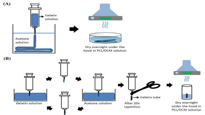
Figure 1: Scheme of the wet spinning setup. (A) Gelatin fibers. (B) Gelatin tube. Please click here to view a larger version of this figure.
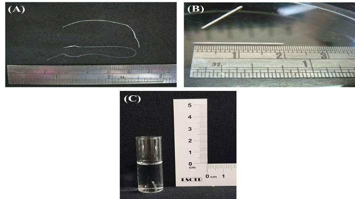
Figure 2: Bright view images of gelatin-based material obtained through the wet spinning method. (A) Gelatin fibers. (B) Gelatin tube. (C) Gelatin tube in PBS solution. Please click here to view a larger version of this figure.
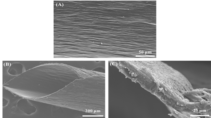
Figure 3: Morphology of the gelatin tube, observed by scanning electron microscopy. (A) A superficial view of the surface of the gelatin tube (Scale bar = 50 µm). (B) Inclined plane view (Scale bar = 200 µm). (C) Vertical view (Scale bar = 25 µm). Please click here to view a larger version of this figure.
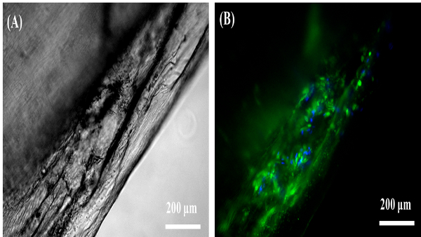
Figure 4: Observation of human adipose stem cell (hASC) growth on the gelatin tube. (A) Bright view image of gelatin tube-hASCs. (B) Under the fluorescence microscope, the staining showed the positive nestin staining marker along with a bunch of nuclei stained with Hoechst 33342.The gelatin tube provided an optimal environment for hASCs to adhere and grow inside the tube (Scale bar = 200 µm; blue color = nuclei; green color = nestin). Please click here to view a larger version of this figure.
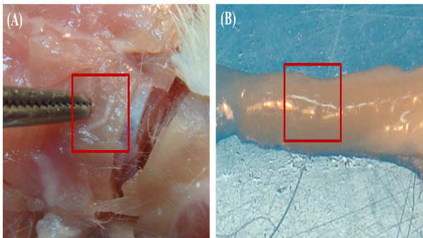
Figure 5: In vivo biocompatibility test of the gelatin tube. (A) The implantation of the gelatin tube into the fascia layer. (B) Observation of the local tissue environment implanted with the gelatin tube (red rectangle area) after 7 days. No redness, swelling, or other inflammation symptoms were observed, and the tube is surrounded by well-developed connective tissues 7 days after implantation. This is indicative of an excellent biocompatibility of the gelatin tube with the local tissue environment. Please click here to view a larger version of this figure.
Discussion
We presented the development of gelatin-based biomaterials by using a simple wet spinning technique that can be applied in the study of natural polymers for tissue regeneration. This work demonstrated the possibility of gelatin fabrication as a great protein source without the addition of other sources, with the aim to optimize the properties of gelatin itself. The development of gelatin-based biomaterials was entirely carried out in room temperature (22 – 26 °C). A gentle solution preparation is a critical step within the protocol, as is maintaining an exact solution concentration and stirring the solution very smoothly to avoid any bubbles. The appearance of bubbles in the solution, especially when loaded into the syringe, will affect the form of gelatin in the coagulation medium.
Gelatin-based biomaterials were obtained through the phase transition of the gelatin solution in a suitable coagulation medium that causes the desolvation of the polymer molecules and precipitates the gelatin solution into a solid polymer phase11. By employing this basic concept of the wet spinning method, we utilized the shape of the gelatin into tube form to increase the functionality of its surface and to mimic the features of human tissues through the molding process. In this case, the adequate times to load, hold, and repeat the steps are necessary to build up an exact tube form. In this protocol, the homogeneity of the tube shape during the process cannot be controlled, considering the asynchronously of the transition phase from the bottom to the up part of the tube. Additionally, the preserved and sterilization methods for this material are limited because of its protein content.
Examined by in vitro and in vivo tests, the current results demonstrated that the gelatin tubes created with this protocol exhibit an excellent biocompatibility and have great potential applicability in tissue engineering. The inner diameter of the gelatin tubes obtained in this study was more than 200 µm (Figure 3B), which allows the cells to pass through and easily adhere to the tubes' inner surface16. In addition, the thickness and mildly asperous surface of the tube provides a proper template for cell attachment (Figure 3A,C). Consistently, the immunocytochemistry results showed that the cells penetrated and adhered to the gelatin tube-particularly the neural stem cells, as observed through the positive staining of the neural cells with the marker nestin (Figure 4B). As the nestin marker was found in the area of the growth cones during the stages of axon extension, the positive staining of nestin is indicative of the cone growth17. Ultimately, this positive gelatin-hASCs combination can be implemented in the treatment of nervous system regeneration. The spinal cord is made up of groups of neuron axons and bundles of nerve fibers along the spinal vertebrae. Thus, the gelatin tube may not only provide appropriate filling gap material but will also guide nerve fiber growth and regeneration through the tube. Moreover, the great biocompatibility observed by the implantation of a gelatin tube into the fascia layer did not demonstrate any harmful and inflammation effect on the local tissues, and the tube was surrounded by well-developed connective tissues (Figure 5A,B).
In conclusion, we successfully developed a simple and useful protocol for the construction of gelatin-based biomaterials with excellent biocompatibility and cell environment that can be used in prospect tissue engineering applications, such as in nervous system regeneration, blood vessel replacement, urinary tract reconstruction, and in the rebuilding of organ parts. This protocol can be implemented by tissue engineers who are currently developing correlative natural polymers without any synthetic polymer addition. The protocol can be easily expanded upon and can be used to customize the design of specific biomaterials. In the future, this protocol can be adapted to produce protein-based biomaterials on a mass scale, thus helping to realize tissue regeneration.
Disclosures
The authors have nothing to disclose.
Acknowledgements
This study was supported by the Ministry of National Defense (MAB-105-070; MAB-106-077; MAB-107-032; MAB-107-065), the Ministry of Science and Technology (MOST 107-2320-B016-016), Tri-Service General Hospital, the National Defense Medical Center, Taiwan (TSGH-C106-046; TSGH-C106-115; TSGH-C107-041), and Cheng-Hsin General Hospital and National Defense Medical Center Cooperation (CH-NDMC-107-8).
Materials
| Solution preparation: | |||
| Gelatin type B (porcine) | Ferak | Art. -Nr. 10733 | 500 g vial |
| Wet spinning process: | |||
| Peristaltic pump | Gilson | Model M312 | Minipuls*3 |
| Plastic tube connector | World Precision Instruments | 14011 | 1 box |
| Syringe | Sterican | 5A06258541 | 26Gx1/2"(0.45 x 12mm) |
| Acetone | Ferak | Art. -Nr. 00010 | 2.5 L vial |
| Polycaprolactone CAPA 6500 | Perstorp | 24980-41-4 | – |
| Dichloromethane | Scharlau | CL03421000 | 1 L vial |
| Glass Pasteur pipette | Fisher Scientific | 13-678-20A | – |
| Hemostat | Shinetec instruments | ST-B021 | – |
| Peripheral venous catheter (Introcan Certo) | B. Braun | 1B03258241 | 24Gx3/4"(0.7 x 19mm) |
| Morphology of the gelatin tube: | |||
| Ion sputter coater machine | Hitachi | e1010 | – |
| Scanning electron microscopy | Hitachi | S-3000N | – |
| Cultivation of cells on the gelatin tube: | |||
| Trypsin-EDTA | Gibco | 488625 | 100 mL vial |
| Fetal bovine serum | Gibco | 923119 | 500 mL vial |
| Dulbecco's modified Eagle's medium | Gibco | 31600-034 | Powder |
| Keratinocyte-SFM medium | Gibco | 10744-019 | 500 mL vial |
| T25 culture flask | TPP | 90025 | VENT type |
| 6-well plate | Falcon | 1209938 | – |
| Immunocytochemistry: | |||
| Phospate-buffered saline | Gibco | 654471 | 500 mL vial |
| Acetic acid glacial | Ferak | Art. -Nr. 00697 | 500 mL vial |
| NP-40 surfactant (Tergitol solution) | Sigma | 056K0151 | 500 mL vial |
| Normal goat serum | Vector Laboratories | S-1000-20 | 20 mL vial, concentrate |
| Nestin (primary antibody) | Santa Cruz Biotechnology | SC-23927 | – |
| Donkey anti-mouse-fluorescein isothiocyanate (secondary antibody) | Santa Cruz Biotechnology | SC-2099 | – |
| Hoechst 33342 | Anaspec | AS-83218 | 5 mL vial |
| In vivo biocompatibility test: | |||
| Tiletamine+zolazepam | Virbac | BC91 | 5 mL vial |
| Xylazine | Bayer korea | KR03227 | 10 mL vial |
| Ketoprofen | Astar | 1406232 | 2 mL vial |
| Povidone-iodine solution | Everstar | HA161202 | 4 L barrel |
| Cefazolin | China Chemical & Pharmaceutical | 18P909 | 1 g vial |
| Scalpel blade | Shinetec instruments | ST-B021 | – |
| Surgical scissor | Shinetec instruments | ST-B021 | – |
References
- Bagnall, A. M., Jones, L., Duffy, S., Riemsima, R. P. Spinal fixation surgery for acute traumatic spinal cord injury. Cochrane Database of Systematic Reviews. 1, 004725 (2008).
- Fehlings, M. G., Perrin, R. G. The role and timing of early decompression for cervical spinal cord injury: update with a review of recent clinical evidence. Injury. 36, 13-26 (2005).
- Yang, L., Jones, N. R., Stoodley, M. A., Blumbergs, P. C., Brown, C. J. Excitotoxic model of post-traumatic syringomyelia in the rat. Spine. 26, 1842-1849 (2001).
- Rolls, A., et al. Two faces of chondroitin sulfate proteoglycan in spinal cord repair: a role in microglia/macrophage activation. PLoS Medicine. 5, 1262-1277 (2008).
- Properzi, F., Asher, R. A., Fawcett, J. W. Chondroitin sulphate proteoglycans in the central nervous system: changes and synthesis after injury. Biochemical Society Transactions. 31, 335-336 (2003).
- Fawcett, J. W., Asher, R. A. The glial scar and central nervous system repair. Brain Research Bulletin. 49, 377-391 (1999).
- Yang, Z., Mo, L., Duan, H., Li, X. Effects of chitosan/collagen substrates on the behavior of rat neural stem cells. Science China Life Sciences. 53, 215-222 (2010).
- Chawla, K. K. . Fibrous Materials. , (1998).
- Pickering, K. L., Aruan Efendy, M. G. A review of recent developments in natural fibre composites and their mechanical performance. Composites Part A-Applied Science and Manufacturing. 83, 98-112 (2016).
- Lundgren, H. P. Synthetic fibers made from proteins. Advances in Protein Chemistry. 5, 305-351 (1954).
- Radishevskii, M. B., Serkov, A. T. Coagulation mechanism in wet spinning of fibres. Fibre Chemistry. 37, 266-271 (2005).
- Yannas, I. V. Collagen and gelatin in the solid state. Journal of Macromolecular Science Part C Polymer Reviews. 7, 49-106 (1972).
- Baer, E., Cassidy, J. J., Hiltner, A. Hierarchical structure of collagen composite Systems: lessons from biology. Pure and Applied Chemistry. 6, 961-973 (2009).
- Harrington, W. F., Von Hippel, P. H. The structure of collagen and gelatin. Advances in Protein Chemistry. 16, 1-138 (1961).
- Veis, A. . The Macromolecular Chemistry of Gelatin. , (1994).
- Freyman, T. M., Yannas, I. V., Gibson, L. J. Cellular materials as porous scaffolds for tissue engineering. Progress in Materials Science. 46, 273-282 (2001).
- Michalczyk, K., Ziman, M. Nestin structure and predicted function in cellular cytoskeletal organization. Histology and Histopathology. 20, 665-671 (2005).
